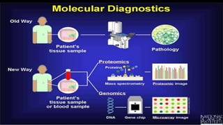
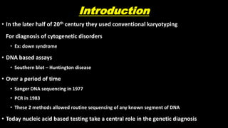
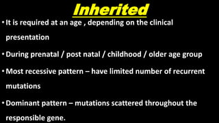
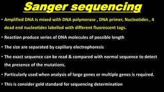
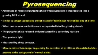
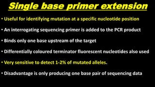
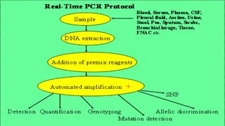
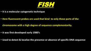
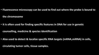
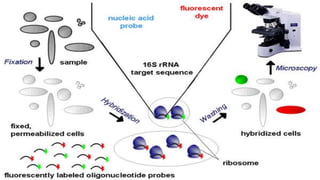
MOLECULARGENETICDIAGNOSIS
Dr.T.Arivazhagan
Post Graduate
Dept. Of Pathology
Introduction
• In the later half of 20th century they used conventional karyotyping
For diagnosis of cytogenetic disorders
• Ex: down syndrome
• DNA based assays
• Southern blot – Huntington disease
• Over a period of time
• Sanger DNA sequencing in 1977
• PCR in 1983
• These 2 methods allowed routine sequencing of any known segment of DNA
• Today nucleic acid based testing take a central role in the genetic diagnosis
Indications
1. Inherited
2. Acquired
Inherited
•It is required at an age , depending on the clinical
presentation
•During prenatal / post natal / childhood / older age group
•Most recessive pattern – have limited number of recurrent
mutations
•Dominant pattern – mutations scattered throughout the
responsible gene.
Prenatal indications
• Advanced maternal age
• Parent known to have balanced chromosomal rearrangements
• Fetal anomalies observed on ultrasound
• Routine maternal screening ( down syndrome)
• Cystic fibrosis
• Spinal muscular atrophy
• At present usually performed cells obtained by amniocentesis, chorionic villus
biopsy , umbilical cord blood
Post natal indications
• Multiple congenital anomalies
• Suspicion of metabolic syndrome
• Unexplained mental retardation
• Suspected aneuploidy
• Suspected monogenic disease
Older patients
• Inherited cancer syndrome
• Atypically mid monogenic disease
• Neurodegenerative disorders ( Familial Alzheimer disease, Huntington
disease)
Acquired conditions
• Diagnosis & management of cancer:
• Detection of tumor specific mutations
• Determination of clonality
• Identification of specific genetic alterations
• Determination of treatment efficacy
• Detection of drug resistant secondary malignancy
• Diagnosis & management of infectious disease:
• Detection of micro organism specific genetic material
• Identification of specific genetic alteration in the microbes
• Determination of treatment efficacy
Tests
• PCR & Detection of DNA sequence alterations:
• Sanger sequencing
• Pyro sequencing
• Single based primer extensions
• Restriction fragment length analysis
• Amplicon length analysis
• Real time PCR
Molecular analysis
• Fluorescence in situ hybridization
• Multiplex ligation dependent probe amplification
• Southern blot
• Cytogenetic array technology
• Array based comparative genomic hybridization
• SNP genotyping arrays
• RNA analysis
• Next generation sequencing
• Bioinformatics
• Clinical applications
• Targeted sequencing
• Whole exome sequencing
• Whole genome sequencing
PCR
• PCR is a technique used in molecular biology to amplify a single copy
or a few copies of a segment of DNA across several orders of
magnitude , generating thousands to millions of copies of a particular
DNA sequence.
• Develop in 1983 by kary mullis
• Using appropriate heat stable DNA polymerase ,Thermal cycling
• The target DNA ( less than 1000 base pairs) are amplified.
Sanger sequencing
• Amplified DNA is mixed with DNA polymerase , DNA primer, Nucleotides , 4
dead end nucleotides labelled with different fluorescent tags.
• Reaction produce series of DNA molecules of possible length
• The size are separated by capillary electrophoresis
• The exact sequence can be read & compared with normal sequence to detect
the presence of the mutations.
• Particularly used when analysis of large genes or multiple genes is required.
• This is consider gold standard for sequencing determination
Pyrosequencing
• Advantage of release of pyrophosphate when nucleotide is incorporated into a
growing DNA strand.
• Similar to sanger sequencing except instead of terminator nucleotides one at a time
• When one or more nucleotides are incorporated into the growing strands
• The pyrophosphate released and participated in a secondary reaction
• That produce light
• Measured by photo detector.
• More sensitive than sanger sequencing for detection of as little as 5% mutated alleles
present in the background of normal alleles.
Single base primer extension
• Useful for identifying mutation at a specific nucleotide position
• An interrogating sequencing primer is added to the PCR product
• Binds only one base upstream of the target
• Differentially coloured terminator fluorescent nucleotides also used
• Very sensitive to detect 1-2% of mutated alleles.
• Disadvantage is only producing one base pair of sequencing data
Restriction fragment length analysis
• Advantage of digestion of DNA with endo nucleases that recognize
and cut DNA at specific sequences.
• Amplified PCR product may be digested and normal and mutated
PCR products yield different size fragments
• These different bands are identified by electrophoresis
• Less comprehensive than other methods
Amplicon length analysis
• Here 2 primers that flank the region containing trinucleotide repeats at
5’end
• Amplification done
• Large difference in the number of repeats from the normal individual and
mutated individual
• This is easily distinguished by gel electrophoresis
Real time PCR
• Variety of PCR based technologies
• Use fluorophore indicators can detect and quantify the presence of
particular nucleic acid sequences in real time
• Used to monitor the frequency of cancer cells bearing characteristic
genetic lesion
• Also used to detect the somatic point mutation in oncogenes
• Advantage is avoiding the need for post PCR analysis
MOLECULAR
ANALYSIS OF
GENOMIC
ALTERATIONS
FISH
• It is a molecular cytogenetic technique
• Here fluorescent probes are used that bind to only those parts of the
chromosome with a high degree of sequence complementarity.
• It was first developed early 1980’s
• Used to detect & localize the presence or absence of specific DNA sequence
• Fluorescence microscopy can be used to find out where the probe is bound to
the chromosome
• It is often used for finding specific features in DNA for use in genetic
counselling, medicine & species identification
• Also used to detect & localize specific RNA targets (mRNA,miRNA) in cells,
circulating tumor cells, tissue samples.
Samples
•Prenatal
•Peripheral blood cells
•Imprint smear from cancer biopsies
•Fixed tissue sections
Types
• Single-molecule RNA FISH
• Multiple short single labelled fluorescence probe are used
• Fiber FISH
• Chromosomes are attached to a slide in such a way that they are stretched out in a
straight line, rather than being tightly coiled, as in conventional FISH
• Q-FISH
• FISH with PNA’s with computer software to quantify the result
• Used in telomere length research
•Flow-FISH
• Flow cytometry used
•MA-FISH
• Microfluidic flow to increase DNA hybridization efficiency
• Detecting the HER2 gene in breast cancer tissues
Medical applications
• Prader-Willi syndrome
• Angelman syndrome
• 22q13 deletion syndrome
• Chronic myelogenous leukemia
• Acute lymphoblastic leukemia
• Velocardiofacial syndrome
• Down syndrome
• Sperm cells to find somatic or meiotic karyotype
• Infection – suspected micro organisms
MULTIPLEX LIGATION DEPENDENT
PROBE AMPLIFICATION
• It is a variation of the multiplex polymerase chain reaction that
permits amplification of multiple targets with only a
single primer pair
• Used to detect deletions & duplications of any size
• Including anomalies that too large detected by PCR & too small
detected by FISH
Uses
• Detection of mutations and single nucleotide polymorphisms
• Analysis of DNA methylation
• Relative mRNA quantification
• Chromosomal characterization of cell lines and tissue samples
• Detection of gene copy number
• Detection of duplications and deletions in human cancer predisposition genes such
as BRCA1, BRCA2, hMLH1 and hMSH2 and
• Aneuploidy determination.
• MLPA has potential application in prenatal diagnosis both invasive and noninvasive
Southern blotting
• Detection of a specific DNA sequence in DNA samples
• Hybridization of radiolabeled sequence specific probes to genomic DNA
• This has been digested by restriction enzyme
• Separated by gel electrophoresis
• Its rarely used but remains useful in detection of large trinucleotide expansion
disease
Next generation sequencing
• The term used to describe several newer DNA sequencing
technologies that are capable of producing large amount of sequence
data in a massively parallel manner
• Differ from sanger sequencing by its input sample requirements
• In sanger – Single simple homogenous template DNA needed
• In NGS – No need of such material
• Any DNA from almost any source can be used.
Basic process
•Spatial separation
•Local amplification
•Parallel sequencing
Types
• Targeted sequencing
• Only targeted gene or panel of gene used to minimize the sequence cost
• Whole exome sequencing
• Type of targeted sequencing
• Used to detect protein coding mutations
• Whole genome sequencing
• When exome sequencing gives negative result but still clinical suspicion is
still high
Molecular genetic diagnosis

Molecular genetic diagnosis

  • 1.
  • 3.
    Introduction • In thelater half of 20th century they used conventional karyotyping For diagnosis of cytogenetic disorders • Ex: down syndrome • DNA based assays • Southern blot – Huntington disease • Over a period of time • Sanger DNA sequencing in 1977 • PCR in 1983 • These 2 methods allowed routine sequencing of any known segment of DNA • Today nucleic acid based testing take a central role in the genetic diagnosis
  • 4.
  • 5.
    Inherited •It is requiredat an age , depending on the clinical presentation •During prenatal / post natal / childhood / older age group •Most recessive pattern – have limited number of recurrent mutations •Dominant pattern – mutations scattered throughout the responsible gene.
  • 6.
    Prenatal indications • Advancedmaternal age • Parent known to have balanced chromosomal rearrangements • Fetal anomalies observed on ultrasound • Routine maternal screening ( down syndrome) • Cystic fibrosis • Spinal muscular atrophy • At present usually performed cells obtained by amniocentesis, chorionic villus biopsy , umbilical cord blood
  • 7.
    Post natal indications •Multiple congenital anomalies • Suspicion of metabolic syndrome • Unexplained mental retardation • Suspected aneuploidy • Suspected monogenic disease
  • 8.
    Older patients • Inheritedcancer syndrome • Atypically mid monogenic disease • Neurodegenerative disorders ( Familial Alzheimer disease, Huntington disease)
  • 9.
    Acquired conditions • Diagnosis& management of cancer: • Detection of tumor specific mutations • Determination of clonality • Identification of specific genetic alterations • Determination of treatment efficacy • Detection of drug resistant secondary malignancy • Diagnosis & management of infectious disease: • Detection of micro organism specific genetic material • Identification of specific genetic alteration in the microbes • Determination of treatment efficacy
  • 10.
    Tests • PCR &Detection of DNA sequence alterations: • Sanger sequencing • Pyro sequencing • Single based primer extensions • Restriction fragment length analysis • Amplicon length analysis • Real time PCR
  • 11.
    Molecular analysis • Fluorescencein situ hybridization • Multiplex ligation dependent probe amplification • Southern blot • Cytogenetic array technology • Array based comparative genomic hybridization • SNP genotyping arrays
  • 12.
    • RNA analysis •Next generation sequencing • Bioinformatics • Clinical applications • Targeted sequencing • Whole exome sequencing • Whole genome sequencing
  • 13.
    PCR • PCR isa technique used in molecular biology to amplify a single copy or a few copies of a segment of DNA across several orders of magnitude , generating thousands to millions of copies of a particular DNA sequence. • Develop in 1983 by kary mullis • Using appropriate heat stable DNA polymerase ,Thermal cycling • The target DNA ( less than 1000 base pairs) are amplified.
  • 15.
    Sanger sequencing • AmplifiedDNA is mixed with DNA polymerase , DNA primer, Nucleotides , 4 dead end nucleotides labelled with different fluorescent tags. • Reaction produce series of DNA molecules of possible length • The size are separated by capillary electrophoresis • The exact sequence can be read & compared with normal sequence to detect the presence of the mutations. • Particularly used when analysis of large genes or multiple genes is required. • This is consider gold standard for sequencing determination
  • 17.
    Pyrosequencing • Advantage ofrelease of pyrophosphate when nucleotide is incorporated into a growing DNA strand. • Similar to sanger sequencing except instead of terminator nucleotides one at a time • When one or more nucleotides are incorporated into the growing strands • The pyrophosphate released and participated in a secondary reaction • That produce light • Measured by photo detector. • More sensitive than sanger sequencing for detection of as little as 5% mutated alleles present in the background of normal alleles.
  • 19.
    Single base primerextension • Useful for identifying mutation at a specific nucleotide position • An interrogating sequencing primer is added to the PCR product • Binds only one base upstream of the target • Differentially coloured terminator fluorescent nucleotides also used • Very sensitive to detect 1-2% of mutated alleles. • Disadvantage is only producing one base pair of sequencing data
  • 21.
    Restriction fragment lengthanalysis • Advantage of digestion of DNA with endo nucleases that recognize and cut DNA at specific sequences. • Amplified PCR product may be digested and normal and mutated PCR products yield different size fragments • These different bands are identified by electrophoresis • Less comprehensive than other methods
  • 22.
    Amplicon length analysis •Here 2 primers that flank the region containing trinucleotide repeats at 5’end • Amplification done • Large difference in the number of repeats from the normal individual and mutated individual • This is easily distinguished by gel electrophoresis
  • 23.
    Real time PCR •Variety of PCR based technologies • Use fluorophore indicators can detect and quantify the presence of particular nucleic acid sequences in real time • Used to monitor the frequency of cancer cells bearing characteristic genetic lesion • Also used to detect the somatic point mutation in oncogenes • Advantage is avoiding the need for post PCR analysis
  • 25.
  • 26.
    FISH • It isa molecular cytogenetic technique • Here fluorescent probes are used that bind to only those parts of the chromosome with a high degree of sequence complementarity. • It was first developed early 1980’s • Used to detect & localize the presence or absence of specific DNA sequence
  • 27.
    • Fluorescence microscopycan be used to find out where the probe is bound to the chromosome • It is often used for finding specific features in DNA for use in genetic counselling, medicine & species identification • Also used to detect & localize specific RNA targets (mRNA,miRNA) in cells, circulating tumor cells, tissue samples.
  • 29.
    Samples •Prenatal •Peripheral blood cells •Imprintsmear from cancer biopsies •Fixed tissue sections
  • 32.
    Types • Single-molecule RNAFISH • Multiple short single labelled fluorescence probe are used • Fiber FISH • Chromosomes are attached to a slide in such a way that they are stretched out in a straight line, rather than being tightly coiled, as in conventional FISH • Q-FISH • FISH with PNA’s with computer software to quantify the result • Used in telomere length research
  • 33.
    •Flow-FISH • Flow cytometryused •MA-FISH • Microfluidic flow to increase DNA hybridization efficiency • Detecting the HER2 gene in breast cancer tissues
  • 34.
    Medical applications • Prader-Willisyndrome • Angelman syndrome • 22q13 deletion syndrome • Chronic myelogenous leukemia • Acute lymphoblastic leukemia • Velocardiofacial syndrome • Down syndrome • Sperm cells to find somatic or meiotic karyotype • Infection – suspected micro organisms
  • 35.
    MULTIPLEX LIGATION DEPENDENT PROBEAMPLIFICATION • It is a variation of the multiplex polymerase chain reaction that permits amplification of multiple targets with only a single primer pair • Used to detect deletions & duplications of any size • Including anomalies that too large detected by PCR & too small detected by FISH
  • 37.
    Uses • Detection ofmutations and single nucleotide polymorphisms • Analysis of DNA methylation • Relative mRNA quantification • Chromosomal characterization of cell lines and tissue samples • Detection of gene copy number • Detection of duplications and deletions in human cancer predisposition genes such as BRCA1, BRCA2, hMLH1 and hMSH2 and • Aneuploidy determination. • MLPA has potential application in prenatal diagnosis both invasive and noninvasive
  • 38.
    Southern blotting • Detectionof a specific DNA sequence in DNA samples • Hybridization of radiolabeled sequence specific probes to genomic DNA • This has been digested by restriction enzyme • Separated by gel electrophoresis • Its rarely used but remains useful in detection of large trinucleotide expansion disease
  • 39.
    Next generation sequencing •The term used to describe several newer DNA sequencing technologies that are capable of producing large amount of sequence data in a massively parallel manner • Differ from sanger sequencing by its input sample requirements • In sanger – Single simple homogenous template DNA needed • In NGS – No need of such material • Any DNA from almost any source can be used.
  • 40.
    Basic process •Spatial separation •Localamplification •Parallel sequencing
  • 42.
    Types • Targeted sequencing •Only targeted gene or panel of gene used to minimize the sequence cost • Whole exome sequencing • Type of targeted sequencing • Used to detect protein coding mutations • Whole genome sequencing • When exome sequencing gives negative result but still clinical suspicion is still high