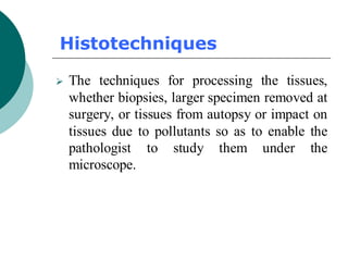
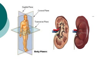
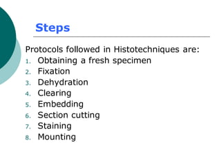
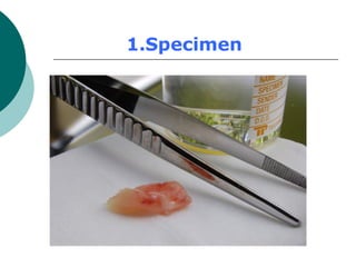
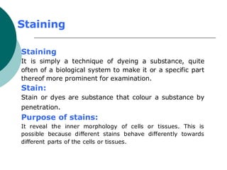
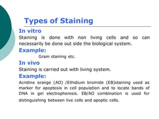
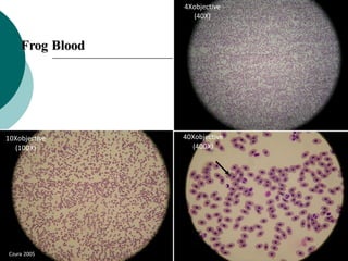
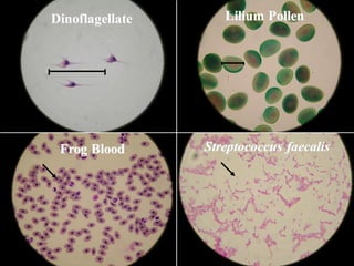
1. Histotechniques involve processing tissues through fixation, dehydration, clearing, embedding, section cutting, and staining to enable pathological examination under a microscope.
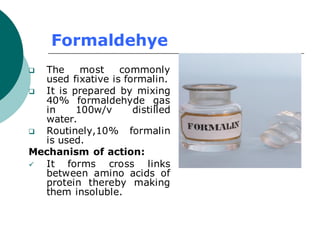
2. Tissues are first fixed in chemicals like formaldehyde to preserve their structure, then dehydrated with graded alcohols, cleared with solvents, and embedded in paraffin wax for section cutting with a microtome.
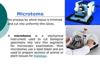
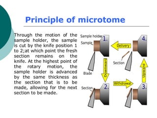
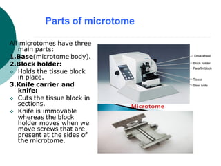
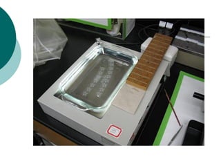
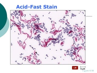
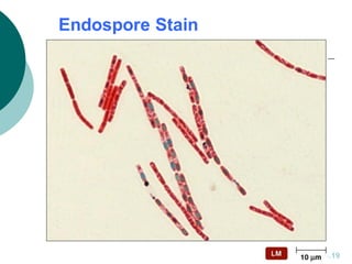
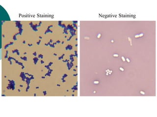
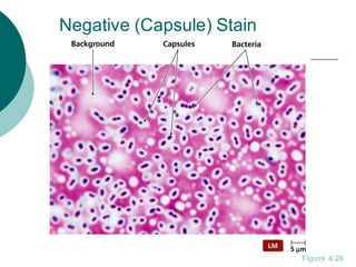
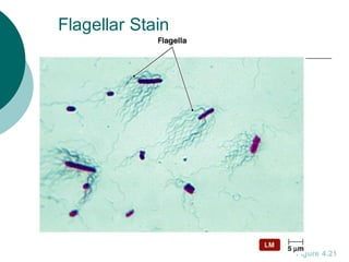
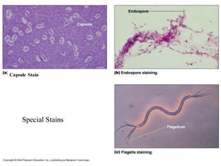
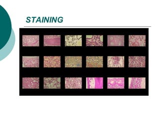
3. The microtome uses a knife to cut extremely thin sections of the wax-embedded tissue, which are then floated in water, mounted on slides, and stained for microscopic examination.


![ANIMAL_CELL_,_TISSUE_AND_ORGAN_CULTURE[1].pptx](https://cdn.slidesharecdn.com/ss_thumbnails/animalcelltissueandorganculture1-260204172026-4462b440-thumbnail.jpg?width=640&height=640&fit=bounds)


