Comprehensive PowerPoint on the Microscope
Slide 1 – Title
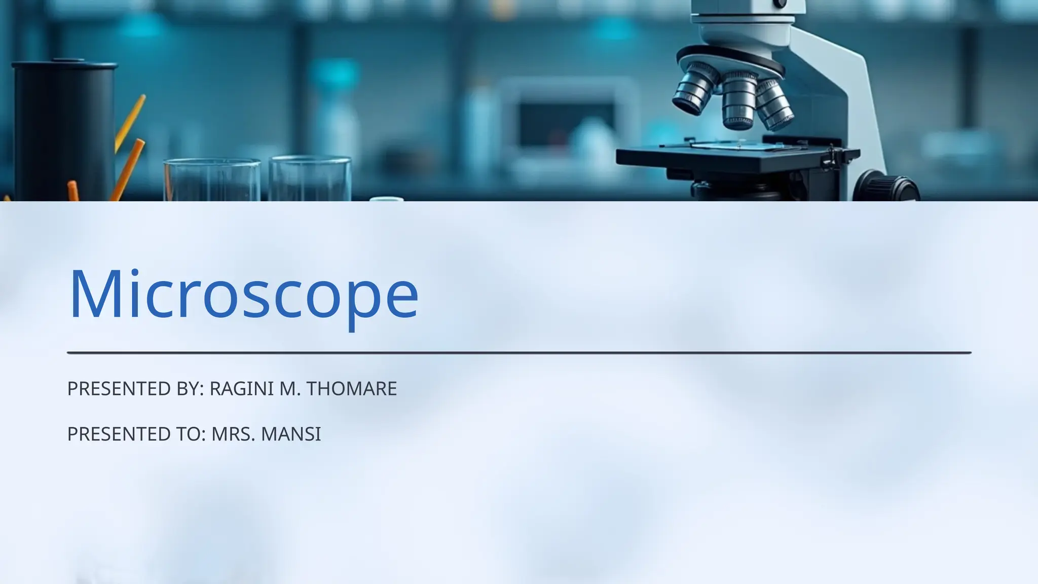
The Microscope
An In-Depth Study of Structure, Function, and Types
Presented by: [Your Name]
Department: [Your Department]
Date: [Insert Date]
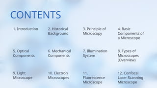
Slide 2 – Introduction
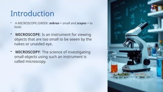
The microscope is an instrument that magnifies small objects invisible to the naked eye.
It has revolutionized science, especially biology, medicine, and materials research.
Enables the observation of cells, microorganisms, tissues, and nano-structures.
Slide 3 – Historical Background
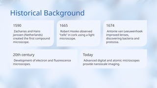
1590: Zacharias and Hans Janssen (Netherlands) created the first compound microscope.
1665: Robert Hooke observed “cells” in cork using a light microscope.
1674: Antonie van Leeuwenhoek improved lenses, discovering bacteria and protozoa.
20th century: Development of electron and fluorescence microscopes.
Today: Advanced digital and atomic microscopes provide nanoscale imaging.
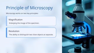
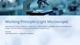
Slide 4 – Principle of Microscopy
Microscopy works on two key principles:
Magnification – enlarging the image of the specimen.
Resolution – the ability to distinguish two close objects as separate.
Formula:
Resolution (d) = 0.61 × λ / NA
Where λ = wavelength of light or electrons, NA = numerical aperture.
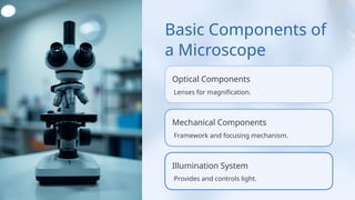
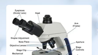
Slide 5 – Basic Components of a Microscope
Optical Components – Lenses for magnification.
Mechanical Components – Framework and focusing mechanism.
Illumination System – Provides and controls light.
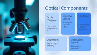
Slide 6 – Optical Components
Ocular (Eyepiece): 10× or 15× magnification.
Objective Lenses: Usually 4×, 10×, 40×, 100×.
Condenser: Concentrates light on specimen.
Diaphragm: Adjusts light intensity.
Mirror/Light Source: Illuminates specimen.
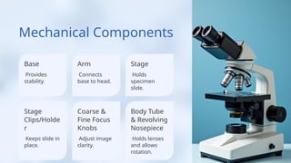
Slide 7 – Mechanical Components
Base: Provides stability.
Arm: Connects base to head.
Stage: Holds specimen slide.
Stage Clips/Holder: Keeps slide in place.
Coarse & Fine Focus Knobs: Adjust image clarity.
Body Tube & Revolving Nosepiece: Holds lenses and allows rotation.
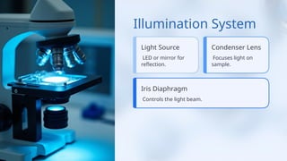
Slide 8 – Illumination System
Light Source: LED or mirror for reflection.
Condenser Lens: Focuses light on sample.
Iris Diaphragm: Controls the light beam.
Köhler Illumination: Ensures even, bright field lighting.
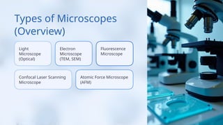
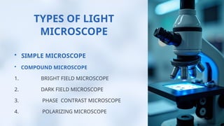
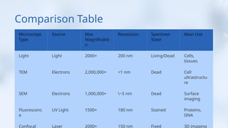
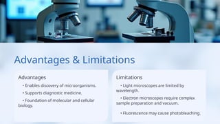
Slide 9 – Types of Microscopes (Overview)
Light Microscope (Optical)
Electron Microscope (TEM, SEM)
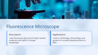
Fluorescence Microscope
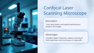
Confocal Laser Scanning Microscope
Phase Contrast Microscope
Polarizing Microscope
Digital Microscope
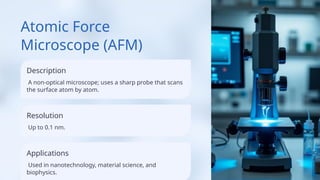
Atomic Force Microscope (AFM)
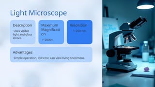
Slide 10 – Light Microscope
Uses visible light and glass lenses.
Maximum magnification: ~2000×.
Resolution: ~200 nm.
Common types:
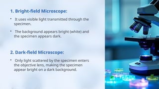
Bright-field microscope
Dark-field microscope
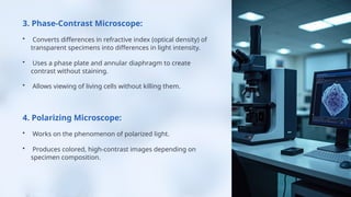
Phase-contrast microscope
Polarizing microscope
Advantages:
Simple operation, low cost, can view living specimens.
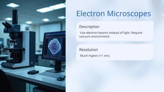
Slide 11 – Electron Microscopes
Use electron beams instead of light.
Require vacuum environment.
Much higher resolution (<1 nm).
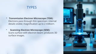
Types:
Transmission Electron Microscope (TEM):
Electrons pass through thin specimen.
Internal details visible.
Magnification: up t