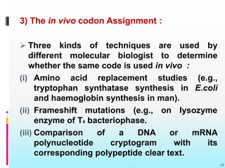
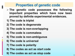
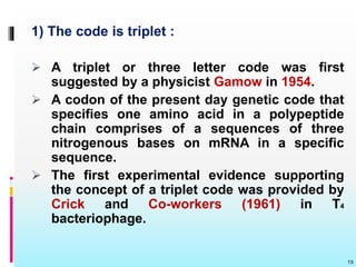
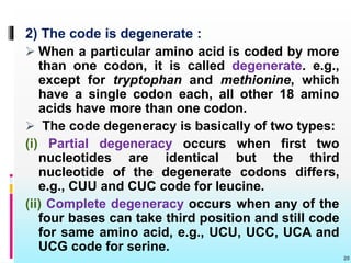
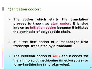
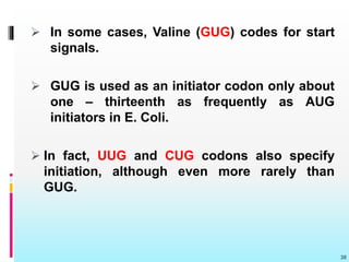
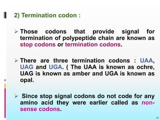
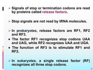
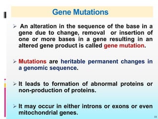
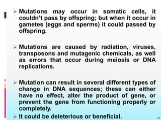
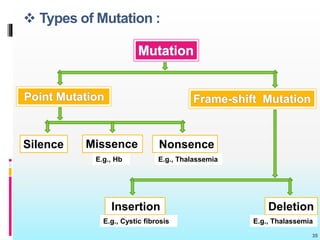
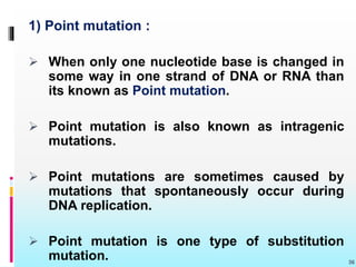
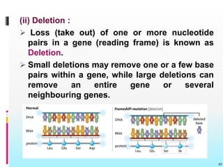
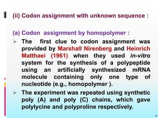
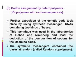
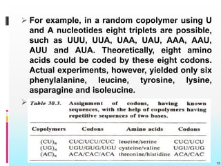
1
Presented by…
Maitri M. Thakor
M.Sc. (Botany)
Department of Life Sciences,
H.N.G.U., Patan.
Contents
 Introduction
 Genetic code
 Deciphering of genetic code
 Properties of genetic code
 Initiation and termination of codons
 Gene mutation 2
Introduction
 All the genetic information is encoded in DNA
molecule and is later transcribed into mRNA.
 However, there are a few cases where DNA is
absent, but the information is contained in
genetic RNA.
 Proteins have 20 different kinds of essential
amino acids, nucleic acids have only 4 different
kinds of bases (A, C, G, U or T).
3
 These four bases have been considered as the
alphabets of the language of nucleic acids and
20 amino acids as the alphabets of the
language of proteins.
 What is the sequence of the nucleotides on
the mRNA will decide the sequence of the
amino acids in a protein chain and this lead to
the discovery of what the genetic code.
 The genetic code works to prepare a
dictionary for translating the language of RNA
into the language of proteins.
4
Genetic code
 The sequence of nitrogenous bases in mRNA
molecule which encloses information for the
synthesis of protein molecules is known as
Genetic code.
 The nucleotide or nucleotide sequence in
mRNA which codes for a particular amino acid
is known as Codon.
 The main problem of genetic code was to
determine the exact number of nucleotides in
a codon which codes for one amino acid.
5
6
 A single code consisting of only one nucleotide
provides for just four codons A, C, G and U.
These are insufficient to code for 20 amino
acids.
 Similarly a combination of two nitrogenous
bases (double code) provides 4 ˣ 4 = 16 codons
still insufficient for 20 amino acids.
 George Gamow (1954) pointed out the
possibility of three letter code, i.e., each codon
consisting of three nitrogenous bases.
● This will give 4 ˣ 4 ˣ 4 = 64 codons, which are
more than enough to code for 20 amino acids.
7
 The triple code is the most acceptable one as
it has found support from experimental
evidences of both genetic and biochemical
nature.
 The genetic code has been experimentally
deciphered and perfected indeoendently by
Marshall Warren Nirenberg, Robert Holley and
Hargovind Khorana for which they were jointly
awarded the Noble prize for medicine and
physiology in 1968.
8
Deciphering of genetic code
 The genetic code has been cracked or
deciphered by the following kinds of
approaches:
1) Theoretical Approach
2) The in vitro codon Assignment
3) The in vivo codon Assignment
1) Theoretical Approach:
 The physicist George Gamow proposed the
diamond code (1954) and the triangle code
(1955) and suggested an exhaustive
theoretical framework to the different aspect
of the genetic code. 9
 Gamow suggested the following properties of
the genetic code.
(i) A triplet codon corresponding to one amino
acid of the polypeptide chain.
(ii) Direct template translation by codon - amino
acid pairing.
(iii) Translation of the code in an overlapping
manner.
(iv) Degeneracy of the code, i.e., an amino acid
being coded by more than one codon.
(v) Colinearity of nucleic acid and the primary
protein synthesized.
(vi) Universality of the code, i.e., the code being
essentially the same for different organisms.
10
2) The in vitro codon Assignment :
 It is divided into three parts:
(i) Discovery and use of polynucleotide
phosphorylase enzyme.
(ii) Codon assignment with unknown sequence
(a) Codon assignment by homopolymer
(b) Codon assignment by heteropolymer
(iii) Assignment of codons with known sequence
11
(i) Discovery and use of polynucleotide
phosphorylase enzyme :
 Marianne Grunberg Manago and Severo Ochoa
isolated an enzyme from the bacteria (e.g.,
Azotobacter vinelandii or Micrococcus
lysodeikticus ) that catalyzes the breakdown of
RNA in bacterial cells. This enzyme is called
polynucleotide phosphorylase enzyme.
 Monago and Ochoa found that outside of the
cell (in vitro), with high concentration Of
rebonucleotides, the reaction could be driven
in reverse and an RNA could be made (see
Burns and Bottino, 1989).
12
(ii) Codon assignment with unknown sequence :
(a) Codon assignment by homopolymer :
 The first clue to codon assignment was
provided by Marshall Nirenberg and Heinrich
Matthaei (1961) when they used in-vitro
system for the synthesis of a polypeptide
using an artificially synthesized mRNA
molecule containing only one type of
nucleotide (e.g., homopolymer ).
 The experiment was repeated using synthetic
poly (A) and poly (C) chains, which gave
polylycine and polyproline respectively.
13
(b) Codon assignment by heteropolymers
( Copolymers with random sequences) :
 Further exposition of the genetic code took
place by using synthetic messenger RNAs
containing two kinds of bases.
 This technique was used in the laboratories
of Ochoa and Nirenberg and lead the
deduction of the composition of codons for
the 20 amino acids.
 The synthetic messengers contained the
bases at random (called Random copolymers).
14
 For example, in a random copolymer using U
and A nucleotides eight triplets are possible,
such as UUU, UUA, UAA, UAU, AAA, AAU,
AUU and AUA. Theoretically, eight amino
acids could be coded by these eight codons.
Actual experiments, however, yielded only six
phenylalanine, leucine, tyrosine, lysine,
asparagine and isoleucine.
 n
15
(iii) Assignment of codons with known sequence:
[Use of trinucleotides or minimessengers in
filter binding (Ribosome – binding technique)]
 Ribosome binding technique of Nirenberg and
Leder (1964) made use of the finding that
aminocyl-tRNA molecules specifically bind to
ribosome – mRNA complex.
 This binding does not require the presence of a
long mRNA molecules; in fact, the association
of a trinucleotide or minimessenger with the
ribosome is sufficient to cause aminoacyl-tRNA
binding.
16
3) The in vivo codon Assignment :
 Three kinds of techniques are used by
different molecular biologist to determine
whether the same code is used in vivo :
(i) Amino acid replacement studies (e.g.,
tryptophan synthatase synthesis in E.coli
and haemoglobin synthesis in man).
(ii) Frameshift mutations (e.g., on lysozyme
enzyme of T4 bacteriophase.
(iii) Comparison of a DNA or mRNA
polynucleotide cryptogram with its
corresponding polypeptide clear text.
17
Properties of genetic code
 The genetic code possesses the following
important properties which have now been
proved by definite experimental evidences.
1) The code is triplet
2) The code is degenerate
3) The code is non-overlapping
4) The code is commaless
5) The code is non-ambiguous
6) The code is universal
7) The code is polarity
8) The codes as act as start code
9) The codes as act as stop code 18
1) The code is triplet :
 A triplet or three letter code was first
suggested by a physicist Gamow in 1954.
 A codon of the present day genetic code that
specifies one amino acid in a polypeptide
chain comprises of a sequences of three
nitrogenous bases on mRNA in a specific
sequence.
 The first experimental evidence supporting
the concept of a triplet code was provided by
Crick and Co-workers (1961) in T4
bacteriophage.
19
2) The code is degenerate :
 When a particular amino acid is coded by more
than one codon, it is called degenerate. e.g.,
except for tryptophan and methionine, which
have a single codon each, all other 18 amino
acids have more than one codon.
 The code degeneracy is basically of two types:
(i) Partial degeneracy occurs when first two
nucleotides are identical but the third
nucleotide of the degenerate codons differs,
e.g., CUU and CUC code for leucine.
(ii) Complete degeneracy occurs when any of the
four bases can take third position and still code
for same amino acid, e.g., UCU, UCC, UCA and
UCG code for serine.
20
 The following table represents the occurrence
of multiple codons for different amino acids
and clearly illustrates the degeneracy of
genetic code.
21
Sr.
No.
Amino acids No. of codons
for each amino
acids
Total no. of
codons
1. Arginine, Leucine, Serine 6 each 18
2. Alanine, Glycine, Proline, Threonine,
Valine.
4 each 20
3. Isoleucine, Stop codons 3 each 06
4. Asparagine, Aspartic acid, Cysteine,
Glutamic acid, Histidine, Lysine,
Phenylalanine and Tyrosine
2 each 18
5. Methionine and Tryptophan 1 each 02
Total 64
3) The code is non-overlapping :
 The code is non-overlapping which means
that the same latter is not used for different
codons.
22
 The evidencies are available to show that
translation of genetic code in an mRNA begins
at the correct point and there is no
overlapping of codons. i.e., genetic code is
non overlapping.
 Although the code is non overlapping but in
the bacteriophage ɸ x 174 there is a
possibility of overlapping genes and codons.
(Barrel and coworkers, 1976; Sanger, et al.,
1977).
23
4) The code is commaless :
 There are no punctuations or comma etc.
between two codons, In the genetic code.
 In other words no codon is reserved for
punctuations.
5) The code is non ambiguous :
 The genetic code inside the cell medium (in
vivo) is said to be non abmiguos because a
particular codon always codes for the same
amino acid may be coded by more than one
codons (degeneracy), but one codon never
codes for two different amino acids.
24
6) The code is universal :
 Same genetic code is found valid for all
organisms ranging from bacteria to man.
 Such universality of the code was
demonstrated by Marshall, Caskey and
Nirenberg (1967) who found that E.coli
(bacterium) and guinea pig (mammal) amino
acyl – tRNA use almost the same code.
7) The code has polarity :
 The code is always read in a fixed direction,
i.e., in the 5’ 3’ direction. In other words, the
codon has a polarity.
 Reading from left to right and right to left will
specify for different amino acids.
25
8) Some codes act as start codons :
 In most organisms, AUG codon is the start or
initiation codons the polypeptide chain starts
either with methionine or formaylmethionine.
 Normally GUG codes for valine but when
normal AUG codon is lost by deletion only
than GUG is use as initiation codon.
9) Some codon act as stop codons :
 Three codons UAG, UAA, and UGA are the
chain stop or termination codon.
 They do not code for any of the amino acids.
26
Initiation and termination of codon
 A reading frame of the genetic message
always has a specific start and stop signals for
protein synthesis.
 The initiator and terminator codons are known
as signals and this phenomenon is known as
punctuation.
 Punctuation helps in delimiting the different
cistrons on a polycistronic mRNA.
 The signals or spacers do not occur between
the codons of a message but initiate and
terminate larger sections of the message
between functional genes.
27
 Signal codons :
 Those codons that code for signals during
protein synthesis are known as signal
codons.
 There are four codons which code for a
signal, these are AUG, UAA, UAG and UGA.
 Signal codons are of two types :
1) Initiation / Start codons
2) Termination / Stop codons
28
1) Initiation codon :
 The codon which starts the translation
process is known as start codon. It is also
known as initiation codon because it initiates
the synthesis of polypeptide chain.
 It is the first codon of a messenger RNA
transcript translated by a ribosome.
 The initiation codon is AUG and it codes for
the amino acid, methionine (in eukaryotes) or
formylmethionine (in prokaryotes).
29
 In some cases, Valine (GUG) codes for start
signals.
 GUG is used as an initiator codon only about
one – thirteenth as frequently as AUG
initiators in E. Coli.
 In fact, UUG and CUG codons also specify
initiation, although even more rarely than
GUG.
30
2) Termination codon :
 Those codons that provide signal for
termination of polypeptide chain are known as
stop codons or termination codons.
 There are three termination codons : UAA,
UAG and UGA. ( The UAA is known as ochre,
UAG is known as amber and UGA is known as
opal.
 Since stop signal codons do not code for any
amino acid they were earlier called as non-
sense codons.
31
 Signals of stop or termination codons are read
by proteins called release factors.
 Stop signals are not read by tRNA molecules.
 In prokaryotes, release factors are RF1, RF2
and RF3.
● The factor RF1 recognizes stop codons UAA
and UAG, while RF2 recognizes UAA and UGA.
● The function of RF3 is to stimulate RF1 and
RF2.
 In eukaryotes, a single release factor (RF)
recognizes all three stop codons.
32
Gene Mutations
 An alteration in the sequence of the base in a
gene due to change, removal or insertion of
one or more bases in a gene resulting in an
altered gene product is called gene mutation.
 Mutations are heritable permanent changes in
a genomic sequence.
 It leads to formation of abnormal proteins or
non-production of proteins.
 It may occur in either introns or exons or even
mitochondrial genes.
33
 Mutations may occur in somatic cells, it
couldn’t pass by offspring; but when it occur in
gametes (eggs and sperms) it could passed by
offspring.
 Mutations are caused by radiation, viruses,
transposons and mutagenic chemicals, as well
as errors that occur during meiosis or DNA
replications.
 Mutation can result in several different types of
change in DNA sequences; these can either
have no effect, alter the product of gene, or
prevent the gene from functioning properly or
completely.
 It could be deleterious or beneficial. 34
 Types of Mutation :
35
Mutation
Point Mutation Frame-shift Mutation
Silence Missence Nonsence
Insertion Deletion
E.g., Hb E.g., Thalassemia
E.g., Cystic fibrosis E.g., Thalassemia
1) Point mutation :
 When only one nucleotide base is changed in
some way in one strand of DNA or RNA than
its known as Point mutation.
 Point mutation is also known as intragenic
mutations.
 Point mutations are sometimes caused by
mutations that spontaneously occur during
DNA replication.
 Point mutation is one type of substitution
mutation. 36
 A substitution mutation occurs when one
base pair is substituted by another.
 For example, This would occur when one
nucleotide containing cytosine is
accidentally substituted for one containing
guanine.
 It may be transition or transversions.
37
 There are three types of point mutations :
(i) Silent mutations
(ii) Missense mutation
(iii) Nonsense mutation 38
Transitions:
● Homologous change
occurs. E.g., purine with
purine and pyrimidine
with pyrimidine.
Trans-versions :
● Non homologous
change occurs. E.g.,
purine with a pyrimidine
and vice versa.
(i) Silent Mutation :
 When the codon
containing the changed
base may code for the
same amino acid its
known as silent mutation.
 This can occur because
multiple codons can code
for the same amino acid.
 E.g., in serine codon UCA,
if A is changed to U giving
the codon UCU, it still
code for Serine.
39
(ii) Missense mutation :
 When the codon
containing the changed
base may code for a
different amino acid than
its known as Missense
mutation.
 E.g., if the serine codon
GAA is changed to be
GAC (A is replaced by C),
it will code for Glutamic
acid not Aspartic acid
leading to insertion of
incorrect amino acid into
polypeptide chain. 40
41
(iii) Nonsense mutation :
 When the codon
containing the changed
base may become a
termination codon than
its known as Nonsense
mutation.
 E.g., serine codon UCA
becomes UAA if C is
changed to A. UAA is a
stop codon leading to
termination of translation
at that point. 42
2) Frame-shift mutation :
 Deletion or addition of one or two base to
message sequence, leading to change in
reading frame (reading sequence) is known as
Frame-shift mutation.
 The resulting amino acid sequence may
become completely different from this point.
 The resulting protein is usually non functional.
 Frame-shift mutation have two types :
(i) Insertion
(ii) Deletion 43
(i) Insertion :
 Addition of one or more nucleotide base
pairs in a gene (reading frame) is known as
Insertion.
 As a result, the protein made by the gene
may not function properly.
44
(ii) Deletion :
 Loss (take out) of one or more nucleotide
pairs in a gene (reading frame) is known as
Deletion.
 Small deletions may remove one or a few base
pairs within a gene, while large deletions can
remove an entire gene or several
neighbouring genes.
45
References
1) Fundamental of Molecular Biology
Author : Veer Bala Rastogi
Edition : 2008 (first edition)
2) Cell Biology, Genetics, Molecular Biology,
Evolution and Ecology
Author : P. S. Verma, V. K. Agarwal
Edition : 2016
3) A textbook of Plant physiology, Biochemistry and
Biotechnology
Author : S. K. Verma, Mohit Verma
Edition : 2007 (Sixth edition) 46
4) Cell and Molecular Biology (International
Edition )
Author : E.D.P. De Robertis,
E.M.F. De Robertis.
Edition : Eighth Edition
5) www.biologydiscussion.com
6) www.slideshare.net
47
48

Genetic code

  • 1.
    1 Presented by… Maitri M.Thakor M.Sc. (Botany) Department of Life Sciences, H.N.G.U., Patan.
  • 2.
    Contents  Introduction  Geneticcode  Deciphering of genetic code  Properties of genetic code  Initiation and termination of codons  Gene mutation 2
  • 3.
    Introduction  All thegenetic information is encoded in DNA molecule and is later transcribed into mRNA.  However, there are a few cases where DNA is absent, but the information is contained in genetic RNA.  Proteins have 20 different kinds of essential amino acids, nucleic acids have only 4 different kinds of bases (A, C, G, U or T). 3
  • 4.
     These fourbases have been considered as the alphabets of the language of nucleic acids and 20 amino acids as the alphabets of the language of proteins.  What is the sequence of the nucleotides on the mRNA will decide the sequence of the amino acids in a protein chain and this lead to the discovery of what the genetic code.  The genetic code works to prepare a dictionary for translating the language of RNA into the language of proteins. 4
  • 5.
    Genetic code  Thesequence of nitrogenous bases in mRNA molecule which encloses information for the synthesis of protein molecules is known as Genetic code.  The nucleotide or nucleotide sequence in mRNA which codes for a particular amino acid is known as Codon.  The main problem of genetic code was to determine the exact number of nucleotides in a codon which codes for one amino acid. 5
  • 6.
    6  A singlecode consisting of only one nucleotide provides for just four codons A, C, G and U. These are insufficient to code for 20 amino acids.  Similarly a combination of two nitrogenous bases (double code) provides 4 ˣ 4 = 16 codons still insufficient for 20 amino acids.  George Gamow (1954) pointed out the possibility of three letter code, i.e., each codon consisting of three nitrogenous bases. ● This will give 4 ˣ 4 ˣ 4 = 64 codons, which are more than enough to code for 20 amino acids.
  • 7.
  • 8.
     The triplecode is the most acceptable one as it has found support from experimental evidences of both genetic and biochemical nature.  The genetic code has been experimentally deciphered and perfected indeoendently by Marshall Warren Nirenberg, Robert Holley and Hargovind Khorana for which they were jointly awarded the Noble prize for medicine and physiology in 1968. 8
  • 9.
    Deciphering of geneticcode  The genetic code has been cracked or deciphered by the following kinds of approaches: 1) Theoretical Approach 2) The in vitro codon Assignment 3) The in vivo codon Assignment 1) Theoretical Approach:  The physicist George Gamow proposed the diamond code (1954) and the triangle code (1955) and suggested an exhaustive theoretical framework to the different aspect of the genetic code. 9
  • 10.
     Gamow suggestedthe following properties of the genetic code. (i) A triplet codon corresponding to one amino acid of the polypeptide chain. (ii) Direct template translation by codon - amino acid pairing. (iii) Translation of the code in an overlapping manner. (iv) Degeneracy of the code, i.e., an amino acid being coded by more than one codon. (v) Colinearity of nucleic acid and the primary protein synthesized. (vi) Universality of the code, i.e., the code being essentially the same for different organisms. 10
  • 11.
    2) The invitro codon Assignment :  It is divided into three parts: (i) Discovery and use of polynucleotide phosphorylase enzyme. (ii) Codon assignment with unknown sequence (a) Codon assignment by homopolymer (b) Codon assignment by heteropolymer (iii) Assignment of codons with known sequence 11
  • 12.
    (i) Discovery anduse of polynucleotide phosphorylase enzyme :  Marianne Grunberg Manago and Severo Ochoa isolated an enzyme from the bacteria (e.g., Azotobacter vinelandii or Micrococcus lysodeikticus ) that catalyzes the breakdown of RNA in bacterial cells. This enzyme is called polynucleotide phosphorylase enzyme.  Monago and Ochoa found that outside of the cell (in vitro), with high concentration Of rebonucleotides, the reaction could be driven in reverse and an RNA could be made (see Burns and Bottino, 1989). 12
  • 13.
    (ii) Codon assignmentwith unknown sequence : (a) Codon assignment by homopolymer :  The first clue to codon assignment was provided by Marshall Nirenberg and Heinrich Matthaei (1961) when they used in-vitro system for the synthesis of a polypeptide using an artificially synthesized mRNA molecule containing only one type of nucleotide (e.g., homopolymer ).  The experiment was repeated using synthetic poly (A) and poly (C) chains, which gave polylycine and polyproline respectively. 13
  • 14.
    (b) Codon assignmentby heteropolymers ( Copolymers with random sequences) :  Further exposition of the genetic code took place by using synthetic messenger RNAs containing two kinds of bases.  This technique was used in the laboratories of Ochoa and Nirenberg and lead the deduction of the composition of codons for the 20 amino acids.  The synthetic messengers contained the bases at random (called Random copolymers). 14
  • 15.
     For example,in a random copolymer using U and A nucleotides eight triplets are possible, such as UUU, UUA, UAA, UAU, AAA, AAU, AUU and AUA. Theoretically, eight amino acids could be coded by these eight codons. Actual experiments, however, yielded only six phenylalanine, leucine, tyrosine, lysine, asparagine and isoleucine.  n 15
  • 16.
    (iii) Assignment ofcodons with known sequence: [Use of trinucleotides or minimessengers in filter binding (Ribosome – binding technique)]  Ribosome binding technique of Nirenberg and Leder (1964) made use of the finding that aminocyl-tRNA molecules specifically bind to ribosome – mRNA complex.  This binding does not require the presence of a long mRNA molecules; in fact, the association of a trinucleotide or minimessenger with the ribosome is sufficient to cause aminoacyl-tRNA binding. 16
  • 17.
    3) The invivo codon Assignment :  Three kinds of techniques are used by different molecular biologist to determine whether the same code is used in vivo : (i) Amino acid replacement studies (e.g., tryptophan synthatase synthesis in E.coli and haemoglobin synthesis in man). (ii) Frameshift mutations (e.g., on lysozyme enzyme of T4 bacteriophase. (iii) Comparison of a DNA or mRNA polynucleotide cryptogram with its corresponding polypeptide clear text. 17
  • 18.
    Properties of geneticcode  The genetic code possesses the following important properties which have now been proved by definite experimental evidences. 1) The code is triplet 2) The code is degenerate 3) The code is non-overlapping 4) The code is commaless 5) The code is non-ambiguous 6) The code is universal 7) The code is polarity 8) The codes as act as start code 9) The codes as act as stop code 18
  • 19.
    1) The codeis triplet :  A triplet or three letter code was first suggested by a physicist Gamow in 1954.  A codon of the present day genetic code that specifies one amino acid in a polypeptide chain comprises of a sequences of three nitrogenous bases on mRNA in a specific sequence.  The first experimental evidence supporting the concept of a triplet code was provided by Crick and Co-workers (1961) in T4 bacteriophage. 19
  • 20.
    2) The codeis degenerate :  When a particular amino acid is coded by more than one codon, it is called degenerate. e.g., except for tryptophan and methionine, which have a single codon each, all other 18 amino acids have more than one codon.  The code degeneracy is basically of two types: (i) Partial degeneracy occurs when first two nucleotides are identical but the third nucleotide of the degenerate codons differs, e.g., CUU and CUC code for leucine. (ii) Complete degeneracy occurs when any of the four bases can take third position and still code for same amino acid, e.g., UCU, UCC, UCA and UCG code for serine. 20
  • 21.
     The followingtable represents the occurrence of multiple codons for different amino acids and clearly illustrates the degeneracy of genetic code. 21 Sr. No. Amino acids No. of codons for each amino acids Total no. of codons 1. Arginine, Leucine, Serine 6 each 18 2. Alanine, Glycine, Proline, Threonine, Valine. 4 each 20 3. Isoleucine, Stop codons 3 each 06 4. Asparagine, Aspartic acid, Cysteine, Glutamic acid, Histidine, Lysine, Phenylalanine and Tyrosine 2 each 18 5. Methionine and Tryptophan 1 each 02 Total 64
  • 22.
    3) The codeis non-overlapping :  The code is non-overlapping which means that the same latter is not used for different codons. 22
  • 23.
     The evidenciesare available to show that translation of genetic code in an mRNA begins at the correct point and there is no overlapping of codons. i.e., genetic code is non overlapping.  Although the code is non overlapping but in the bacteriophage ɸ x 174 there is a possibility of overlapping genes and codons. (Barrel and coworkers, 1976; Sanger, et al., 1977). 23
  • 24.
    4) The codeis commaless :  There are no punctuations or comma etc. between two codons, In the genetic code.  In other words no codon is reserved for punctuations. 5) The code is non ambiguous :  The genetic code inside the cell medium (in vivo) is said to be non abmiguos because a particular codon always codes for the same amino acid may be coded by more than one codons (degeneracy), but one codon never codes for two different amino acids. 24
  • 25.
    6) The codeis universal :  Same genetic code is found valid for all organisms ranging from bacteria to man.  Such universality of the code was demonstrated by Marshall, Caskey and Nirenberg (1967) who found that E.coli (bacterium) and guinea pig (mammal) amino acyl – tRNA use almost the same code. 7) The code has polarity :  The code is always read in a fixed direction, i.e., in the 5’ 3’ direction. In other words, the codon has a polarity.  Reading from left to right and right to left will specify for different amino acids. 25
  • 26.
    8) Some codesact as start codons :  In most organisms, AUG codon is the start or initiation codons the polypeptide chain starts either with methionine or formaylmethionine.  Normally GUG codes for valine but when normal AUG codon is lost by deletion only than GUG is use as initiation codon. 9) Some codon act as stop codons :  Three codons UAG, UAA, and UGA are the chain stop or termination codon.  They do not code for any of the amino acids. 26
  • 27.
    Initiation and terminationof codon  A reading frame of the genetic message always has a specific start and stop signals for protein synthesis.  The initiator and terminator codons are known as signals and this phenomenon is known as punctuation.  Punctuation helps in delimiting the different cistrons on a polycistronic mRNA.  The signals or spacers do not occur between the codons of a message but initiate and terminate larger sections of the message between functional genes. 27
  • 28.
     Signal codons:  Those codons that code for signals during protein synthesis are known as signal codons.  There are four codons which code for a signal, these are AUG, UAA, UAG and UGA.  Signal codons are of two types : 1) Initiation / Start codons 2) Termination / Stop codons 28
  • 29.
    1) Initiation codon:  The codon which starts the translation process is known as start codon. It is also known as initiation codon because it initiates the synthesis of polypeptide chain.  It is the first codon of a messenger RNA transcript translated by a ribosome.  The initiation codon is AUG and it codes for the amino acid, methionine (in eukaryotes) or formylmethionine (in prokaryotes). 29
  • 30.
     In somecases, Valine (GUG) codes for start signals.  GUG is used as an initiator codon only about one – thirteenth as frequently as AUG initiators in E. Coli.  In fact, UUG and CUG codons also specify initiation, although even more rarely than GUG. 30
  • 31.
    2) Termination codon:  Those codons that provide signal for termination of polypeptide chain are known as stop codons or termination codons.  There are three termination codons : UAA, UAG and UGA. ( The UAA is known as ochre, UAG is known as amber and UGA is known as opal.  Since stop signal codons do not code for any amino acid they were earlier called as non- sense codons. 31
  • 32.
     Signals ofstop or termination codons are read by proteins called release factors.  Stop signals are not read by tRNA molecules.  In prokaryotes, release factors are RF1, RF2 and RF3. ● The factor RF1 recognizes stop codons UAA and UAG, while RF2 recognizes UAA and UGA. ● The function of RF3 is to stimulate RF1 and RF2.  In eukaryotes, a single release factor (RF) recognizes all three stop codons. 32
  • 33.
    Gene Mutations  Analteration in the sequence of the base in a gene due to change, removal or insertion of one or more bases in a gene resulting in an altered gene product is called gene mutation.  Mutations are heritable permanent changes in a genomic sequence.  It leads to formation of abnormal proteins or non-production of proteins.  It may occur in either introns or exons or even mitochondrial genes. 33
  • 34.
     Mutations mayoccur in somatic cells, it couldn’t pass by offspring; but when it occur in gametes (eggs and sperms) it could passed by offspring.  Mutations are caused by radiation, viruses, transposons and mutagenic chemicals, as well as errors that occur during meiosis or DNA replications.  Mutation can result in several different types of change in DNA sequences; these can either have no effect, alter the product of gene, or prevent the gene from functioning properly or completely.  It could be deleterious or beneficial. 34
  • 35.
     Types ofMutation : 35 Mutation Point Mutation Frame-shift Mutation Silence Missence Nonsence Insertion Deletion E.g., Hb E.g., Thalassemia E.g., Cystic fibrosis E.g., Thalassemia
  • 36.
    1) Point mutation:  When only one nucleotide base is changed in some way in one strand of DNA or RNA than its known as Point mutation.  Point mutation is also known as intragenic mutations.  Point mutations are sometimes caused by mutations that spontaneously occur during DNA replication.  Point mutation is one type of substitution mutation. 36
  • 37.
     A substitutionmutation occurs when one base pair is substituted by another.  For example, This would occur when one nucleotide containing cytosine is accidentally substituted for one containing guanine.  It may be transition or transversions. 37
  • 38.
     There arethree types of point mutations : (i) Silent mutations (ii) Missense mutation (iii) Nonsense mutation 38 Transitions: ● Homologous change occurs. E.g., purine with purine and pyrimidine with pyrimidine. Trans-versions : ● Non homologous change occurs. E.g., purine with a pyrimidine and vice versa.
  • 39.
    (i) Silent Mutation:  When the codon containing the changed base may code for the same amino acid its known as silent mutation.  This can occur because multiple codons can code for the same amino acid.  E.g., in serine codon UCA, if A is changed to U giving the codon UCU, it still code for Serine. 39
  • 40.
    (ii) Missense mutation:  When the codon containing the changed base may code for a different amino acid than its known as Missense mutation.  E.g., if the serine codon GAA is changed to be GAC (A is replaced by C), it will code for Glutamic acid not Aspartic acid leading to insertion of incorrect amino acid into polypeptide chain. 40
  • 41.
  • 42.
    (iii) Nonsense mutation:  When the codon containing the changed base may become a termination codon than its known as Nonsense mutation.  E.g., serine codon UCA becomes UAA if C is changed to A. UAA is a stop codon leading to termination of translation at that point. 42
  • 43.
    2) Frame-shift mutation:  Deletion or addition of one or two base to message sequence, leading to change in reading frame (reading sequence) is known as Frame-shift mutation.  The resulting amino acid sequence may become completely different from this point.  The resulting protein is usually non functional.  Frame-shift mutation have two types : (i) Insertion (ii) Deletion 43
  • 44.
    (i) Insertion : Addition of one or more nucleotide base pairs in a gene (reading frame) is known as Insertion.  As a result, the protein made by the gene may not function properly. 44
  • 45.
    (ii) Deletion : Loss (take out) of one or more nucleotide pairs in a gene (reading frame) is known as Deletion.  Small deletions may remove one or a few base pairs within a gene, while large deletions can remove an entire gene or several neighbouring genes. 45
  • 46.
    References 1) Fundamental ofMolecular Biology Author : Veer Bala Rastogi Edition : 2008 (first edition) 2) Cell Biology, Genetics, Molecular Biology, Evolution and Ecology Author : P. S. Verma, V. K. Agarwal Edition : 2016 3) A textbook of Plant physiology, Biochemistry and Biotechnology Author : S. K. Verma, Mohit Verma Edition : 2007 (Sixth edition) 46
  • 47.
    4) Cell andMolecular Biology (International Edition ) Author : E.D.P. De Robertis, E.M.F. De Robertis. Edition : Eighth Edition 5) www.biologydiscussion.com 6) www.slideshare.net 47
  • 48.