This document provides information about osteoarthritis (OA), including its definition, prevalence, risk factors, pathology, diagnosis, natural history, differential diagnosis, and treatment. It notes that OA is the most common form of arthritis, affecting over 20 million people in the US. Risk factors include age, obesity, family history, and previous joint injury or disorder. Diagnosis is typically based on symptoms like pain and stiffness, physical exam findings, and x-ray evidence of cartilage loss, bone spurs, and bone changes. Treatment includes conservative options like medications, exercise, and weight loss, as well as intra-articular injections or surgery for advanced cases.
Charcot joint or neuropathic joint are destructed joint occurs in Diabetes, syphilis, syringomyelia , leprosy, AMLS, Peripheral neuropathy and any condition leads to impair sensation of peripheral part of body
“Don’t touch” lesions new version Dr Ahmed EsawyAHMED ESAWY
“Don’t touch” lesions new version dr ahmed esawy
CALCANEAL PSEUDOCYST
INTRAOSSEOUS LIPOMA
BIPARTITE PATELLA
MYOSITIS OSSIFICANS
AVULSION INJURY
CORTICAL DESMIOD
GEODES
DORSAL DEFECT OF THE PATELLA
PSEUDOCYST OF THE HUMURUS
OS ODONTOIDEUM
NON OSSIFYING FIBROMA
BONE ISLANDS
UNICAMERAL BONY CYST
EARLY BONE INFARCT
MELORHEOSTOSIS
HYPERTROPHIC PULMONARY OSTEOARTHROPATHY
ACHONDROPLASIA
AVASCULAR NECROSIS
HURLER SYNDROME
TRANSIENT OSTEOPOROSIS OF THE HIP
DIAPHYSEAL ACLASIA
MULTIPLE HEREDITARY EXOSTOSIS
OSTEOID OSTEOMA
OSTEPATHIA STRIATA
OSTEOPIKILOSIS
SARCIOD
OS STYLOIDEUM
OS TRIGONUM
The Indian Dental Academy is the Leader in continuing dental education , training dentists in all aspects of dentistry and
offering a wide range of dental certified courses in different formats.
The prostate is an exocrine gland of the male mammalian reproductive system
It is a walnut-sized gland that forms part of the male reproductive system and is located in front of the rectum and just below the urinary bladder
Function is to store and secrete a clear, slightly alkaline fluid that constitutes 10-30% of the volume of the seminal fluid that along with the spermatozoa, constitutes semen
A healthy human prostate measures (4cm-vertical, by 3cm-horizontal, 2cm ant-post ).
It surrounds the urethra just below the urinary bladder. It has anterior, median, posterior and two lateral lobes
It’s work is regulated by androgens which are responsible for male sex characteristics
Generalised disease of the prostate due to hormonal derangement which leads to non malignant enlargement of the gland (increase in the number of epithelial cells and stromal tissue)to cause compression of the urethra leading to symptoms (LUTS
TEST BANK for Operations Management, 14th Edition by William J. Stevenson, Ve...kevinkariuki227
TEST BANK for Operations Management, 14th Edition by William J. Stevenson, Verified Chapters 1 - 19, Complete Newest Version.pdf
TEST BANK for Operations Management, 14th Edition by William J. Stevenson, Verified Chapters 1 - 19, Complete Newest Version.pdf
These simplified slides by Dr. Sidra Arshad present an overview of the non-respiratory functions of the respiratory tract.
Learning objectives:
1. Enlist the non-respiratory functions of the respiratory tract
2. Briefly explain how these functions are carried out
3. Discuss the significance of dead space
4. Differentiate between minute ventilation and alveolar ventilation
5. Describe the cough and sneeze reflexes
Study Resources:
1. Chapter 39, Guyton and Hall Textbook of Medical Physiology, 14th edition
2. Chapter 34, Ganong’s Review of Medical Physiology, 26th edition
3. Chapter 17, Human Physiology by Lauralee Sherwood, 9th edition
4. Non-respiratory functions of the lungs https://academic.oup.com/bjaed/article/13/3/98/278874
Title: Sense of Smell
Presenter: Dr. Faiza, Assistant Professor of Physiology
Qualifications:
MBBS (Best Graduate, AIMC Lahore)
FCPS Physiology
ICMT, CHPE, DHPE (STMU)
MPH (GC University, Faisalabad)
MBA (Virtual University of Pakistan)
Learning Objectives:
Describe the primary categories of smells and the concept of odor blindness.
Explain the structure and location of the olfactory membrane and mucosa, including the types and roles of cells involved in olfaction.
Describe the pathway and mechanisms of olfactory signal transmission from the olfactory receptors to the brain.
Illustrate the biochemical cascade triggered by odorant binding to olfactory receptors, including the role of G-proteins and second messengers in generating an action potential.
Identify different types of olfactory disorders such as anosmia, hyposmia, hyperosmia, and dysosmia, including their potential causes.
Key Topics:
Olfactory Genes:
3% of the human genome accounts for olfactory genes.
400 genes for odorant receptors.
Olfactory Membrane:
Located in the superior part of the nasal cavity.
Medially: Folds downward along the superior septum.
Laterally: Folds over the superior turbinate and upper surface of the middle turbinate.
Total surface area: 5-10 square centimeters.
Olfactory Mucosa:
Olfactory Cells: Bipolar nerve cells derived from the CNS (100 million), with 4-25 olfactory cilia per cell.
Sustentacular Cells: Produce mucus and maintain ionic and molecular environment.
Basal Cells: Replace worn-out olfactory cells with an average lifespan of 1-2 months.
Bowman’s Gland: Secretes mucus.
Stimulation of Olfactory Cells:
Odorant dissolves in mucus and attaches to receptors on olfactory cilia.
Involves a cascade effect through G-proteins and second messengers, leading to depolarization and action potential generation in the olfactory nerve.
Quality of a Good Odorant:
Small (3-20 Carbon atoms), volatile, water-soluble, and lipid-soluble.
Facilitated by odorant-binding proteins in mucus.
Membrane Potential and Action Potential:
Resting membrane potential: -55mV.
Action potential frequency in the olfactory nerve increases with odorant strength.
Adaptation Towards the Sense of Smell:
Rapid adaptation within the first second, with further slow adaptation.
Psychological adaptation greater than receptor adaptation, involving feedback inhibition from the central nervous system.
Primary Sensations of Smell:
Camphoraceous, Musky, Floral, Pepperminty, Ethereal, Pungent, Putrid.
Odor Detection Threshold:
Examples: Hydrogen sulfide (0.0005 ppm), Methyl-mercaptan (0.002 ppm).
Some toxic substances are odorless at lethal concentrations.
Characteristics of Smell:
Odor blindness for single substances due to lack of appropriate receptor protein.
Behavioral and emotional influences of smell.
Transmission of Olfactory Signals:
From olfactory cells to glomeruli in the olfactory bulb, involving lateral inhibition.
Primitive, less old, and new olfactory systems with different path
Ozempic: Preoperative Management of Patients on GLP-1 Receptor Agonists Saeid Safari
Preoperative Management of Patients on GLP-1 Receptor Agonists like Ozempic and Semiglutide
ASA GUIDELINE
NYSORA Guideline
2 Case Reports of Gastric Ultrasound
These lecture slides, by Dr Sidra Arshad, offer a quick overview of physiological basis of a normal electrocardiogram.
Learning objectives:
1. Define an electrocardiogram (ECG) and electrocardiography
2. Describe how dipoles generated by the heart produce the waveforms of the ECG
3. Describe the components of a normal electrocardiogram of a typical bipolar leads (limb II)
4. Differentiate between intervals and segments
5. Enlist some common indications for obtaining an ECG
Study Resources:
1. Chapter 11, Guyton and Hall Textbook of Medical Physiology, 14th edition
2. Chapter 9, Human Physiology - From Cells to Systems, Lauralee Sherwood, 9th edition
3. Chapter 29, Ganong’s Review of Medical Physiology, 26th edition
4. Electrocardiogram, StatPearls - https://www.ncbi.nlm.nih.gov/books/NBK549803/
5. ECG in Medical Practice by ABM Abdullah, 4th edition
6. ECG Basics, http://www.nataliescasebook.com/tag/e-c-g-basics
- Video recording of this lecture in English language: https://youtu.be/lK81BzxMqdo
- Video recording of this lecture in Arabic language: https://youtu.be/Ve4P0COk9OI
- Link to download the book free: https://nephrotube.blogspot.com/p/nephrotube-nephrology-books.html
- Link to NephroTube website: www.NephroTube.com
- Link to NephroTube social media accounts: https://nephrotube.blogspot.com/p/join-nephrotube-on-social-media.html
Factory Supply Best Quality Pmk Oil CAS 28578–16–7 PMK Powder in Stockrebeccabio
Factory Supply Best Quality Pmk Oil CAS 28578–16–7 PMK Powder in Stock
Telegram: bmksupplier
signal: +85264872720
threema: TUD4A6YC
You can contact me on Telegram or Threema
Communicate promptly and reply
Free of customs clearance, Double Clearance 100% pass delivery to USA, Canada, Spain, Germany, Netherland, Poland, Italy, Sweden, UK, Czech Republic, Australia, Mexico, Russia, Ukraine, Kazakhstan.Door to door service
Hot Selling Organic intermediates
Flu Vaccine Alert in Bangalore Karnatakaaddon Scans
As flu season approaches, health officials in Bangalore, Karnataka, are urging residents to get their flu vaccinations. The seasonal flu, while common, can lead to severe health complications, particularly for vulnerable populations such as young children, the elderly, and those with underlying health conditions.
Dr. Vidisha Kumari, a leading epidemiologist in Bangalore, emphasizes the importance of getting vaccinated. "The flu vaccine is our best defense against the influenza virus. It not only protects individuals but also helps prevent the spread of the virus in our communities," he says.
This year, the flu season is expected to coincide with a potential increase in other respiratory illnesses. The Karnataka Health Department has launched an awareness campaign highlighting the significance of flu vaccinations. They have set up multiple vaccination centers across Bangalore, making it convenient for residents to receive their shots.
To encourage widespread vaccination, the government is also collaborating with local schools, workplaces, and community centers to facilitate vaccination drives. Special attention is being given to ensuring that the vaccine is accessible to all, including marginalized communities who may have limited access to healthcare.
Residents are reminded that the flu vaccine is safe and effective. Common side effects are mild and may include soreness at the injection site, mild fever, or muscle aches. These side effects are generally short-lived and far less severe than the flu itself.
Healthcare providers are also stressing the importance of continuing COVID-19 precautions. Wearing masks, practicing good hand hygiene, and maintaining social distancing are still crucial, especially in crowded places.
Protect yourself and your loved ones by getting vaccinated. Together, we can help keep Bangalore healthy and safe this flu season. For more information on vaccination centers and schedules, residents can visit the Karnataka Health Department’s official website or follow their social media pages.
Stay informed, stay safe, and get your flu shot today!
Pulmonary Thromboembolism - etilogy, types, medical- Surgical and nursing man...VarunMahajani
Disruption of blood supply to lung alveoli due to blockage of one or more pulmonary blood vessels is called as Pulmonary thromboembolism. In this presentation we will discuss its causes, types and its management in depth.
6. Types of Arthritis
l Osteoarthritis (OA)
l Rheumatoid arthritis (RA)
l Sero-negative arthritis
l Ankylosing spondylitis
l Reiter’s disease
l Crystal arthropathies
7.
8. Articular Cartilage
Hyaline cartilage covers the
bone ends in every
diarthrodeal joint, is
supremely adapted to
transmit load and
movement.
It increases the area of
the articular surfaces
It distributes compressive
forces widely to the
subarticular bone;
It is more slippery than any
man-made material,
offering very little frictional
resistance to movement and
surface gliding.
9. Hyaline cartilage is specialized
connective tissue which has a gel-like
matrix consisting of a
proteoglycan ground substance
which are embedded in an
architecturally structured collagen
network and a relatively few scattered
specialized cells, the
chondrocytes, which are
responsible for producing all the
structural components of this tissue.
It has a high water content (60-
80%), most of which is exchangeable
with the synovial fluid.
10. Definition
A chronic joint disorder
in which there is progressive
softening and disintegration of
articular cartilage
accompanied by
new growth of cartilage and bone
at the joint margins
(osteophytes) ,
cyst formation
and sclerosis in the subchondral
bone,
mild synovitis and capsular
fibrosis.
11. It is asymmetrically
distributed and often
localized to only one part
of a joint; and it is related
to abnormal loading
rather than frictional wear.
In its most common form,
It is unaccompanied by
any systemic illness and
although there are
sometimes local signs of
inflammation, it is not
primarily an
inflammatory disorder .
12. The most common articular
disease
Affecting over 20 million individuals in the USA
Its high prevalence entails significant costs
to society.
Direct costs: clinician visits, medications, and
surgical intervention
Indirect costs: time lost from work.
As the populations of developed
nations age over the next few
decades, the need for better
understanding of osteoarthritis
and for improved therapeutic
alternatives will continue to grow.
13. Prevalence
OA is the commonest of all joint
diseases.
Autopsy studies show OA changes
in everyone over the age of 65
years.
OA of the finger joints is
particularly common in elderly
women, affecting more than 70% of
those over 70 years.
Men and women are equally likely
to develop OA, but more joints are
affected in women than in men.
14. OA is much more common in some joints
Knees (41%)
Hands (30%)
Hips (19%)
Spine (cervical and
lumbar regions)
Toes (first metatarso-
phalangeal joint)
than in others (the elbow, wrist and ankle).
DIP and PIP joint involvement that results in Heberden and
Bouchard nodes is more common in women.
15. Classification
Primary or
idiopathic:
when there is no obvious
antecedant factor
Secondary ; when it
follows a demonstrable
abnormality e.g.
- infection
- dysplasia
- Perthes’
- SUFE ( Slipped
Upper Femoral
Epiphysis)
- trauma
- AVN (Avascular
Necrosis)
16. Aetiology
Genetic:
- Studies have demonstrated a significant increase in the prevalence of
generalized OA in first-degree relatives of patients with OA as
compared with controls (Kellgren, 1963)
Metabolic Hormonal
Mechanical Ageing
Disparity between:-
stress applied to articular cartilage
&
strength of articular cartilage
17. Increased stress
(F/A)
Increased load eg BW or
activity
Decreased area eg varus knee
or dysplastic hip
Weak cartilage
age
abnormal bony support eg
AVN
18. Obesity: Obesity causes increased joint loading and
therefore predisposes to OA.
Family history Women with generalized OA are likely
to see the same condition developing in their daughters.
The particular trait for this is not known.
19. Pathology:
The cardinal features are:
(1) Progressive cartilage destruction; softening then fibrillation
and fraying
(2)Subarticular cyst formation; focal trabecular degeneration with
small tufts of fibrocartilage seen growing into the bony surface
(3)sclerosis of the surrounding bone; underlying bone becomes
exposed and some areas may be polished to ivory-like smoothness
(eburnation).
(4) Osteophyte formation; osteophytes appear to arise from
cartilage hyperplasia and ossification at the edge of the articular
surface.
(5) Capsular fibrosis and the synovial lining is mildly inflammed.
20. Clinical features:
-Usually middle age and
older.
-A family history is
common in patients with
polyarticular OA.
- Typically, the symptoms
of OA follow an
insidiuous onset with
intermittent course,
with periods of remission
sometimes lasting for
months.
21. -Pain:
is the usual presenting
symptom.
It is often quite-widespread,
or it may be referred to a
distant site e.g. pain in the
knee from OA of the hip.
It is aggravated by exertion
and by change of weather and
relieved by rest.
In the late stage the patient
may have pain in bed at night.
22. - Stiffness is common;
characteristically it occurs after
periods of inactivity, but with time
it becomes constant and
progressive.
Swelling may be intermittent (due
to effusion) or continuos (with
capsular thickening or large
osteophytes).
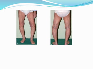
-Deformity result from capsular
contracture or joint instability.
-Loss of function e.g. a limp,
difficulty in climbing stairs,
restriction of walking distance or
progressive inability to perform
everyday tasks or enjoy recreation.
23.
24. Local tenderness is common, and is accompanied by
crepitus.
-In the late stages, joint instability may occur for any
of three reasons; loss of cartilage and bone;
asymmetrical capsular contracture; and muscle
weakness
25. Other joints should always
be examined;
they may show signs of a
more generalized disorder.
Also it may add to the
difficulties in the one
complained of e.g. a stiff LSS
or an unstable knee making it
more difficult to cope with
restricted movement in an
arthritic hip.
X ray appearances don’t
always correlate with either
the degree of pain or the
patient’s actual functional
capacity.
26. affected joints:
Malalignment with a bony
enlargement may occur
No erythema or warmth over the
affected joint(s); however, an
effusion may be present.
Limitation of joint motion or
muscle atrophy
Heberden nodes in DIP joints, and
Bouchard nodes in PIP joints
27. Imaging:
-Asymmetric loss of cartilage
(narrowing of the 'joint
space'),
-Sclerosis of the subchondral .
-Cysts
-Osteophytes at the margin of
the joint and
-Remodeling of the bone ends
on either side of the joint.
Sclerosis and osteophytes
distinguish OA from RA in
which the predominant
radiological feature is bone loss.
28.
29.
30.
31.
32.
33.
34.
35. CT & MRI:
sometimes needed to
elucidate a specific problem
e.g avascular necrosis. Also
used for grading of severity in
clinical trials.
Arthroscopy:
Arthroscopy may show cartilage
damage long before x-ray
changes appear.
Outerbridge classified articular cartilage damage based on the
arthroscopic findings in patients affected with osteoarthritis
4 grades:
Grade I - Softening and swelling
Grade II - Fragmentation and fissuring of less than 0.5
inches
Grade III - Fragmentation and fissuring of greater than
0.5 inches
Grade IV - Erosion down to the subchondral bone
36. Natural history
OA usually evolves as a slowly progressive disorder.
However, symptoms may wax and wane in intensity,
sometimes disappearing for several months.
Complications
Loose bodies: leading to episodes of locking.
Capsular herniation e.g. baker’s cyst in the knee.
Rotator cuff dysfunction: OA of the ACJ my cause RC
impingement, tendinitis or tears.
Spinal stenosis.
Sondyloleisthesis: destructive OA of the apophyseal joints
may result in segmental instability and spondyloleithesis ; the
so called degenerative spondyloleithesis, mostly at L4/5.
37. Differential Diagnoses
Rheumatoid arthritis
predominately affects the wrists and
the metacarpophalangeal (MCP)
and PIP joints.
Rheumatoid arthritis rarely, if ever,
involves the DIP joints or lumbosacral
spine.
Rheumatoid arthritis is associated with
prominent prolonged (>1 h) morning
stiffness.
Radiographic findings of rheumatoid
arthritis include bone erosion (eg,
periarticular osteopenia, marginal
erosions of bone) rather than
formation.
Laboratory findings that further
differentiate rheumatoid arthritis
include systemic inflammation,
positive rheumatoid factor results, joint
fluid with polymorphonuclear cell
predominance, and a substantially
elevated WBC count.
38. Clinical history and characteristic radiographic
findings differentiate spondyloarthropathy
from sacroiliac and lumbosacral spine
involvement.
Neuropathic [Charcot] joint
Reactive Arthritis
39. Treatment:
1-Conservative Treatment:
The main line of treatment
in the majority of patients
Analgesics are prescribed for
pain
Warmth (e.g. radiant heat or
shortwave diathermy) is
soothing.
Physiotherapy; ROM and
Muscle strengthening exercises.
A simple elastic support may
do wonders, probably by
improving proprioception in an
unstable knee.
41. 2-Intra-articular
Injections:
Intra-articular
corticosteroid injections will
often relieve pain, although
repeated injections may
permit (or even predispose
to) progressive cartilage and
bone destruction.
Intra-articular Hyaluronic
acid injections: can provide
temporary symptomatic
improvement specially early
cases
42. 3-Operative Ttreatment:
Indications:
Persistent pain unresponsive to conservative treatment
Progressive deformity and
Instability are the usual for operative treatment.
43. Procedures:
Arthroscopic washouts, with trimming of degenerate
meniscal tissue and osteophytes, may give temporary
relief; this is a useful measure when there are
contraindications to replacement surgery.
44. Realignment osteotomy:
Usually indicated in a relatively
'young' patient (under 50 years)
with a varus knee and
osteoarthritis confined to the
medial compartment:' a high
tibial valgus osteotomy will
redistribute weight to the lateral
side of the joint.
This is often successful in
relieving symptoms and saving
patient from replacement surgery.
45. Replacement arthroplasty: is indicated
in older patients with progressive joint destruction.
Joint Replacement
ideal
l painless joint
l full range of movement
l stable
l permanent
46. Types:
Total Knee Replacement:
Most common procedure
in advanced arthritis
Unicompartmental
replacement: If the disease
is largely confined to one
compartment, a can be
done as an alternative to
osteotomy.
.
helps to improve their adaptability and stability;
covered by a film of synovial fluid,
Articular Cartilage Comprises:
Chondrocytes (5%)
Extracellular Matrix (95%)
Water (75%)
Collagen (mainly type 2) (5%)
Proteoglycans (20%)
Enzymes
Growth Factors (PDGF, TGF beta, FGF, IGF-1)
Lipids
Adhesives (fibronectin, chondronectin)
Proteoglycans have a strong affinity for water, resulting in the collagen network being subjected to considerable tensile stresses.
Genetic:
and others have published similar observations for OA of the hip (Lanyon et al., 2000). However, one should bear in mind that OA of large joints is often attributable to anatomical variations, e.g. acetabular dysplasia and other forms of epiphyseal dysplasia, and it is these that are inherited rather than any tendency to develop OA as a primary abnormality.
Sources of pain in osteoarthritis include the following:
Joint effusion
stretching of the joint capsule
Increased vascular pressure in subchondral bone
Torn menisci
Inflammation of periarticular bursae
Periarticular muscle spasm
Psychological factors
X-rays: so characteristic so that other forms of imaging are seldom necessary for ordinary clinical assessement
=In the late stage, displacement of the joint is common and bone destruction may be severe
=There may be evidence of previous disorders (congenital defects, old fractures, rheumatoid arthritis, chondrocalcinosis).
Radionuclide scanning with 99mTc_HDP shows increased activity during the bone phase in the subchondral regions of affected joints. This is due to increased vascularity and new bone formation
painless joint
full range of movement
stable
Permanent
With modem techniques, and meticulous attention to anatomical alignment of the knee, the results of replacement arthroplasty are excellent
Joint Replacement knee complications
limited ROM
patellar instability 3-5%
loosening > 90% 10y survival
DVT / PE
infection - 2%