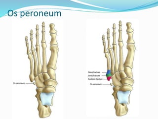
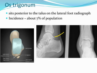
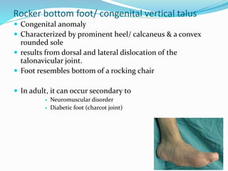
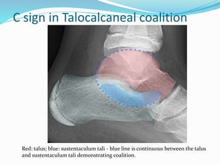
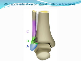
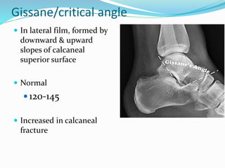
SlideShare a Scribd company logo
Dr. Manmohan Bir Shrestha
FOR RADIOLOGY
Contents
 Accessory ossicles of the foot
 Rocker bottom foot
 Tarsal coalition
 Fractures around the ankle
 Plantar fascia
 Brodie’s abscess in distal tibia
 Soft tissue masses around ankle
 Retrocalcaneal bursitis
 Haglund syndrome
 Achilles tendinopathy
 Achilles tendon tears
Accessory ossicles of the foot
 are secondary ossification centers that are separate from
the adjacent bone
 Usually round or ovoid
 have well defined smooth cortical margins on all sides.
 Clinical importance
 May fracture
 Can cause pain
 May be involved in rheumatoid arthritis, osteoarthritis, infections
& hyperparathyroidism
Accessory ossicles of the foot
 Os tibiale externum
 Os trigonum
 Os peroneum
 Os vesalianum
 Os subtibiale
 Os subfibulare
 Os supratalare
 Os cancaneus secundarius
Os tibiale externum
 Also called accessory navicular
 is present adjacent to the medial side of navicular
 Can cause painful tendinosis of tibialis posterior
tendon
 Imaging
 best visualized on the lateral-oblique view
 may appear as a 'hot spot' on bone scan
 on MRI, bone marrow oedema can be seen
Bilateral Os tibiale externum
Os peroneum
 Commonly found – 8%
 located at the lateral plantar aspect of the cuboid
within the substance of the peroneus longus tendon
 D/D
 Os vesalianum
 Apophysis of the 5th metatarsal
 Avulsion fracture
Os peroneum
Os peroneum
Fractured Os peroneum
Os vesalianum
 situated at the base of the fifth metatarsal in the
peroneus brevis tendon
 Less common than os peroneum
 D/D
 Os peroneum
 Apophysis of 5th metatarsal – in children
 Avulsion fracture
Os vesalianum
Os trigonum
 sits posterior to the talus on the lateral foot radiograph
 Incidence – about 7% of population
Less common accessory ossicles
 Os subtibiale
 related to the posterior colliculus of the medial malleolus
 Os subfibulare
 lies at the tip of the lateral malleolus
 Os supratalare
 located at the superior aspect of the talar head or neck
 Os cancaneus secundarius
 Located at anterior calcaneal process
Rocker bottom foot
Rocker bottom foot/ congenital vertical talus
 Congenital anomaly
 Characterized by prominent heel/ calcaneus & a convex
rounded sole
 results from dorsal and lateral dislocation of the
talonavicular joint.
 Foot resembles bottom of a rocking chair
 In adult, it can occur secondary to
 Neuromuscular disorder
 Diabetic foot (charcot joint)
Imaging findings
 fixed equinus: plantarflexion of the calcaneus
 vertical talus: plantarflexion of the talus
 irreducible dorsal subluxation or dislocation of the
navicular
 forefoot valgus: divergence of bases of the metatarsal heads
on AP and superimposition of the metatarsal bones on the
lateral view
 long axis of the talus passes plantar to metatarsal axis on
lateral view and medial to the first metatarsal on AP view
Rocker bottom foot
AP view shows calcaneus valgus
& metatarsal valgus. The long
axis of talus is much medial to
st
On lateral view, there is equinus of the
calcaneus and vertical orientation of talus.
Navicular has not yet ossified
Tarsal coalition
Tarsal coalition
 complete or partial union between two or more bones
in the midfoot & hindfoot
 Incidence – about 5 % of population
 Patient usually present in adolescence
 refers to developmental fusion rather than fusion that
is acquired secondary to conditions such
as rheumatoid arthritis, trauma or post-surgical.
Types
 They may be of 3 types, depending on the tissue which
bridges between the two bones. The three types are 1:
I. bony: synostosis
II. cartilaginous: synchondrosis
III. fibrous: syndesmosis
Tarsal coalition
 Calcaneonavicular – about 45%
 Talocalcaneal - about 45%
 Less common are
 Calcaneocuboid
 Talonavicular
 cubonavicular
Calcaneonavicular coalition
 Oblique view x-ray is best
 MRI - more helpful in assessing and characterising
cartilaginous and fibrous coalition
Calcaneonavicular coalition
Talocalcaneal coalition
 Although all three facets of the talocalcaneal joint can be
involved, the middle facet is most commonly involved.
 Often requires cross-sectional imaging for accurate diagnosis
 Plain radiograph
 C-sign(lateral film) – posterior continuity of talus & sustentaculum
tali
 Talar beak sign (latearal film) – prominent beak at the anterior end
of talus
 non-visualisation of the middle articular facet
 Sclerosis around the articular margins of the talocalcaneal joint
 CT – coronal reformats are best
 MRI
C sign in Talocalcaneal coalition
Red: talus; blue: sustentaculum tali - blue line is continuous between the talus
and sustentaculum tali demonstrating coalition.
A case of Talocalcaneal coalition
Fractures around the ankle
 Lateral malleolus fracture
 Fracture through the Tibial plafond
 Talus fracture
 Calcaneus fracture
Lateral malleolus fracture
 commonly the result of twisting injury of the talus in
ankle mortise.
 Radiographs are usually sufficient for the management
of what are typically simple fractures.
 CT axial images through both ankles are useful when
the integrity of the syndesmosis is questioned.
 “Weber” staging system for ankle fractures – to
understand mechanism of syndesmotic injury.
 syndesmotic injuries usually require screw fixation
Weber classification of lateral malleolar fractures
 Type A
 Below talar dome
 Usually transverse
 Syndesmosis intact
 Deltoid ligament intact
 Type B
 distal extent at the level of the talar dome
 Usually spiral
 syndesmosis usually intact, but widening of the distal tibiofibular joint
(especially on stressed views) indicates syndesmotic injury
 deltoid ligament may be torn, indicated by widening of the space between
the medial malleolus and talar dome
 Type C
 above the level of the ankle joint
 tibiofibular syndesmosis disruption with widening of the distal tibiofibular
articulation
 medial malleolus fracture or deltoid ligament injury often present
Weber classification of lateral malleolar fractures
Weber A
Transverse fracture through lateral malleolus below ankle joint – “Weber A”
Weber B
There is oblique fracture of distal fibula.
fracture extends distally to the level of the ankle joint.
There is no significant widening of the tibiofibular joint to suggest syndesmosis tear.
Weber C
Widened
syndesmosis
Fracture through the Tibial plafond
 In adult –
 Pilon fracture
 In adolescents
 Tillaux fracture
 Triplane fracture
Pilon Fracture
 any tibial fracture that involves the distal articular
plafond and are typically the result of an axial loading
force
 can produce significant comminution with multiple
displaced fracture fragments
 X-ray & CT
 fractures lines are seen extending into the tibiotalar
articular surface

Pilon fracture
28 yrs. after Road
Traffic Accident
There is a comminuted
distal tibial fracture
extending into
the tibial plafond
Juvenile Tillaux fracture
 Salter-Harris type 3 fracture
 Have characteristic appearance on CT
 Mechanism - an external rotation force pulling on the
anterior tibiofibular ligament, causing avulsion of the
anterolateral corner of the distal tibial epiphysis with
variable amount of displacement
 Why always laterally?
 because the distal tibial physis fuses from medial to lateral as
a child matures
 Age
 Adolescents in whom, lateral growth plate has not fused
12-15 years
Tillaux fracture – 15 years male
Tillaux fracture, i.e. Salter-Harris III fracture of anterolateral
aspect of distal tibial epiphysis, minimally displaced.
Triplane fracture
 Salter-Harris type 4
 Multiplanar CT scans are ideally suited to visualize
these fractures in all planes
 The name is due to the fact of the fracture
expanding both in frontal and lateral as well as
transverse planes
 It comprises of
 a vertical fracture through the epiphysis
 a horizontal fracture through the physis
 an oblique fracture through the metaphysis
 Age
 adolescents
Triplane fracture in adolescent
Tibial physis closed medially. Fracture in lateral tibia extending into epiphysis
and metaphysis consistent with Triplane fracture. No major dislocations.
Review of Salter-Harris classification of
physeal fractures
 1 – Slipped
 2 – Above
 3 – Lower
 4 – Together
 (epi, metaphysis &
growth plate)
 5 - Rammed
Talar Fracture
Talar fracture
 Location
 Head
 Neck
 Body
 Talar dome osteochondral fracture
 Posterior talar process fracture
 Lateral talar process fracture
Talar dome osteochondral defect/injury
 These are are focal areas of articular damage with
cartilage damage and injury of the adjacent
subchondral bone
 Plain x-ray findings can be normal in early stages
 MRI more sensitive and specific
Anderson staging of osteochondral
defect
 Stage I
 “subchondral trabecular compression”
 Plain film & CT negative
 Bone marrow edema in MRI
 Stage II
 “incomplete separation of the fragment,”
 X-ray – only thin sclerotic rim
 IIa – if subchondral cyst
 Stage III
 “unattached, undisplaced fragment,”
 Presence of synovial fluid in T2 around fragment
 Stage IV
 Displaced fragment
Anderson Stage I defect
Anderson Stage II defect
Anderson Stage IIa defect
Anderson Stage III defect
Anderson Stage IV defect
Frequently missed in plain films
Bohler/Tuber/Calcaneal angle
 Angle between 2 lines in
lateral film
 Line 1 – vertex to
postero-superior edge
 Line 2 – vertex to
anterior horn of
calcaneum
 Normal
 20-40
 Less than 20 degrees in
calcaneal fracture
Gissane/critical angle
 In lateral film, formed by
downward & upward
slopes of calcaneal
superior surface
 Normal
 120-145
 Increased in calcaneal
fracture
Chopart fracture/injury
 Fracture/dislocation of the mid-
tarsal joint i.e. talonavicular &
calcaneocuboid joints, which
separates hindfoot from midfoot
 The commonly fractured bones are
calcaneum, cuboid & navicular
 The foot is usually dislocated
medially & superiorly as it is
plantar flexed & inverted, usually
as a result of high energy impact
 Where the foot is everted, lateral
displacement occurs
Lisfranc injury/fracture
 are the most common type of
dislocation involving
the foot and correspond to the
dislocation of the articulation of
the tarsus with the metatarsal
bases
 Displacement can also occur
from fractures at distal
metatarsals
A case of Lisfranc fracture/dislocation
Figure - Divergent Lisfranc dislocation in a 34-year-old who was the front passenger in
a motor vehicle accident. Radiographs were obtained in the emergency department.
Anteroposterior (A) and oblique (B) views of the foot reveal lateral dislocation of the
second through fifth metatarsals. The white arrow points to a fragment fractured off
the base of the second metatarsal.
Less obvious on the lateral view
Plantar fascia
Plantar fasciitis
Plantar fascia tear
Plantar fascia calcification
Plantar fascia
 dense collection of collagen fibres on the sole (plantar
surface) of the foot. These fibres are mostly longitudinal
but also transverse
 Attachments
 Posteriorly it attaches to the medial process of the tuberosity
of the calcaneus, proximal to flexor digitorum brevis. It is
narrow and thick at this attachment and becomes more broad
and thin distally and anteriorly.
 Anteriorly it divides into five heads, one for each toe, just
proximal to the heads of the metatarsals. The superficial
layers of these fibres insert into the dermis at the ball of the
foot and the crease between the ball and the toes via the
retinacula cutis (skin ligaments). The deep layers of each digit
become septa that separate the digital flexor tendons from
the lumbricals and the digital vessels and nerves.
 Laterally it covers abductor digiti minimi.
 Medially it covers abductor hallucis and merges with
the flexor retinaculum and dorsalis paedis fascia.
X-RAY
MRI
HYPOINTENSE
Plantar fascitis
 Most common cause of pain in heel
 is a stress reaction occurring at the origin of the
plantar aponeurosis from the calcaneus, typically at
the medial calcaneal tubercle.
 Etiology - degenerative changes from repetitive
microtrauma in the origin of the plantar fascia cause
traction periostitis and microtears
Imaging Plantar Fascitis
 Plain radiograph
 Non-specific
 Associated calcaneal spur can be found
 MRI
 Thickened >4.5 mm
 Edema around the origin of the aponeurosis
 there may be edema in the underlying calcaneal
bone marrow
A case of plantar fasciitis –
Thick fascia near origin with high signal intensity
Plantar fascial tear
 refer to disruption of plantar fascial fibres which
can occur in associated with longstanding plantar
fascitis or those treated with steroid injections.
 The tears can be complete (i.e. rupture) or
incomplete.
 MRI – T1WI
 absence of T1-weighted low signal intensity at the site
of complete rupture or partial loss of T1-weighted low
signal intensity.

Plantar fascia calcification
Brodie’s abscess in distal tibia
 chronic intraosseous abscess resulting from
incomplete resolution of acute osteomyelitis
 Distal tibial metaphysis – 1 of the common location of
brodie’s abscess.
 Rarely cross the growth plate & epiphysis in children
 Age- children with unfused epiphyseal plates
 Pathology – Staphylococcus aureus
Imaging
 Plain radiograph
 lytic lesion often oval that is oriented along the long axis of
the bone
 surrounded by a thick dense rim of reactive sclerosis
 Periosteal reaction - +-
 CT
 central intramedullary hypodense cystic lesion with thick rim
ossification
 Extensive periosteal reaction and bone sclerosis around the
lesion
 MRI
 T2 & STIR hyperintense rim with surrounding hypointense
sclerotic rim
 Adjacent bone marrow edema +-
Case of 13 yrs. Old with Brodie’s abscess in distal tibia
Lytic lesion in
metadiaphyseal
region of distal tibia
MRI
Bilobed T1 hypointense and T2 hyperintense lesion
with surrounding sclerosis.
Adjacent marrow edema is also present
Soft tissue masses/tumors around Ankle
I. Synovial cysts or ganglia
II. Schwannomas
III. Plantar fibromas
IV. Giant cell tumor of the tendon sheath
Synovial cysts or ganglia
 Most common around ankle & foot
 are para-articular fluid-filled sacs or pouch-
like structures containing synovial fluid and lined by
synovial membrane
 MRI – uniformly bright on fluid-sensitive images
Synovial cyst in 51 year old
Lateral radiograph shows a
round soft tissue mass
dorsal to the metatarsals
T1 – Iso
T2 – Bright
Post contrast – peripheral
enhancement
Plantar fibromas
 Plantar fibromas can have variable signal
characteristics but are typically dark on all sequences
 These are usually found in the plantar fat adjacent to
the aponeurosis, usually close to the calcaneus
A case of Plantar fibroma in a 44-year-old
Coronal T1-weighted (A), proton-density–weighted
(B), and T2-weighted (C) images reveal that the lesion (arrows) is relatively dark on
all sequences and confined to the fat of the plantar heel pad
Giant cell tumor of tendon sheath
 is a localized form of pigmented villonodular synovitis
 usually benign lesions that arise from the tendon
sheath
 localized solitary subcutaneous soft tissue nodules
Plain radiograph
 they may cause pressure erosions on the underlying
bone in 10-20% of cases
 more commonly these masses arise from the palmar
tendons
 the mass itself is of soft tissue density
 periosteal reaction and calcification are uncommon
MRI
 T1WI
 low signal
 T2WI
 low signal
 Post contrast
 moderate enhancement
 GRE
 low and may demonstrate blooming
Case of Giant cell tumor of tendon sheath in 18 year-old
A well defined oval shape mass is seen anterior to the talus, extra articular in
position and underneath the tendon of the Extensor hallucis longus. The mass
displays low signal on T1, lower signal on T2 likely from hemosiderin deposition,
heterogeneous high signal on STIR, and avid enhancement on post contrast study.
No evidence of infiltration of the adjacent structures.
Retrocalcaneal Bursitis
 refers to inflammation of the retrocalcaneal bursa,
which lies between the antero-inferior calcaneal
tendon and posterosuperior calcaneus.
 It forms part of Haglund syndrome.
 Note – there is another 1 bursa i.e. subcutaneous
calcaneal bursa (between the tendon and the skin)
 rarely occurs in isolation and is almost always
associated with calcaneal tendinitis and/or Haglund
deformity
 Bursae - are small fluid-filled sacs lined by synovial
membrane with an inner capillary layer of synovial
fluid
causes
 calcaneal tendon injury: rupture or tendinitis
 inflammatory arthropathies: Reiter
syndrome, ankylosing spondylitis, rheumatoid
arthritis
 calcaneal fractures
 infectious bursitis
Imaging
 Plain film
 prominence of the posterosuperior calcanum can be
frequently seen 1
 decreased lucency of the retrocalcaneal soft tissue (Kager
triangle)
 Ultrasound
 bursa distension by a hypoechogenic fluid collection: > 1 mm
anteroposteriorly, > 7 mm craniocaudally, or > 11 mm
transversely is considered abnormal
 MRI
 fluid collection:
 T1: low signal
 T2: high signal
 STIR: high signal
Case of Retrocalcaneal Bursitis
Normal achilles tendon.
Fluid collection deep to tendon
Haglund syndrome
 Refers to Haglund triad of
1) insertional Achilles
tendinopathy
2) retrocalcaneal bursitis
3) posterosuperior
calcaneal exostosis
 Associated with calcaneal spurs
 Wearing high heels
 Stiffed backed shoes
Imaging
 Plain film
 loss of the Kager triangle due to retrocalcaneal bursitis
 Achilles tendon measuring over 9 mm in thickness 2
cm above the bursal projection due to Achilles
tendinopathy
 Postero-superior calcaneal spur
 MRI
 focal enlargement and abnormal signal at Achilles
tendon insertion segment
 retrocalcaneal and retroachilles bursal fluid collection
 calcaneal bony spur better appreciated on T1 sagittal
images
 marrow oedema of the posterior calcaneal tuberosity
Case of Haglund syndrome in 20 yrs. male
High signal near
achilles insertion
Achilles tendinopathy
 Macroscopically, tendinopathy results in
enlargement, disruption of fibrillar pattern and an
increase in tendon vascularity
 Ultrasound
 shows thickening and rounding of the affected portion of the
tendon. The cutoff value of 1 cm in anteroposterior diameter
is usually used for diagnosis.
 Additional signs include increased Kager’s fat pad
echogenicity.
 MRI
 shows increased intratendinous signal and tendon
enlargement, with oedema in Kager's fat pad in cases of
tendinosis.
Case of Achilles tendinopathy.
Patient with pain on dorsiflexion
Thickening of the achilles tendon
on the lateral view of the ankle can
be seen 5-6 cm above its insertion
into the calcaneum
Achilles tendon tears
 Pathology
 interstitial tears (parallel to the long axis of
the Achilles)
 partial tears
 complete tears.
 Location
 ruptures in the 'critical zone', which is a region
of relative watershed hypovascularity 2-6 cm
proximal to insertion
Kuwada classification of Achilles
tendon tear
 type I: partial ruptures ≤50%
 typically treated with conservative management
 type II: complete rupture with tendinous gap ≤3 cm
 typically treated with end-end anastomosis
 type III: complete rupture with tendinous gap 3 to 6 cm
 often requires tendon/synthetic graft
 type IV: complete rupture with defect of >6 cm (neglected
ruptures)
 often requires tendon/synthetic graft and gastrocnemius
recession
Case of Kuwada type I tear
thickened and hypoechoic right Achilles tendon with discontinuation of its deep
fibers at its insertion site at calcaneum with adjacent fluid.
Findings are suggestive of tendinopathy with partial thickness tear
involving less than 50% fibres
Case of Kuwada Type II tear
Achilles tear, just above the insertion

More Related Content

What's hot

Imaging of shoulder - Dr. Vishal Sankpal
Imaging of shoulder - Dr. Vishal SankpalImaging of shoulder - Dr. Vishal Sankpal
Imaging of shoulder - Dr. Vishal Sankpal
Vishal Sankpal
 
MRI KNEE OF ORTHOPEDIC IMPORTANCE
MRI KNEE OF ORTHOPEDIC IMPORTANCEMRI KNEE OF ORTHOPEDIC IMPORTANCE
MRI KNEE OF ORTHOPEDIC IMPORTANCE
Benthungo Tungoe
 
Presentation1.pptx, ultrasound of the hand and fingers.
Presentation1.pptx, ultrasound of the hand and fingers.Presentation1.pptx, ultrasound of the hand and fingers.
Presentation1.pptx, ultrasound of the hand and fingers.
Abdellah Nazeer
 
MR Imaging of shoulder and knee joints
MR Imaging of shoulder and knee jointsMR Imaging of shoulder and knee joints
MR Imaging of shoulder and knee joints
Sahil Chaudhry
 
Presentation1.pptx mri of elbow joint
Presentation1.pptx mri of elbow jointPresentation1.pptx mri of elbow joint
Presentation1.pptx mri of elbow joint
Abdellah Nazeer
 
Mri shoulder joint with common pathologies
Mri shoulder joint with common pathologiesMri shoulder joint with common pathologies
Mri shoulder joint with common pathologies
Gobardhan Thapa
 
HOW TO READ SHOULDER MRI
HOW TO READ SHOULDER MRI HOW TO READ SHOULDER MRI
HOW TO READ SHOULDER MRI
Arjun Kouloth
 
Diagnostic Imaging of Bone Tumors
Diagnostic Imaging of Bone TumorsDiagnostic Imaging of Bone Tumors
Diagnostic Imaging of Bone Tumors
Mohamed M.A. Zaitoun
 
Presentation1.pptx. imaging of the cartilage.
Presentation1.pptx. imaging of the cartilage.Presentation1.pptx. imaging of the cartilage.
Presentation1.pptx. imaging of the cartilage.
Abdellah Nazeer
 
Role of ultrasound in clinical evaluation of shoulder Dr. Muhammad Bin Zulfiqar
Role of ultrasound in clinical evaluation of shoulder Dr. Muhammad Bin ZulfiqarRole of ultrasound in clinical evaluation of shoulder Dr. Muhammad Bin Zulfiqar
Role of ultrasound in clinical evaluation of shoulder Dr. Muhammad Bin Zulfiqar
Dr. Muhammad Bin Zulfiqar
 
Presentation1, radiological imaging of anterior knee pain.
Presentation1, radiological imaging of anterior knee pain.Presentation1, radiological imaging of anterior knee pain.
Presentation1, radiological imaging of anterior knee pain.
Abdellah Nazeer
 
Lecture 3 shah radiology in foot and ankle
Lecture 3 shah radiology in foot and ankleLecture 3 shah radiology in foot and ankle
Lecture 3 shah radiology in foot and ankle
Selene G. Parekh, MD, MBA
 
Mri anatomy of ankle
Mri anatomy of ankleMri anatomy of ankle
Mri anatomy of ankle
Shrikant Nagare
 
Knee mri
Knee mriKnee mri
Knee mri
Sitanshu Barik
 
Presentation2.pptx wrist joint.
Presentation2.pptx wrist joint.Presentation2.pptx wrist joint.
Presentation2.pptx wrist joint.
Abdellah Nazeer
 
Ankle joint radiography
Ankle joint radiographyAnkle joint radiography
Ankle joint radiography
Nikhil Murkey
 
74-Dr Ahmed Esawy imaging oral board MRI ankle & foot part I
74-Dr Ahmed Esawy imaging oral board MRI ankle & foot part I74-Dr Ahmed Esawy imaging oral board MRI ankle & foot part I
74-Dr Ahmed Esawy imaging oral board MRI ankle & foot part I
AHMED ESAWY
 
Imaging in arthritis
Imaging in arthritisImaging in arthritis
Imaging in arthritis
Navni Garg
 
MRI of the shoulder
MRI of the shoulderMRI of the shoulder
MRI of the shoulder
Thorsang Chayovan
 
Systemic approach to bone tumor radiology
Systemic approach to bone tumor radiologySystemic approach to bone tumor radiology
Systemic approach to bone tumor radiology
Ramin Sadeghi
 

What's hot (20)

Imaging of shoulder - Dr. Vishal Sankpal
Imaging of shoulder - Dr. Vishal SankpalImaging of shoulder - Dr. Vishal Sankpal
Imaging of shoulder - Dr. Vishal Sankpal
 
MRI KNEE OF ORTHOPEDIC IMPORTANCE
MRI KNEE OF ORTHOPEDIC IMPORTANCEMRI KNEE OF ORTHOPEDIC IMPORTANCE
MRI KNEE OF ORTHOPEDIC IMPORTANCE
 
Presentation1.pptx, ultrasound of the hand and fingers.
Presentation1.pptx, ultrasound of the hand and fingers.Presentation1.pptx, ultrasound of the hand and fingers.
Presentation1.pptx, ultrasound of the hand and fingers.
 
MR Imaging of shoulder and knee joints
MR Imaging of shoulder and knee jointsMR Imaging of shoulder and knee joints
MR Imaging of shoulder and knee joints
 
Presentation1.pptx mri of elbow joint
Presentation1.pptx mri of elbow jointPresentation1.pptx mri of elbow joint
Presentation1.pptx mri of elbow joint
 
Mri shoulder joint with common pathologies
Mri shoulder joint with common pathologiesMri shoulder joint with common pathologies
Mri shoulder joint with common pathologies
 
HOW TO READ SHOULDER MRI
HOW TO READ SHOULDER MRI HOW TO READ SHOULDER MRI
HOW TO READ SHOULDER MRI
 
Diagnostic Imaging of Bone Tumors
Diagnostic Imaging of Bone TumorsDiagnostic Imaging of Bone Tumors
Diagnostic Imaging of Bone Tumors
 
Presentation1.pptx. imaging of the cartilage.
Presentation1.pptx. imaging of the cartilage.Presentation1.pptx. imaging of the cartilage.
Presentation1.pptx. imaging of the cartilage.
 
Role of ultrasound in clinical evaluation of shoulder Dr. Muhammad Bin Zulfiqar
Role of ultrasound in clinical evaluation of shoulder Dr. Muhammad Bin ZulfiqarRole of ultrasound in clinical evaluation of shoulder Dr. Muhammad Bin Zulfiqar
Role of ultrasound in clinical evaluation of shoulder Dr. Muhammad Bin Zulfiqar
 
Presentation1, radiological imaging of anterior knee pain.
Presentation1, radiological imaging of anterior knee pain.Presentation1, radiological imaging of anterior knee pain.
Presentation1, radiological imaging of anterior knee pain.
 
Lecture 3 shah radiology in foot and ankle
Lecture 3 shah radiology in foot and ankleLecture 3 shah radiology in foot and ankle
Lecture 3 shah radiology in foot and ankle
 
Mri anatomy of ankle
Mri anatomy of ankleMri anatomy of ankle
Mri anatomy of ankle
 
Knee mri
Knee mriKnee mri
Knee mri
 
Presentation2.pptx wrist joint.
Presentation2.pptx wrist joint.Presentation2.pptx wrist joint.
Presentation2.pptx wrist joint.
 
Ankle joint radiography
Ankle joint radiographyAnkle joint radiography
Ankle joint radiography
 
74-Dr Ahmed Esawy imaging oral board MRI ankle & foot part I
74-Dr Ahmed Esawy imaging oral board MRI ankle & foot part I74-Dr Ahmed Esawy imaging oral board MRI ankle & foot part I
74-Dr Ahmed Esawy imaging oral board MRI ankle & foot part I
 
Imaging in arthritis
Imaging in arthritisImaging in arthritis
Imaging in arthritis
 
MRI of the shoulder
MRI of the shoulderMRI of the shoulder
MRI of the shoulder
 
Systemic approach to bone tumor radiology
Systemic approach to bone tumor radiologySystemic approach to bone tumor radiology
Systemic approach to bone tumor radiology
 

Similar to Ankle joint pathology imaging

Fai and open surgery
Fai and open surgeryFai and open surgery
Fai and open surgery
Saikrishna Katragadda
 
Nof fracture
Nof fractureNof fracture
Nof fracture
Dr Chinmoy Mazumder
 
Msk signs edited
Msk signs   editedMsk signs   edited
Msk signs edited
Dalia El Said
 
Skeletal trauma imaging
Skeletal trauma imagingSkeletal trauma imaging
Skeletal trauma imaging
radiologyoffice
 
Role of sonography in knee joint diseases
Role of sonography in knee joint diseasesRole of sonography in knee joint diseases
Role of sonography in knee joint diseases
REKHAKHARE
 
Injuries of the ankle joint which can occur
Injuries of the ankle joint which can occurInjuries of the ankle joint which can occur
Injuries of the ankle joint which can occur
mdjstf48sq
 
Fracture talus
Fracture talusFracture talus
Fracture talus
orthoprince
 
appendicular trauma in radiology. .pptx
appendicular trauma in radiology.  .pptxappendicular trauma in radiology.  .pptx
appendicular trauma in radiology. .pptx
yashovrattiwari1
 
Ankle and Foot
Ankle and FootAnkle and Foot
Ankle and Foot
Tarique Ajij
 
Msk Lecture2 1st Hospital
Msk Lecture2 1st HospitalMsk Lecture2 1st Hospital
Msk Lecture2 1st Hospital
Sumit Prajapati
 
lowerlimbfractures-141119052341-conversion-gate01.pdf
lowerlimbfractures-141119052341-conversion-gate01.pdflowerlimbfractures-141119052341-conversion-gate01.pdf
lowerlimbfractures-141119052341-conversion-gate01.pdf
DominicLaibuni
 
Orthopedics 5th year, 4th lecture (Dr. Ali A.Nabi)
Orthopedics 5th year, 4th lecture (Dr. Ali A.Nabi)Orthopedics 5th year, 4th lecture (Dr. Ali A.Nabi)
Orthopedics 5th year, 4th lecture (Dr. Ali A.Nabi)
College of Medicine, Sulaymaniyah
 
Kuis 20 November.pdf
Kuis 20 November.pdfKuis 20 November.pdf
Kuis 20 November.pdf
AlfonsusCiptaRaya
 
fractures-of-lower-limb.ppt-1-Copyجاسم.ppt
fractures-of-lower-limb.ppt-1-Copyجاسم.pptfractures-of-lower-limb.ppt-1-Copyجاسم.ppt
fractures-of-lower-limb.ppt-1-Copyجاسم.ppt
RajveerYadav40
 
Lower limb fractures
Lower limb fracturesLower limb fractures
Lower limb fractures
airwave12
 
Ankle fracture
Ankle fractureAnkle fracture
Ankle fracture
Dr Prajith
 
Ankle injuries - Copy.pptx
Ankle injuries - Copy.pptxAnkle injuries - Copy.pptx
Ankle injuries - Copy.pptx
SwetaUpadhyay18
 
radiology.Spine 1.(dr.ameer)
radiology.Spine 1.(dr.ameer)radiology.Spine 1.(dr.ameer)
radiology.Spine 1.(dr.ameer)
student
 
Femur fracture and it management and cases
Femur fracture and it management and casesFemur fracture and it management and cases
Femur fracture and it management and cases
onkosurgery
 
Femur fracture
Femur fractureFemur fracture
Femur fracture
muhammad bilal
 

Similar to Ankle joint pathology imaging (20)

Fai and open surgery
Fai and open surgeryFai and open surgery
Fai and open surgery
 
Nof fracture
Nof fractureNof fracture
Nof fracture
 
Msk signs edited
Msk signs   editedMsk signs   edited
Msk signs edited
 
Skeletal trauma imaging
Skeletal trauma imagingSkeletal trauma imaging
Skeletal trauma imaging
 
Role of sonography in knee joint diseases
Role of sonography in knee joint diseasesRole of sonography in knee joint diseases
Role of sonography in knee joint diseases
 
Injuries of the ankle joint which can occur
Injuries of the ankle joint which can occurInjuries of the ankle joint which can occur
Injuries of the ankle joint which can occur
 
Fracture talus
Fracture talusFracture talus
Fracture talus
 
appendicular trauma in radiology. .pptx
appendicular trauma in radiology.  .pptxappendicular trauma in radiology.  .pptx
appendicular trauma in radiology. .pptx
 
Ankle and Foot
Ankle and FootAnkle and Foot
Ankle and Foot
 
Msk Lecture2 1st Hospital
Msk Lecture2 1st HospitalMsk Lecture2 1st Hospital
Msk Lecture2 1st Hospital
 
lowerlimbfractures-141119052341-conversion-gate01.pdf
lowerlimbfractures-141119052341-conversion-gate01.pdflowerlimbfractures-141119052341-conversion-gate01.pdf
lowerlimbfractures-141119052341-conversion-gate01.pdf
 
Orthopedics 5th year, 4th lecture (Dr. Ali A.Nabi)
Orthopedics 5th year, 4th lecture (Dr. Ali A.Nabi)Orthopedics 5th year, 4th lecture (Dr. Ali A.Nabi)
Orthopedics 5th year, 4th lecture (Dr. Ali A.Nabi)
 
Kuis 20 November.pdf
Kuis 20 November.pdfKuis 20 November.pdf
Kuis 20 November.pdf
 
fractures-of-lower-limb.ppt-1-Copyجاسم.ppt
fractures-of-lower-limb.ppt-1-Copyجاسم.pptfractures-of-lower-limb.ppt-1-Copyجاسم.ppt
fractures-of-lower-limb.ppt-1-Copyجاسم.ppt
 
Lower limb fractures
Lower limb fracturesLower limb fractures
Lower limb fractures
 
Ankle fracture
Ankle fractureAnkle fracture
Ankle fracture
 
Ankle injuries - Copy.pptx
Ankle injuries - Copy.pptxAnkle injuries - Copy.pptx
Ankle injuries - Copy.pptx
 
radiology.Spine 1.(dr.ameer)
radiology.Spine 1.(dr.ameer)radiology.Spine 1.(dr.ameer)
radiology.Spine 1.(dr.ameer)
 
Femur fracture and it management and cases
Femur fracture and it management and casesFemur fracture and it management and cases
Femur fracture and it management and cases
 
Femur fracture
Femur fractureFemur fracture
Femur fracture
 

More from macshrestha

Cerebral/Pial Arteriovenous Malformation (AVM)
Cerebral/Pial Arteriovenous Malformation (AVM)Cerebral/Pial Arteriovenous Malformation (AVM)
Cerebral/Pial Arteriovenous Malformation (AVM)
macshrestha
 
Hepatic peliosis
Hepatic peliosisHepatic peliosis
Hepatic peliosis
macshrestha
 
Cns tuberculoma
Cns tuberculomaCns tuberculoma
Cns tuberculoma
macshrestha
 
Benign bone tumors
Benign bone tumorsBenign bone tumors
Benign bone tumors
macshrestha
 
Dural venous sinus thrombosis for Radiology & Imaging
Dural venous sinus thrombosis for Radiology & ImagingDural venous sinus thrombosis for Radiology & Imaging
Dural venous sinus thrombosis for Radiology & Imaging
macshrestha
 
Empyema necessitans
Empyema necessitansEmpyema necessitans
Empyema necessitans
macshrestha
 
Extraskeletal Mesenchymal Chondrosarcoma
Extraskeletal Mesenchymal ChondrosarcomaExtraskeletal Mesenchymal Chondrosarcoma
Extraskeletal Mesenchymal Chondrosarcoma
macshrestha
 
Ct head
Ct headCt head
Ct head
macshrestha
 
Brain abscess
Brain abscessBrain abscess
Brain abscess
macshrestha
 
Choledochal cyst
Choledochal cystCholedochal cyst
Choledochal cyst
macshrestha
 
Pulmonary metastases
Pulmonary metastasesPulmonary metastases
Pulmonary metastases
macshrestha
 
Simple bone cyst
Simple bone cystSimple bone cyst
Simple bone cyst
macshrestha
 

More from macshrestha (12)

Cerebral/Pial Arteriovenous Malformation (AVM)
Cerebral/Pial Arteriovenous Malformation (AVM)Cerebral/Pial Arteriovenous Malformation (AVM)
Cerebral/Pial Arteriovenous Malformation (AVM)
 
Hepatic peliosis
Hepatic peliosisHepatic peliosis
Hepatic peliosis
 
Cns tuberculoma
Cns tuberculomaCns tuberculoma
Cns tuberculoma
 
Benign bone tumors
Benign bone tumorsBenign bone tumors
Benign bone tumors
 
Dural venous sinus thrombosis for Radiology & Imaging
Dural venous sinus thrombosis for Radiology & ImagingDural venous sinus thrombosis for Radiology & Imaging
Dural venous sinus thrombosis for Radiology & Imaging
 
Empyema necessitans
Empyema necessitansEmpyema necessitans
Empyema necessitans
 
Extraskeletal Mesenchymal Chondrosarcoma
Extraskeletal Mesenchymal ChondrosarcomaExtraskeletal Mesenchymal Chondrosarcoma
Extraskeletal Mesenchymal Chondrosarcoma
 
Ct head
Ct headCt head
Ct head
 
Brain abscess
Brain abscessBrain abscess
Brain abscess
 
Choledochal cyst
Choledochal cystCholedochal cyst
Choledochal cyst
 
Pulmonary metastases
Pulmonary metastasesPulmonary metastases
Pulmonary metastases
 
Simple bone cyst
Simple bone cystSimple bone cyst
Simple bone cyst
 

Recently uploaded

5 Effective Homeopathic Medicines for Irregular Periods
5 Effective Homeopathic Medicines for Irregular Periods5 Effective Homeopathic Medicines for Irregular Periods
5 Effective Homeopathic Medicines for Irregular Periods
Dr. Deepika's Homeopathy - Gaur City
 
Artificial Intelligence Symposium (THAIS)
Artificial Intelligence Symposium (THAIS)Artificial Intelligence Symposium (THAIS)
Artificial Intelligence Symposium (THAIS)
Josep Vidal-Alaball
 
How to Control Your Asthma Tips by gokuldas hospital.
How to Control Your Asthma Tips by gokuldas hospital.How to Control Your Asthma Tips by gokuldas hospital.
How to Control Your Asthma Tips by gokuldas hospital.
Gokuldas Hospital
 
Hemodialysis: Chapter 5, Dialyzers Overview - Dr.Gawad
Hemodialysis: Chapter 5, Dialyzers Overview - Dr.GawadHemodialysis: Chapter 5, Dialyzers Overview - Dr.Gawad
Hemodialysis: Chapter 5, Dialyzers Overview - Dr.Gawad
NephroTube - Dr.Gawad
 
Call Girls In Mumbai +91-7426014248 High Profile Call Girl Mumbai
Call Girls In Mumbai +91-7426014248 High Profile Call Girl MumbaiCall Girls In Mumbai +91-7426014248 High Profile Call Girl Mumbai
Call Girls In Mumbai +91-7426014248 High Profile Call Girl Mumbai
Mobile Problem
 
Ageing, the Elderly, Gerontology and Public Health
Ageing, the Elderly, Gerontology and Public HealthAgeing, the Elderly, Gerontology and Public Health
Ageing, the Elderly, Gerontology and Public Health
phuakl
 
Local anesthetics 2024/ Medicinal Chemistry pdf
Local anesthetics 2024/ Medicinal Chemistry pdfLocal anesthetics 2024/ Medicinal Chemistry pdf
Local anesthetics 2024/ Medicinal Chemistry pdf
NarminHamaaminHussen
 
Foundation of Yoga, YCB Level-3, Unit-1
Foundation of Yoga, YCB Level-3, Unit-1 Foundation of Yoga, YCB Level-3, Unit-1
Foundation of Yoga, YCB Level-3, Unit-1
Jyoti Bhaghasra
 
K CỔ TỬ CUNG.pdf tự ghi chép, chữ hơi xấu
K CỔ TỬ CUNG.pdf tự ghi chép, chữ hơi xấuK CỔ TỬ CUNG.pdf tự ghi chép, chữ hơi xấu
K CỔ TỬ CUNG.pdf tự ghi chép, chữ hơi xấu
HongBiThi1
 
Demystifying Fallopian Tube Blockage- Grading the Differences and Implication...
Demystifying Fallopian Tube Blockage- Grading the Differences and Implication...Demystifying Fallopian Tube Blockage- Grading the Differences and Implication...
Demystifying Fallopian Tube Blockage- Grading the Differences and Implication...
FFragrant
 
Top Travel Vaccinations in Manchester
Top Travel Vaccinations in ManchesterTop Travel Vaccinations in Manchester
Top Travel Vaccinations in Manchester
NX Healthcare
 
Pollen and Fungal allergy: aeroallergy.pdf
Pollen and Fungal allergy: aeroallergy.pdfPollen and Fungal allergy: aeroallergy.pdf
Pollen and Fungal allergy: aeroallergy.pdf
Chulalongkorn Allergy and Clinical Immunology Research Group
 
STUDIES IN SUPPORT OF SPECIAL POPULATIONS: GERIATRICS E7
STUDIES IN SUPPORT OF SPECIAL POPULATIONS: GERIATRICS E7STUDIES IN SUPPORT OF SPECIAL POPULATIONS: GERIATRICS E7
STUDIES IN SUPPORT OF SPECIAL POPULATIONS: GERIATRICS E7
shruti jagirdar
 
How to choose the best dermatologists in Indore.
How to choose the best dermatologists in Indore.How to choose the best dermatologists in Indore.
How to choose the best dermatologists in Indore.
Gokuldas Hospital
 
June 2024 Oncology Cartoons By Dr Kanhu Charan Patro
June 2024 Oncology Cartoons By Dr Kanhu Charan PatroJune 2024 Oncology Cartoons By Dr Kanhu Charan Patro
June 2024 Oncology Cartoons By Dr Kanhu Charan Patro
Kanhu Charan
 
Computer in pharmaceutical research and development-Mpharm(Pharmaceutics)
Computer in pharmaceutical research and development-Mpharm(Pharmaceutics)Computer in pharmaceutical research and development-Mpharm(Pharmaceutics)
Computer in pharmaceutical research and development-Mpharm(Pharmaceutics)
MuskanShingari
 
What is Obesity? How to overcome Obesity?
What is Obesity? How to overcome Obesity?What is Obesity? How to overcome Obesity?
What is Obesity? How to overcome Obesity?
Healthmedsrx.com
 
Full Handwritten notes of RA by Ayush Kumar M pharm - Al ameen college of pha...
Full Handwritten notes of RA by Ayush Kumar M pharm - Al ameen college of pha...Full Handwritten notes of RA by Ayush Kumar M pharm - Al ameen college of pha...
Full Handwritten notes of RA by Ayush Kumar M pharm - Al ameen college of pha...
ayushrajshrivastava7
 
Histololgy of Female Reproductive System.pptx
Histololgy of Female Reproductive System.pptxHistololgy of Female Reproductive System.pptx
Histololgy of Female Reproductive System.pptx
AyeshaZaid1
 
Breast cancer: Post menopausal endocrine therapy
Breast cancer: Post menopausal endocrine therapyBreast cancer: Post menopausal endocrine therapy
Breast cancer: Post menopausal endocrine therapy
Dr. Sumit KUMAR
 

Recently uploaded (20)

5 Effective Homeopathic Medicines for Irregular Periods
5 Effective Homeopathic Medicines for Irregular Periods5 Effective Homeopathic Medicines for Irregular Periods
5 Effective Homeopathic Medicines for Irregular Periods
 
Artificial Intelligence Symposium (THAIS)
Artificial Intelligence Symposium (THAIS)Artificial Intelligence Symposium (THAIS)
Artificial Intelligence Symposium (THAIS)
 
How to Control Your Asthma Tips by gokuldas hospital.
How to Control Your Asthma Tips by gokuldas hospital.How to Control Your Asthma Tips by gokuldas hospital.
How to Control Your Asthma Tips by gokuldas hospital.
 
Hemodialysis: Chapter 5, Dialyzers Overview - Dr.Gawad
Hemodialysis: Chapter 5, Dialyzers Overview - Dr.GawadHemodialysis: Chapter 5, Dialyzers Overview - Dr.Gawad
Hemodialysis: Chapter 5, Dialyzers Overview - Dr.Gawad
 
Call Girls In Mumbai +91-7426014248 High Profile Call Girl Mumbai
Call Girls In Mumbai +91-7426014248 High Profile Call Girl MumbaiCall Girls In Mumbai +91-7426014248 High Profile Call Girl Mumbai
Call Girls In Mumbai +91-7426014248 High Profile Call Girl Mumbai
 
Ageing, the Elderly, Gerontology and Public Health
Ageing, the Elderly, Gerontology and Public HealthAgeing, the Elderly, Gerontology and Public Health
Ageing, the Elderly, Gerontology and Public Health
 
Local anesthetics 2024/ Medicinal Chemistry pdf
Local anesthetics 2024/ Medicinal Chemistry pdfLocal anesthetics 2024/ Medicinal Chemistry pdf
Local anesthetics 2024/ Medicinal Chemistry pdf
 
Foundation of Yoga, YCB Level-3, Unit-1
Foundation of Yoga, YCB Level-3, Unit-1 Foundation of Yoga, YCB Level-3, Unit-1
Foundation of Yoga, YCB Level-3, Unit-1
 
K CỔ TỬ CUNG.pdf tự ghi chép, chữ hơi xấu
K CỔ TỬ CUNG.pdf tự ghi chép, chữ hơi xấuK CỔ TỬ CUNG.pdf tự ghi chép, chữ hơi xấu
K CỔ TỬ CUNG.pdf tự ghi chép, chữ hơi xấu
 
Demystifying Fallopian Tube Blockage- Grading the Differences and Implication...
Demystifying Fallopian Tube Blockage- Grading the Differences and Implication...Demystifying Fallopian Tube Blockage- Grading the Differences and Implication...
Demystifying Fallopian Tube Blockage- Grading the Differences and Implication...
 
Top Travel Vaccinations in Manchester
Top Travel Vaccinations in ManchesterTop Travel Vaccinations in Manchester
Top Travel Vaccinations in Manchester
 
Pollen and Fungal allergy: aeroallergy.pdf
Pollen and Fungal allergy: aeroallergy.pdfPollen and Fungal allergy: aeroallergy.pdf
Pollen and Fungal allergy: aeroallergy.pdf
 
STUDIES IN SUPPORT OF SPECIAL POPULATIONS: GERIATRICS E7
STUDIES IN SUPPORT OF SPECIAL POPULATIONS: GERIATRICS E7STUDIES IN SUPPORT OF SPECIAL POPULATIONS: GERIATRICS E7
STUDIES IN SUPPORT OF SPECIAL POPULATIONS: GERIATRICS E7
 
How to choose the best dermatologists in Indore.
How to choose the best dermatologists in Indore.How to choose the best dermatologists in Indore.
How to choose the best dermatologists in Indore.
 
June 2024 Oncology Cartoons By Dr Kanhu Charan Patro
June 2024 Oncology Cartoons By Dr Kanhu Charan PatroJune 2024 Oncology Cartoons By Dr Kanhu Charan Patro
June 2024 Oncology Cartoons By Dr Kanhu Charan Patro
 
Computer in pharmaceutical research and development-Mpharm(Pharmaceutics)
Computer in pharmaceutical research and development-Mpharm(Pharmaceutics)Computer in pharmaceutical research and development-Mpharm(Pharmaceutics)
Computer in pharmaceutical research and development-Mpharm(Pharmaceutics)
 
What is Obesity? How to overcome Obesity?
What is Obesity? How to overcome Obesity?What is Obesity? How to overcome Obesity?
What is Obesity? How to overcome Obesity?
 
Full Handwritten notes of RA by Ayush Kumar M pharm - Al ameen college of pha...
Full Handwritten notes of RA by Ayush Kumar M pharm - Al ameen college of pha...Full Handwritten notes of RA by Ayush Kumar M pharm - Al ameen college of pha...
Full Handwritten notes of RA by Ayush Kumar M pharm - Al ameen college of pha...
 
Histololgy of Female Reproductive System.pptx
Histololgy of Female Reproductive System.pptxHistololgy of Female Reproductive System.pptx
Histololgy of Female Reproductive System.pptx
 
Breast cancer: Post menopausal endocrine therapy
Breast cancer: Post menopausal endocrine therapyBreast cancer: Post menopausal endocrine therapy
Breast cancer: Post menopausal endocrine therapy
 

Ankle joint pathology imaging

  • 1. Dr. Manmohan Bir Shrestha FOR RADIOLOGY
  • 2. Contents  Accessory ossicles of the foot  Rocker bottom foot  Tarsal coalition  Fractures around the ankle  Plantar fascia  Brodie’s abscess in distal tibia  Soft tissue masses around ankle  Retrocalcaneal bursitis  Haglund syndrome  Achilles tendinopathy  Achilles tendon tears
  • 3. Accessory ossicles of the foot  are secondary ossification centers that are separate from the adjacent bone  Usually round or ovoid  have well defined smooth cortical margins on all sides.  Clinical importance  May fracture  Can cause pain  May be involved in rheumatoid arthritis, osteoarthritis, infections & hyperparathyroidism
  • 4. Accessory ossicles of the foot  Os tibiale externum  Os trigonum  Os peroneum  Os vesalianum  Os subtibiale  Os subfibulare  Os supratalare  Os cancaneus secundarius
  • 5. Os tibiale externum  Also called accessory navicular  is present adjacent to the medial side of navicular  Can cause painful tendinosis of tibialis posterior tendon  Imaging  best visualized on the lateral-oblique view  may appear as a 'hot spot' on bone scan  on MRI, bone marrow oedema can be seen
  • 8. Os peroneum  Commonly found – 8%  located at the lateral plantar aspect of the cuboid within the substance of the peroneus longus tendon  D/D  Os vesalianum  Apophysis of the 5th metatarsal  Avulsion fracture
  • 12. Os vesalianum  situated at the base of the fifth metatarsal in the peroneus brevis tendon  Less common than os peroneum  D/D  Os peroneum  Apophysis of 5th metatarsal – in children  Avulsion fracture
  • 14. Os trigonum  sits posterior to the talus on the lateral foot radiograph  Incidence – about 7% of population
  • 15. Less common accessory ossicles  Os subtibiale  related to the posterior colliculus of the medial malleolus  Os subfibulare  lies at the tip of the lateral malleolus  Os supratalare  located at the superior aspect of the talar head or neck  Os cancaneus secundarius  Located at anterior calcaneal process
  • 17. Rocker bottom foot/ congenital vertical talus  Congenital anomaly  Characterized by prominent heel/ calcaneus & a convex rounded sole  results from dorsal and lateral dislocation of the talonavicular joint.  Foot resembles bottom of a rocking chair  In adult, it can occur secondary to  Neuromuscular disorder  Diabetic foot (charcot joint)
  • 18. Imaging findings  fixed equinus: plantarflexion of the calcaneus  vertical talus: plantarflexion of the talus  irreducible dorsal subluxation or dislocation of the navicular  forefoot valgus: divergence of bases of the metatarsal heads on AP and superimposition of the metatarsal bones on the lateral view  long axis of the talus passes plantar to metatarsal axis on lateral view and medial to the first metatarsal on AP view
  • 19. Rocker bottom foot AP view shows calcaneus valgus & metatarsal valgus. The long axis of talus is much medial to st On lateral view, there is equinus of the calcaneus and vertical orientation of talus. Navicular has not yet ossified
  • 21. Tarsal coalition  complete or partial union between two or more bones in the midfoot & hindfoot  Incidence – about 5 % of population  Patient usually present in adolescence  refers to developmental fusion rather than fusion that is acquired secondary to conditions such as rheumatoid arthritis, trauma or post-surgical.
  • 22. Types  They may be of 3 types, depending on the tissue which bridges between the two bones. The three types are 1: I. bony: synostosis II. cartilaginous: synchondrosis III. fibrous: syndesmosis
  • 23. Tarsal coalition  Calcaneonavicular – about 45%  Talocalcaneal - about 45%  Less common are  Calcaneocuboid  Talonavicular  cubonavicular
  • 24. Calcaneonavicular coalition  Oblique view x-ray is best  MRI - more helpful in assessing and characterising cartilaginous and fibrous coalition
  • 26. Talocalcaneal coalition  Although all three facets of the talocalcaneal joint can be involved, the middle facet is most commonly involved.  Often requires cross-sectional imaging for accurate diagnosis  Plain radiograph  C-sign(lateral film) – posterior continuity of talus & sustentaculum tali  Talar beak sign (latearal film) – prominent beak at the anterior end of talus  non-visualisation of the middle articular facet  Sclerosis around the articular margins of the talocalcaneal joint  CT – coronal reformats are best  MRI
  • 27. C sign in Talocalcaneal coalition Red: talus; blue: sustentaculum tali - blue line is continuous between the talus and sustentaculum tali demonstrating coalition.
  • 28. A case of Talocalcaneal coalition
  • 29. Fractures around the ankle  Lateral malleolus fracture  Fracture through the Tibial plafond  Talus fracture  Calcaneus fracture
  • 30. Lateral malleolus fracture  commonly the result of twisting injury of the talus in ankle mortise.  Radiographs are usually sufficient for the management of what are typically simple fractures.  CT axial images through both ankles are useful when the integrity of the syndesmosis is questioned.  “Weber” staging system for ankle fractures – to understand mechanism of syndesmotic injury.  syndesmotic injuries usually require screw fixation
  • 31. Weber classification of lateral malleolar fractures  Type A  Below talar dome  Usually transverse  Syndesmosis intact  Deltoid ligament intact  Type B  distal extent at the level of the talar dome  Usually spiral  syndesmosis usually intact, but widening of the distal tibiofibular joint (especially on stressed views) indicates syndesmotic injury  deltoid ligament may be torn, indicated by widening of the space between the medial malleolus and talar dome  Type C  above the level of the ankle joint  tibiofibular syndesmosis disruption with widening of the distal tibiofibular articulation  medial malleolus fracture or deltoid ligament injury often present
  • 32. Weber classification of lateral malleolar fractures
  • 33. Weber classification of lateral malleolar fractures
  • 34. Weber A Transverse fracture through lateral malleolus below ankle joint – “Weber A”
  • 35. Weber B There is oblique fracture of distal fibula. fracture extends distally to the level of the ankle joint. There is no significant widening of the tibiofibular joint to suggest syndesmosis tear.
  • 37. Fracture through the Tibial plafond
  • 38. Fracture through the Tibial plafond  In adult –  Pilon fracture  In adolescents  Tillaux fracture  Triplane fracture
  • 39. Pilon Fracture  any tibial fracture that involves the distal articular plafond and are typically the result of an axial loading force  can produce significant comminution with multiple displaced fracture fragments  X-ray & CT  fractures lines are seen extending into the tibiotalar articular surface 
  • 40. Pilon fracture 28 yrs. after Road Traffic Accident There is a comminuted distal tibial fracture extending into the tibial plafond
  • 41. Juvenile Tillaux fracture  Salter-Harris type 3 fracture  Have characteristic appearance on CT  Mechanism - an external rotation force pulling on the anterior tibiofibular ligament, causing avulsion of the anterolateral corner of the distal tibial epiphysis with variable amount of displacement  Why always laterally?  because the distal tibial physis fuses from medial to lateral as a child matures  Age  Adolescents in whom, lateral growth plate has not fused 12-15 years
  • 42. Tillaux fracture – 15 years male Tillaux fracture, i.e. Salter-Harris III fracture of anterolateral aspect of distal tibial epiphysis, minimally displaced.
  • 43. Triplane fracture  Salter-Harris type 4  Multiplanar CT scans are ideally suited to visualize these fractures in all planes  The name is due to the fact of the fracture expanding both in frontal and lateral as well as transverse planes  It comprises of  a vertical fracture through the epiphysis  a horizontal fracture through the physis  an oblique fracture through the metaphysis  Age  adolescents
  • 44. Triplane fracture in adolescent Tibial physis closed medially. Fracture in lateral tibia extending into epiphysis and metaphysis consistent with Triplane fracture. No major dislocations.
  • 45. Review of Salter-Harris classification of physeal fractures  1 – Slipped  2 – Above  3 – Lower  4 – Together  (epi, metaphysis & growth plate)  5 - Rammed
  • 47. Talar fracture  Location  Head  Neck  Body  Talar dome osteochondral fracture  Posterior talar process fracture  Lateral talar process fracture
  • 48. Talar dome osteochondral defect/injury  These are are focal areas of articular damage with cartilage damage and injury of the adjacent subchondral bone  Plain x-ray findings can be normal in early stages  MRI more sensitive and specific
  • 49. Anderson staging of osteochondral defect  Stage I  “subchondral trabecular compression”  Plain film & CT negative  Bone marrow edema in MRI  Stage II  “incomplete separation of the fragment,”  X-ray – only thin sclerotic rim  IIa – if subchondral cyst  Stage III  “unattached, undisplaced fragment,”  Presence of synovial fluid in T2 around fragment  Stage IV  Displaced fragment
  • 55. Frequently missed in plain films
  • 56. Bohler/Tuber/Calcaneal angle  Angle between 2 lines in lateral film  Line 1 – vertex to postero-superior edge  Line 2 – vertex to anterior horn of calcaneum  Normal  20-40  Less than 20 degrees in calcaneal fracture
  • 57. Gissane/critical angle  In lateral film, formed by downward & upward slopes of calcaneal superior surface  Normal  120-145  Increased in calcaneal fracture
  • 58. Chopart fracture/injury  Fracture/dislocation of the mid- tarsal joint i.e. talonavicular & calcaneocuboid joints, which separates hindfoot from midfoot  The commonly fractured bones are calcaneum, cuboid & navicular  The foot is usually dislocated medially & superiorly as it is plantar flexed & inverted, usually as a result of high energy impact  Where the foot is everted, lateral displacement occurs
  • 59. Lisfranc injury/fracture  are the most common type of dislocation involving the foot and correspond to the dislocation of the articulation of the tarsus with the metatarsal bases  Displacement can also occur from fractures at distal metatarsals
  • 60. A case of Lisfranc fracture/dislocation Figure - Divergent Lisfranc dislocation in a 34-year-old who was the front passenger in a motor vehicle accident. Radiographs were obtained in the emergency department. Anteroposterior (A) and oblique (B) views of the foot reveal lateral dislocation of the second through fifth metatarsals. The white arrow points to a fragment fractured off the base of the second metatarsal. Less obvious on the lateral view
  • 61. Plantar fascia Plantar fasciitis Plantar fascia tear Plantar fascia calcification
  • 62. Plantar fascia  dense collection of collagen fibres on the sole (plantar surface) of the foot. These fibres are mostly longitudinal but also transverse  Attachments  Posteriorly it attaches to the medial process of the tuberosity of the calcaneus, proximal to flexor digitorum brevis. It is narrow and thick at this attachment and becomes more broad and thin distally and anteriorly.  Anteriorly it divides into five heads, one for each toe, just proximal to the heads of the metatarsals. The superficial layers of these fibres insert into the dermis at the ball of the foot and the crease between the ball and the toes via the retinacula cutis (skin ligaments). The deep layers of each digit become septa that separate the digital flexor tendons from the lumbricals and the digital vessels and nerves.  Laterally it covers abductor digiti minimi.  Medially it covers abductor hallucis and merges with the flexor retinaculum and dorsalis paedis fascia.
  • 63.
  • 65. Plantar fascitis  Most common cause of pain in heel  is a stress reaction occurring at the origin of the plantar aponeurosis from the calcaneus, typically at the medial calcaneal tubercle.  Etiology - degenerative changes from repetitive microtrauma in the origin of the plantar fascia cause traction periostitis and microtears
  • 66. Imaging Plantar Fascitis  Plain radiograph  Non-specific  Associated calcaneal spur can be found  MRI  Thickened >4.5 mm  Edema around the origin of the aponeurosis  there may be edema in the underlying calcaneal bone marrow
  • 67. A case of plantar fasciitis – Thick fascia near origin with high signal intensity
  • 68. Plantar fascial tear  refer to disruption of plantar fascial fibres which can occur in associated with longstanding plantar fascitis or those treated with steroid injections.  The tears can be complete (i.e. rupture) or incomplete.  MRI – T1WI  absence of T1-weighted low signal intensity at the site of complete rupture or partial loss of T1-weighted low signal intensity. 
  • 70. Brodie’s abscess in distal tibia  chronic intraosseous abscess resulting from incomplete resolution of acute osteomyelitis  Distal tibial metaphysis – 1 of the common location of brodie’s abscess.  Rarely cross the growth plate & epiphysis in children  Age- children with unfused epiphyseal plates  Pathology – Staphylococcus aureus
  • 71. Imaging  Plain radiograph  lytic lesion often oval that is oriented along the long axis of the bone  surrounded by a thick dense rim of reactive sclerosis  Periosteal reaction - +-  CT  central intramedullary hypodense cystic lesion with thick rim ossification  Extensive periosteal reaction and bone sclerosis around the lesion  MRI  T2 & STIR hyperintense rim with surrounding hypointense sclerotic rim  Adjacent bone marrow edema +-
  • 72. Case of 13 yrs. Old with Brodie’s abscess in distal tibia Lytic lesion in metadiaphyseal region of distal tibia MRI Bilobed T1 hypointense and T2 hyperintense lesion with surrounding sclerosis. Adjacent marrow edema is also present
  • 73. Soft tissue masses/tumors around Ankle I. Synovial cysts or ganglia II. Schwannomas III. Plantar fibromas IV. Giant cell tumor of the tendon sheath
  • 74. Synovial cysts or ganglia  Most common around ankle & foot  are para-articular fluid-filled sacs or pouch- like structures containing synovial fluid and lined by synovial membrane  MRI – uniformly bright on fluid-sensitive images
  • 75. Synovial cyst in 51 year old Lateral radiograph shows a round soft tissue mass dorsal to the metatarsals T1 – Iso T2 – Bright Post contrast – peripheral enhancement
  • 76. Plantar fibromas  Plantar fibromas can have variable signal characteristics but are typically dark on all sequences  These are usually found in the plantar fat adjacent to the aponeurosis, usually close to the calcaneus
  • 77. A case of Plantar fibroma in a 44-year-old Coronal T1-weighted (A), proton-density–weighted (B), and T2-weighted (C) images reveal that the lesion (arrows) is relatively dark on all sequences and confined to the fat of the plantar heel pad
  • 78. Giant cell tumor of tendon sheath  is a localized form of pigmented villonodular synovitis  usually benign lesions that arise from the tendon sheath  localized solitary subcutaneous soft tissue nodules
  • 79. Plain radiograph  they may cause pressure erosions on the underlying bone in 10-20% of cases  more commonly these masses arise from the palmar tendons  the mass itself is of soft tissue density  periosteal reaction and calcification are uncommon
  • 80. MRI  T1WI  low signal  T2WI  low signal  Post contrast  moderate enhancement  GRE  low and may demonstrate blooming
  • 81. Case of Giant cell tumor of tendon sheath in 18 year-old A well defined oval shape mass is seen anterior to the talus, extra articular in position and underneath the tendon of the Extensor hallucis longus. The mass displays low signal on T1, lower signal on T2 likely from hemosiderin deposition, heterogeneous high signal on STIR, and avid enhancement on post contrast study. No evidence of infiltration of the adjacent structures.
  • 82. Retrocalcaneal Bursitis  refers to inflammation of the retrocalcaneal bursa, which lies between the antero-inferior calcaneal tendon and posterosuperior calcaneus.  It forms part of Haglund syndrome.  Note – there is another 1 bursa i.e. subcutaneous calcaneal bursa (between the tendon and the skin)  rarely occurs in isolation and is almost always associated with calcaneal tendinitis and/or Haglund deformity  Bursae - are small fluid-filled sacs lined by synovial membrane with an inner capillary layer of synovial fluid
  • 83. causes  calcaneal tendon injury: rupture or tendinitis  inflammatory arthropathies: Reiter syndrome, ankylosing spondylitis, rheumatoid arthritis  calcaneal fractures  infectious bursitis
  • 84. Imaging  Plain film  prominence of the posterosuperior calcanum can be frequently seen 1  decreased lucency of the retrocalcaneal soft tissue (Kager triangle)  Ultrasound  bursa distension by a hypoechogenic fluid collection: > 1 mm anteroposteriorly, > 7 mm craniocaudally, or > 11 mm transversely is considered abnormal  MRI  fluid collection:  T1: low signal  T2: high signal  STIR: high signal
  • 85. Case of Retrocalcaneal Bursitis Normal achilles tendon. Fluid collection deep to tendon
  • 86. Haglund syndrome  Refers to Haglund triad of 1) insertional Achilles tendinopathy 2) retrocalcaneal bursitis 3) posterosuperior calcaneal exostosis  Associated with calcaneal spurs  Wearing high heels  Stiffed backed shoes
  • 87. Imaging  Plain film  loss of the Kager triangle due to retrocalcaneal bursitis  Achilles tendon measuring over 9 mm in thickness 2 cm above the bursal projection due to Achilles tendinopathy  Postero-superior calcaneal spur  MRI  focal enlargement and abnormal signal at Achilles tendon insertion segment  retrocalcaneal and retroachilles bursal fluid collection  calcaneal bony spur better appreciated on T1 sagittal images  marrow oedema of the posterior calcaneal tuberosity
  • 88. Case of Haglund syndrome in 20 yrs. male High signal near achilles insertion
  • 89. Achilles tendinopathy  Macroscopically, tendinopathy results in enlargement, disruption of fibrillar pattern and an increase in tendon vascularity  Ultrasound  shows thickening and rounding of the affected portion of the tendon. The cutoff value of 1 cm in anteroposterior diameter is usually used for diagnosis.  Additional signs include increased Kager’s fat pad echogenicity.  MRI  shows increased intratendinous signal and tendon enlargement, with oedema in Kager's fat pad in cases of tendinosis.
  • 90. Case of Achilles tendinopathy. Patient with pain on dorsiflexion Thickening of the achilles tendon on the lateral view of the ankle can be seen 5-6 cm above its insertion into the calcaneum
  • 91. Achilles tendon tears  Pathology  interstitial tears (parallel to the long axis of the Achilles)  partial tears  complete tears.  Location  ruptures in the 'critical zone', which is a region of relative watershed hypovascularity 2-6 cm proximal to insertion
  • 92. Kuwada classification of Achilles tendon tear  type I: partial ruptures ≤50%  typically treated with conservative management  type II: complete rupture with tendinous gap ≤3 cm  typically treated with end-end anastomosis  type III: complete rupture with tendinous gap 3 to 6 cm  often requires tendon/synthetic graft  type IV: complete rupture with defect of >6 cm (neglected ruptures)  often requires tendon/synthetic graft and gastrocnemius recession
  • 93. Case of Kuwada type I tear thickened and hypoechoic right Achilles tendon with discontinuation of its deep fibers at its insertion site at calcaneum with adjacent fluid. Findings are suggestive of tendinopathy with partial thickness tear involving less than 50% fibres
  • 94. Case of Kuwada Type II tear Achilles tear, just above the insertion