SlideShare a Scribd company logo
Radiological film reading of knee pathology.
Dr/ ABD ALLAH NAZEER. MD.
Meniscal pathology.
Grade 1 myxoid degeneration of the Meniscus.
Grade 11 myxoid degeneration of the Meniscus.
Intrameniscal signal intensity into a medial meniscal tear over 48 months. Images
show intrameniscal signal intensity grade 2 (arrowhead) at baseline, having progressed
to grade 3 (arrowhead) at 24-month follow-up. At 48 months, the signal is visible on
several sections and indicates a horizontal meniscal tear in the posterior horn (arrow).
Normal Anatomy Meniscal Tear.
Normal meniscal anatomy. (a) Three-dimensional model (left) and cross-sectional diagram (right) of the semi lunar
meniscus highlight the concave surface, which conforms to the morphology of the femoral condyles. The result is
increased contact area and a tapered central free edge. Circumferentially oriented collagen bundles (blue cylinders)
provide hoop strength and course parallel to the long axis of the meniscus, while radial fibers form a lattice and
provide additional structural support. (b) Three-dimensional model shows the meniscus subdivided into thirds (the
anterior horn [AH], body [B], and posterior horn [PH]) and attached to the tibia via the anterior and posterior root
ligaments. Note the proximity of the roots to the tibial attachment sites for the anterior cruciate ligament (ACL)
(black *) and posterior cruciate ligament (PCL) (white *). (c) Sagittal PD-weighted MR image shows the striated
appearance of the anterior horn of the LM (arrow) due to contributing insertional fibers that originate from the ACL.
Normal meniscal anatomy. (a) Three-
dimensional model (left) and cross-sectional
diagram (right) of the semilunar meniscus
highlight the concave surface, which conforms
to the morphology of the femoral condyles.
The result is increased contact area and a
tapered central free edge. Circumferentially
oriented collagen bundles (blue cylinders)
provide hoop strength and course parallel to
the long axis of the meniscus, while radial
fibers form a lattice and provide additional
structural support. (b) Three-dimensional
model shows the meniscus subdivided into
thirds (the anterior horn [AH], body [B], and
posterior horn [PH]) and attached to the tibia
via the anterior and posterior root ligaments.
Note the proximity of the roots to the tibial
attachment sites for the anterior cruciate
ligament (ACL) (black *) and posterior cruciate
ligament (PCL) (white *). (c) Sagittal PD-
weighted MR image shows the striated
appearance of the anterior horn of the LM
(arrow) due to contributing insertional fibers
that originate from the ACL.
Normal MR imaging appearance of the menisci. (a) Sagittal PD-weighted MR images show the
typical bow-tie configuration of the meniscal body (left) and the opposing triangles of the horns
(center and right). The posterior horn of the MM (arrow) is larger than the anterior horn, whereas
the horns of the LM are similar in size and shape. (b) Coronal PD-weighted MR images demonstrate
the triangular morphology of the meniscal body (left) and the elongated wedge shape of the
posterior horn (right), which is continuous with the crescentic posterior meniscal root.
Normal MR imaging appearance of the menisci. (a) Sagittal PD-weighted MR images show the typical bow-tie
configuration of the meniscal body (left) and the opposing triangles of the horns (center and right). The posterior horn
of the MM (arrow) is larger than the anterior horn, whereas the horns of the LM are similar in size and
shape. (b) Coronal PD-weighted MR images demonstrate the triangular morphology of the meniscal body (left) and the
elongated wedge shape of the posterior horn (right), which is continuous with the crescentic posterior meniscal root.
Surrounding anatomy. (a) Sagittal PD-weighted MR image (left) shows an apparent anterior root tear (black arrow), which, when
followed on multiple imaging sections and better depicted on the axial fluid-sensitive reconstructed MR image (right), is shown to be the
normal transverse meniscal ligament (white arrow). (b) Sagittal PD-weighted MR image shows the Humphry (white arrow) and
Wrisberg (black arrow) ligaments, which are located anterior and posterior to the PCL, respectively. (c) Sagittal T2-weighted MR image
shows the posterosuperior (arrow) and anteroinferior (arrowhead) fascicles, which form the roof and floor of the popliteal hiatus,
respectively. (d) Sagittal T2-weighted MR image obtained at a more medial level than c depicts the posteroinferior fascicle (arrow). Note
the adjacent MFL (arrowhead). (e) Sagittal T2-weighted MR image shows a torn posterosuperior fascicle (arrow), which was found to be
associated with an arthroscopically proved peripheral longitudinal LM tear. Note the “kissing” bone contusions from an ACL tear.
Anatomic variants. (a) Coronal fat-suppressed PD-weighted MR image through the body
of the menisci shows a discoid LM (arrow) that measures 19 mm (normally <15
mm). (b) Sagittal PD-weighted MR image shows the typical rippled appearance of a
meniscal flounce (arrowhead). (c) Sagittal PD-weighted MR image (left) shows marrow
signal intensity within the MM posterior horn (black arrow), a finding that corresponds
to a meniscal ossicle (white arrow) seen on the lateral radiograph (right).
Expected MR imaging appearance of a horizontal tear. (a) Three-dimensional model (center)
shows a horizontal tear (arrow), and diagrams show the expected appearance of the tear on
sagittal (left) and coronal (right) MR images. Dashed lines = orientation of imaging
planes. (b) Sagittal T2-weighted MR image shows a tear of the posterior horn (arrow) and an
associated multiloculated parameniscal cyst (arrowhead). (c) Coronal PD-weighted MR image
shows a horizontal tear of the meniscal body (arrow) that contacts the superior articular surface.
Expected MR imaging appearance of a horizontal
tear. (a) Three-dimensional model (center) shows
a horizontal tear (arrow), and diagrams show the
expected appearance of the tear on sagittal (left)
and coronal (right) MR images. Dashed lines =
orientation of imaging planes. (b) Sagittal T2-
weighted MR image shows a tear of the posterior
horn (arrow) and an associated multiloculated
parameniscal cyst (arrowhead). (c) Coronal PD-
weighted MR image shows a horizontal tear of
the meniscal body (arrow) that contacts the
superior articular surface.
Fat-suppressed proton density-weighted (4a) sagittal and (4b) coronal images reveal a horizontal
tear of the posterior horn of the medial meniscus (arrows), extending to the tibial surface.
Horizontal tear of the posterior horn of the medial meniscus.
Horizontal tear of the posterior horn of the medial meniscus, extending to the tibial surface.
Horizontal tear of the lateral meniscus.
Horizontal flap
tear: Graphic
demonstrating
displaced
horizontal tear.
Coronal and
sagittal PD FS MR
show displaced
fragments in the
posterior aspect
of the joint near
the PCL.
Longitudinal tear. (a) Three-dimensional model (left) and cross-sectional diagram (right) show a
longitudinal tear (black arrows) extending to both articular surfaces, running along the long axis
of the meniscus and dissecting it between the longitudinal collagen bundles (blue cylinders). A
longitudinal tear separates the free edge from the periphery. (b) Arthroscopic image from Movie
1 shows a longitudinal tear parallel to the free edge. Inset shows the arthroscopic field of view.
Expected MR imaging appearance of a longitudinal
tear. (a) Three-dimensional model (center) shows a
typical longitudinal tear (arrow) that divides the
meniscus into inner and outer halves, and diagrams
show the expected appearance of the tear on sagittal
(left) and coronal (right) MR images. Dashed lines =
orientation of imaging planes. (b) Axial fluid-sensitive
reformatted MR image shows a peripheral longitudinal
tear involving the posterior body and posterior horn
that extends into the posterior root arrows). (c) Sagittal
PD-weighted MR image shows a peripheral
longitudinal tear with increased intrasubstance signal
intensity that unequivocally contacts the articular
surface in a vertical orientation (arrow). This type of
tear should not extend to the free edge.
Peripheral longitudinal tear extending to the tibial
surface within the posterior horn of the medial meniscus.
Peripheral
longitudinal tear
extending to the
articular surface
within the
posterior horn
of the medial
meniscus.
Vertical flap tars
(Parrot-beak tears):
Graphic and axial PD FS
MR show an oblique
radial (D) or
longitudinal tear
entering the free edge
of the meniscus with
displacement of the
edge (A), resulting in
a defect shaped like
a parrot's beak or the
letter "V” (B,C).
Radial (Root)tear. (a) Three-dimensional model (left) and cross-sectional diagram (right) show a
radial tear (black arrows) that involves the free edge and is perpendicular to the long axis of the
meniscus. The circumferential fibers responsible for resisting hoop strength are sequentially
torn. Blue cylinders = longitudinal collagen bundles. (b) Arthroscopic image from Movie 2 shows
a radial tear that involves the free edge. Inset shows the arthroscopic field of view.
MR imaging signs of a radial tear. Sagittal PD-weighted MR images demonstrate the cleft sign (arrowhead
in a), the truncated triangle sign of a partial-thickness tear (arrowhead in b), and the ghost meniscus sign
associated with a full-thickness tear (arrow in c). Each tear involves the free edge of the meniscus.
Marching cleft sign of a radial tear. (a) Sagittal PD-weighted contiguous MR images show a
vertically oriented cleft (arrows) “marching” from the free edge into the substance of the
meniscus at the junction of the body and anterior horn. (b) Axial fluid-sensitive reconstructed
MR image shows the oblique course of a radial tear (arrow) with respect to the sagittal plane,
which accounts for the MR imaging appearance of a tear propagating out of plane.
Complete root tear. (a, b) Coronal PD-weighted (a) and axial fluid-sensitive
reconstructed (b) MR images show a complete posterior root tear (arrow).
Meniscal root tear.
(12a) A radial tear (arrow) truncates the central attachment/root of the posterior horn of the medial meniscus on this
fat suppressed proton density-weighted coronal image. Associated medial subluxation of the meniscal body(arrow)
Radial tear with truncation of the posterior root of the medial meniscus. Axial, coronal and sagittal
PD FS MR show meniscal extrusion (B) and the "ghost meniscus" sign of absent meniscal tissue (C).
Small radial tear of the lateral meniscus.
Wrisberg ligament
Radial tear of the posterior horn of the medial meniscus (small arrow).
Wrisberg ligament
(16a) Sagittal and (16b) axial proton density weighted images reveal a very large radial tear
(arrows) that extends broadly across the entire width of the anterior body of the lateral meniscus.
Complex tear. Sagittal PD-weighted MR image shows both vertical (arrowhead)
and horizontal (arrow) components. Complex tears typically appear fragmented.
A GRE T2*-weighted sagittal image reveals a complex tear of the posterior horn of the
medial meniscus, having horizontal (arrows) and longitudinal (arrowhead) components.
Bucket-handle tear. (a) Diagram shows a longitudinal tear (white arrow), with central
displacement (red arrows) of the inner segment or “handle.” (b) Axial fluid-sensitive
reconstructed MR image shows a rare bucket-handle tear that involves only about one-third
of the meniscus (arrow). Typically this type of tear involves the majority of the meniscus.
Displaced tear. (a) Coronal T2-weighted MR image shows a flipped fragment within the intercondylar
notch (arrow) from a complex tear of the MM. (b) Sagittal PD-weighted MR image shows a large
flipped fragment (arrow) in the popliteal recess from a torn LM. (c) Coronal PD-weighted MR image
(left) and corresponding anteroposterior radiograph (right) show a laterally displaced meniscal
fragment (arrowheads) extending into the superior recess, with central areas of chondrocalcinosis.
MR imaging signs of a bucket-handle tear. (a) Sagittal PD-weighted MR image shows the absent bow-tie sign, with non-
visualization of the meniscal body (arrows). (b) Sagittal PD-weighted MR image shows the fragment within the intercondylar
notch sign, with a centrally displaced meniscal fragment (arrow) eccentric to the PCL. This finding is commonly seen with lateral
tears because an intact ACL impedes the fragment from reaching the level of the PCL. (c) Sagittal PD-weighted MR image shows
the double PCL sign, with a displaced fragment from the MM anterior and parallel to the PCL (arrow). (d) Sagittal PD-weighted
MR image shows the double anterior horn sign, with a meniscal fragment (white arrow) posterior to and displacing the native
anterior horn (black arrow) and a markedly diminutive posterior horn (arrowhead).
Bucket handle tear.
Fat-suppressed coronal images demonstrate before and after images following repair of a
bucket handle tear. In (17a), the preoperative study, a large displaced "handle" (arrow) from the
body of the lateral meniscus is seen near the intercondylar notch. Only a small peripheral rim of
meniscal tissue (arrowhead) is present at the native site of the lateral meniscus.
Bucket-handle tear
(BHT): Graphic, axial,
coronal and sagittal
PD FS MR show large
intercondylar notch
fragments (A-E),
creating a "double
PCL" sign anterior to
the posterior cruciate
ligament (D) and a
“double delta” sign
(D).Broken bucket-
handle tear (E).
Flipped meniscus:
Graphic, axial, coronal
and sagittal PD FS MR
show a lateral flipped
meniscus. In this tear, a
large portion of the
posterior horn is
displaced anteriorly and
lies adjacent to the
anterior horn. Notice
the "double meniscus"
sign (D,E). Severely
truncated posterior horn
(C, D, E).
Lateral Displaced Meniscal Bucket Handle Tear: MRI
Lateral Displaced Meniscal Bucket Handle Tear.
(8a) The curvilinear course of oblique tears often results in abnormal vertical signal (arrows) that
progresses towards or away from the free edge of the meniscus on consecutive images, as seen in
these sequential images of an oblique tear (arrows) of the posterior horn of the medial meniscus.
Discoid lateral meniscus.
Discoid lateral meniscus
Normal intrameniscal signal evolution in the site of surgery, with very good final result.
Normal healing sequence. Typical worsening in the first scans - intrameniscal
signal after surgery is worse then before surgery. No healing disturbance.
Normal healing sequence. Typical worsening in the first scans - intrameniscal
signal after surgery is worse then before surgery. Any of these scans without
sequentional imaging would be difficult to differentiate from disturbed healing.
Abnormal healing of degenerated meniscus.
Anterior cruciate ligament.
Partial tear of the proximal anteromedial band of the anterior
cruciate ligament and tear of the medial collateral ligament.
Anterior cruciate ligament.
A fat-suppressed proton-density sagittal view of an acute partial ACL tear depicts focal increased signal
of the ACL (arrowheads) and a wavy contour of posterior fibers, which remain in continuity (arrow).
Partial ACL tear.
Partial ACL tear.
Partial ACL tear.
Partial tear of anterior cruciate ligament. Oblique sagittal T2-weighted
MR shows diffuse high signal intensity within the ligament itself.
Sagittal T2 weighted image of partial anterior cruciate ligament
(ACL) tear. The ACL appears lax and concave in appearance.
Partial ACL tear.
Partial tear of the ACL.
Partial ACL tear.
Sagittal T2W MRI demonstrates typical appearance of ACL
tear at the mid-substance with fibres discontinuity of ACL.
Sagittal T2W MRI image demonstrates avulsion of ACL from tibial attachment.
LEFT: Acute ACL-tear. ACL fibers too flat compared to condylar roof. RIGHT: Discontinuity of fibers.
Primary signs of anterior cruciate ligament tear. Sagittal intermediate-weighted images of three
different patients showing different patterns of anterior cruciate ligament (ACL) tear. A: Typical appearance of ACL
tear at the mid-substance with fibres discontinuity of ACL (arrowheads). Residual stumps on femoral (asterisk) and
tibial sides (white arrow) are lax, thickened and increased in signal intensity; B: Chronic ACL tear with absence of
normal ACL fibres compatible with complete resorption of fibres. PCL (Curved black arrow); C: Acute high grade
intrasubstance tear as characterized by thickening and edematous change of ACL fibres which show increased signal
intensity (white arrows). The fibres are still in continuity suggestive of partial ACL tear.
(a) Normal sagittal image of ACL seen
on 2 slices. The ligament is intact
(green line) and parallels Blumesaat
line (white line). (b-e) Sag PD images
of different patients with complete
ACL tear (red line). (b) ACL fibres are
discontinuous, (c-d) different patients
with mid ACL complete tear with
flattened distal fibres. (e) Complete
ACL tear with small bunched distal
fibres. There is also complete tear of
the proximal PCL (blue line).
(a) Sagittal T2-weighted MRI, complete disruption of the ACL fibres. (b)
Avulsion of the tibial attachment of the ACL (arrows). (c) Same patient on CT.
PCL.
Partial PCL tear.
PCL.
Partial PCL tear.
Multi-planar images showing partial high grade tear of the mid-distal PCL (red arrow).
Complete ACL tear and Partial PCL tear.
Sagittal PD & T2 FS MRI image demonstrates complete tear of PCL.
Complete PCL tear.
Complete PCL tear.
Complete PCL tear.
Sag PD images of 6 different patients with complete PCL tears
(red line). Note is also made of complete ACL tears in all cases.
ACL Mucoid degeneration with cyst-formation (intra-osseous ganglion).
Mucoid material is squeezed from between the ACL-fibers into the bone.
ACL Mucoid degeneration.
ACL ganglion cyst.
A 3-D cutaway at the intercondylar notch in the sagittal plane reveals normal positioning
for a patellar tendon ACL autograft. The tibial tunnel should lie posterior to the line
drawn parallel to the intercondylar notch (red) and the femoral attachment should lie
posterior to a line drawn parallel to the cortex of the distal femoral diaphysis (blue).
The sagittal image reveals diffuse edema and abnormal laxity (arrows)
along the course of the ACL graft, compatible with graft rupture. A
displaced femoral fixation pin (arrowhead) is also apparent.
Femoral tunnel is normally positioned at
the junction of the physeal scar and
posterior intercondylar roof (asterisk).
Abnormal placement of the femoral tunnel
(asterisk), which lies significantly anterior to
a line drawn along the posterior cortex of the
femoral diaphysis with graft degeneration.
Tibial tunnel cyst after ACL graft reconstruction.
Cyclops lesion (arrowheads) attached to the ACL (arrow) with a head-like
appearance, showing a focal area of discoloration resembling an eye (curved arrow)
Hypointense to isointense nodule (arrowhead) attached to the anterior surface of the
anterior cruciate ligament (ACL) graft (arrow). Sagittal T1W MRI image (C) shows a
hypointense nodule (arrowhead) in the anterior intercondylar notch, related to Cyclops.
ACL reconstruction are provided. Diffuse abnormal fluid signal
intensity is seen along the course of the graft (arrows) on the
sagittal image, compatible with extensive graft ganglion formation.
ACL Surgery Failure with stem cells injection.
Collateral ligaments.
Grade 1 MCL Sprain.
Collateral ligaments.
Grade II MCL Sprain
Cor PD images of 3 patients showing thickened MCL with some
disruption of fibres proximally in keeping with partial tears.
Cor PD images of
different patients with
complete distal MCL tear.
Top left image shows
complete disruption of
distal fibres (yellow
arrow). Top right and
bottom left images show
complete MCL tear with
retracted and coiled up
MCL (red arrow), tear of
the medial meniscus is
also seen (black arrow).
Bottom right image
shows complete
discontinuity of MCL
with hemorrhage
(orange arrow) with
muscle tears and
hematoma in vastus
lateralis (yellow star).
Superficial MCL-tear (yellow arrow) and deep MCL tear (orange arrow).
LEFT: distal rupture of fibular collateral ligament. RIGHT: biceps
femoris tendon and collateral ligament do not attach.
Posterolateral corner injury with complete tear of the lateral collateral ligament (blue
arrow) and biceps tendon (white arrow). There is also avulsion of the popliteal tendon (not
shown) with muscle edema (red star). There is fluid decompressed out of the lateral side of
the knee joint into the subcutaneous tissues and interfacial plane (yellow star).
Patellar retinaculum
Complete tear of the medial patellar retinaculum with defect
(white arrow) with retracted fibres in the patellofemoral
compartment (yellow arrow). There is also a partial tear of the
lateral patellar retinaculum (orange arrow) with lateral femoral
condyle contusion (red star) and fascial edema (blue arrow).
Patellar retinaculum
Ax PD FS images (a) with complete medial patellar retinaculum tear (red arrow) and
capsular disruption (orange star). (b) Shows complete lateral patellar retinaculum and
iliotibial band tear (orange arrow) with capsular disruption (orange star). Bony contusions
are seen on the contralateral femoral condyle (yellow star).
Iliotibial Band Friction syndrome: no fat between
iliotibial band (yellow arrow) and the lateral condyle.
Cor PD images from 3 different patients showing complete iliotibial
band tear (red arrow) with capsular disruption (orange star)
Patellar tendinosis.
A sagittal T2 fat sat image depicts a high grade partial tear of the quadriceps tendon. The distal edge (red arrow) of
the tendon is well seen, as is the large hematoma (green arrow). The patella (pink arrow) is positioned more
inferiorly than normal, and the patellar tendon (white arrow) is lax, suggesting that this is functionally a complete
tear. The deep layer of the quadriceps tendon, composed of the vastus intermedius (yellow arrow), remains intact.
Partial quadriceps tendon tear: T2W-images.LEFT: Abnormal attachment of tendon. RIGHT:
Most of tendon is retracted (red arrow) deep part (vastus intermedius) is still intact.
LEFT: Torn tendon with pre-existing tendinopathy (red
arrow). RIGHT: Intact vastus intermedius tendon.
LEFT: Partial quadriceps tear. Only rectus femoris tendon is torn (blue
arrow). RIGHT: Pre-existing tendinopathy (yellow circle) on axial image.
The quadriceps tendon attaches to the superior aspect of the patella and, together with the quadriceps muscles of the thigh and the
patellar ligament, allows knee extension. Strains and overuse injuries to the quadriceps tendon are common and result in microscopic
tears and inflammation. Following a quadriceps tendon injury, patients may or may not be able to ambulate. Tendon rupture can be
manifested as abnormal thickening of the tendon (left image) or as complete, revealing retraction of the quadriceps tendon (right image).
Individuals with a complete tear will be unable to extend the knee and often have a palpable defect at the site of tendon rupture. Initial
management consists of immobilization, crutches, rest, ice, and elevation. Complete tendon rupture requires surgical repair.
A, B, Sagittal MRI scans demonstrating a quadriceps tendon tear at the osteotendinous junction.
Simultaneous Bilateral Quadriceps Tendon Rupture in Patient with Chronic Renal Failure.
Complete quadriceps tear.
Complete quadriceps tear. Sag T2W-images. No continuity. Hematoma in between.
Patellar tendinopathy and Partial patellar tendon tear.
Complete Patellar tendon tear. Image on the right shows hemorrhagic bursitis ( low signal in bursa).
Patellar sleeve avulsion. Fracture line only visible on the coronal images.
LEFT: Bone bruise lateral condyle (yellow circle). Normal MCL (green arrow) but missing patellar femoral
ligament anterior to it. RIGHT: Medial patellar femoral ligament thorn from femoral attachment.
LEFT: Bone bruise medial patella (green arrow).RIGHT: Cartilage fracture.
Hoffa's disease(Hoffitis).
Localized nodular synovitis.
Pigmented villonodular synovitis.
Intra-articular chondroma.
Infra-patellar plica syndrome.
Osgood-Schlatter disease.
Osgood-Schlutter disease. A, Lateral radiograph. B, Proton density sagittal image of the knee
demonstrates tibial tuberosity fragmentation. C, A T2-weighted fat suppression sagittal image
demonstrates edema of the tibial fragmentation (arrow) and adjacent Hoffa’s fat pad.
Unstable Osteochondritis Dissecans.
Stable Osteochondritis Dissecans.
Stable Osteochondritis Dissecans
with arthrogram.
Prepatellar bursitis.
Prepatellar bursitis
Superficial infrapatellar bursitis. A 42-year-old female presented with an anterior
knee swelling. Sagittal proton-density fat saturated (A) and sagittal gradient-echo
T2W (B) images show a distended superficial infrapatellar bursa (arrows)
Deep infrapatellar bursitis.
Suprapatellar bursitis. A 29-year-old male presented with internal derangement of the knee: sagittal
proton density fat saturated (A) and axial T2W gradient-echo (B) images show a distended suprapatellar
bursa (arrows) and in addition, a partial tear of the anterior cruciate ligament (thick arrow in A)
Medial collateral ligament bursitis located between the deep MCL
(yellow arrow) and the superficial MCL (green arrow).
Medial collateral ligament bursitis. A 54-year-old female presented with knee swelling: sagittal true inversion
recovery magnitude (TIRM) (A) and axial T2W (B) images show a distended medial collateral ligament burse (arrows)
Pes anserine bursitis. A 32-year-old female presented with pain along the posteromedial aspect of
the knee: Coronal proton density (A) and axial T2W (B) images show a distended pes anserine
bursa (arrows). The pes anserinus tendons (arrowhead in B) are seen inferior to the bursa
Ilio-tibial bursitis with distended ilio-tibial bursa.
Baker's cyst. A 48-year-old male presented with swelling in the popliteal fossa: sagittal (A) and axial
(B) T2W gradient-echo images show a distended gastrocnemius--semimembranosus bursa (arrows).
Infrapatellar (Hoffa’s fat pad) ganglion cyst arising from the anterior cruciate ligament.
Large infrapatellar ganglionic cyst of the knee fat pad.
T1-weighted sagittal and (9b) proton density-weighted axial images demonstrate a large, fluid signal intensity mass (arrows) which
fills the infrapatellar fat pad in this patient who presented with palpable anterior knee swelling. The well-defined, homogeneous
appearance of the lesion is typical for a benign ganglion, but in light of lesion size, intravenous contrast was administered
Ganglion cyst of the ACL.
Ganglion cyst of the PCL.
Patella Alta and Baja.
Patella Alta. Measurement of the Insall-Salvatti Index on Sagittal 2D GE MRI images of the left (6a) and right
(6b) knees of the patient in the unknown case demonstrates ISI values which are diagnostic of patella alta.
Patella Baja. Demonstration of an abnormal MRI Insall-Salvatti Index measurement on a patient with
patella baja (7a). The patient in 7b and 7c is post operative tibial tubercle transfer and demonstrates
thickening of the patellar tendon insertion (arrows) and scarring in the infrapatellar fat pad (asterisk)
(7b) and an abnormal MRI Insall-Salvatti Index of 0.51 (7c) which is diagnostic of patella baja.
Thank You.

More Related Content

What's hot

MRI KNEE OF ORTHOPEDIC IMPORTANCE
MRI KNEE OF ORTHOPEDIC IMPORTANCEMRI KNEE OF ORTHOPEDIC IMPORTANCE
MRI KNEE OF ORTHOPEDIC IMPORTANCEBenthungo Tungoe
 
Presentation1.pptx. shoulder joint.
Presentation1.pptx. shoulder joint.Presentation1.pptx. shoulder joint.
Presentation1.pptx. shoulder joint.Abdellah Nazeer
 
Presentation1, radiological imaging of artifact and pitfalls in shoulder join...
Presentation1, radiological imaging of artifact and pitfalls in shoulder join...Presentation1, radiological imaging of artifact and pitfalls in shoulder join...
Presentation1, radiological imaging of artifact and pitfalls in shoulder join...Abdellah Nazeer
 
MRI of Knee joint-- hossam massoud
MRI of Knee joint-- hossam massoudMRI of Knee joint-- hossam massoud
MRI of Knee joint-- hossam massoudHossam Massoud
 
Presentation1.pptx, radiological imaging of sacroiliac joint diseases.
Presentation1.pptx, radiological imaging of sacroiliac joint diseases.Presentation1.pptx, radiological imaging of sacroiliac joint diseases.
Presentation1.pptx, radiological imaging of sacroiliac joint diseases.Abdellah Nazeer
 
Presentation1, radiological imaging of anterior knee pain.
Presentation1, radiological imaging of anterior knee pain.Presentation1, radiological imaging of anterior knee pain.
Presentation1, radiological imaging of anterior knee pain.Abdellah Nazeer
 
Mri anatomy of ankle radiology ppt pk
Mri anatomy of ankle radiology ppt pkMri anatomy of ankle radiology ppt pk
Mri anatomy of ankle radiology ppt pkDr pradeep Kumar
 
Transitional vertebrae radiology
Transitional vertebrae radiologyTransitional vertebrae radiology
Transitional vertebrae radiologyDr. Mohit Goel
 
Presentation1, artifacts and pitfalls of the wrist and elbow joints.
Presentation1, artifacts and pitfalls of the wrist and elbow joints.Presentation1, artifacts and pitfalls of the wrist and elbow joints.
Presentation1, artifacts and pitfalls of the wrist and elbow joints.Abdellah Nazeer
 
Presentation1, radiological film reading of the hip joint.
Presentation1, radiological film reading of the hip joint.Presentation1, radiological film reading of the hip joint.
Presentation1, radiological film reading of the hip joint.Abdellah Nazeer
 
Presentation1.pptx mri of elbow joint
Presentation1.pptx mri of elbow jointPresentation1.pptx mri of elbow joint
Presentation1.pptx mri of elbow jointAbdellah Nazeer
 
Presentation1.pptx, diagnostic pitfalls mimicking meniscal tear and post oper...
Presentation1.pptx, diagnostic pitfalls mimicking meniscal tear and post oper...Presentation1.pptx, diagnostic pitfalls mimicking meniscal tear and post oper...
Presentation1.pptx, diagnostic pitfalls mimicking meniscal tear and post oper...Abdellah Nazeer
 
Mr imaging for diagnosis of meniscal tears
Mr imaging for diagnosis of meniscal tearsMr imaging for diagnosis of meniscal tears
Mr imaging for diagnosis of meniscal tearsSumiya Arshad
 
Presentation1.pptx, radiological anatomy of the knee joint.
Presentation1.pptx, radiological anatomy of the knee joint.Presentation1.pptx, radiological anatomy of the knee joint.
Presentation1.pptx, radiological anatomy of the knee joint.Abdellah Nazeer
 
Elbow MRI
Elbow MRIElbow MRI
Elbow MRIfagr s
 

What's hot (20)

MRI KNEE OF ORTHOPEDIC IMPORTANCE
MRI KNEE OF ORTHOPEDIC IMPORTANCEMRI KNEE OF ORTHOPEDIC IMPORTANCE
MRI KNEE OF ORTHOPEDIC IMPORTANCE
 
Knee mri
Knee mriKnee mri
Knee mri
 
Presentation1.pptx. shoulder joint.
Presentation1.pptx. shoulder joint.Presentation1.pptx. shoulder joint.
Presentation1.pptx. shoulder joint.
 
MRI of Shoulder anatomy
MRI of Shoulder anatomyMRI of Shoulder anatomy
MRI of Shoulder anatomy
 
Mri anatomy of ankle
Mri anatomy of ankleMri anatomy of ankle
Mri anatomy of ankle
 
Presentation1, radiological imaging of artifact and pitfalls in shoulder join...
Presentation1, radiological imaging of artifact and pitfalls in shoulder join...Presentation1, radiological imaging of artifact and pitfalls in shoulder join...
Presentation1, radiological imaging of artifact and pitfalls in shoulder join...
 
MRI of Knee joint-- hossam massoud
MRI of Knee joint-- hossam massoudMRI of Knee joint-- hossam massoud
MRI of Knee joint-- hossam massoud
 
Presentation1.pptx, radiological imaging of sacroiliac joint diseases.
Presentation1.pptx, radiological imaging of sacroiliac joint diseases.Presentation1.pptx, radiological imaging of sacroiliac joint diseases.
Presentation1.pptx, radiological imaging of sacroiliac joint diseases.
 
Presentation1, radiological imaging of anterior knee pain.
Presentation1, radiological imaging of anterior knee pain.Presentation1, radiological imaging of anterior knee pain.
Presentation1, radiological imaging of anterior knee pain.
 
Mri anatomy of ankle radiology ppt pk
Mri anatomy of ankle radiology ppt pkMri anatomy of ankle radiology ppt pk
Mri anatomy of ankle radiology ppt pk
 
Transitional vertebrae radiology
Transitional vertebrae radiologyTransitional vertebrae radiology
Transitional vertebrae radiology
 
Presentation1, artifacts and pitfalls of the wrist and elbow joints.
Presentation1, artifacts and pitfalls of the wrist and elbow joints.Presentation1, artifacts and pitfalls of the wrist and elbow joints.
Presentation1, artifacts and pitfalls of the wrist and elbow joints.
 
Hip joint
Hip jointHip joint
Hip joint
 
Presentation1, radiological film reading of the hip joint.
Presentation1, radiological film reading of the hip joint.Presentation1, radiological film reading of the hip joint.
Presentation1, radiological film reading of the hip joint.
 
Mri knee trauma
Mri knee traumaMri knee trauma
Mri knee trauma
 
Presentation1.pptx mri of elbow joint
Presentation1.pptx mri of elbow jointPresentation1.pptx mri of elbow joint
Presentation1.pptx mri of elbow joint
 
Presentation1.pptx, diagnostic pitfalls mimicking meniscal tear and post oper...
Presentation1.pptx, diagnostic pitfalls mimicking meniscal tear and post oper...Presentation1.pptx, diagnostic pitfalls mimicking meniscal tear and post oper...
Presentation1.pptx, diagnostic pitfalls mimicking meniscal tear and post oper...
 
Mr imaging for diagnosis of meniscal tears
Mr imaging for diagnosis of meniscal tearsMr imaging for diagnosis of meniscal tears
Mr imaging for diagnosis of meniscal tears
 
Presentation1.pptx, radiological anatomy of the knee joint.
Presentation1.pptx, radiological anatomy of the knee joint.Presentation1.pptx, radiological anatomy of the knee joint.
Presentation1.pptx, radiological anatomy of the knee joint.
 
Elbow MRI
Elbow MRIElbow MRI
Elbow MRI
 

Similar to Presentation1, radiological film reading of knee joint.

Presentation1, artifact and pitfalls of the knee, hip and ankle joints.
Presentation1, artifact and pitfalls of the knee, hip and ankle joints.Presentation1, artifact and pitfalls of the knee, hip and ankle joints.
Presentation1, artifact and pitfalls of the knee, hip and ankle joints.Abdellah Nazeer
 
Presentation1, radiological film reading of elbow joint.
Presentation1, radiological film reading of elbow joint.Presentation1, radiological film reading of elbow joint.
Presentation1, radiological film reading of elbow joint.Abdellah Nazeer
 
Presentation1.pptx. shoulder joint.
Presentation1.pptx. shoulder joint.Presentation1.pptx. shoulder joint.
Presentation1.pptx. shoulder joint.Abdellah Nazeer
 
Presentation1 radiological film reading of wrist joint.
Presentation1 radiological film reading of wrist joint.Presentation1 radiological film reading of wrist joint.
Presentation1 radiological film reading of wrist joint.Abdellah Nazeer
 
Presentation1, radiological film reading of the ankle joint.
Presentation1, radiological film reading of the ankle joint.Presentation1, radiological film reading of the ankle joint.
Presentation1, radiological film reading of the ankle joint.Abdellah Nazeer
 
August 2013: NYU MSK Ultrasound case of the month
August 2013:  NYU MSK Ultrasound case of the monthAugust 2013:  NYU MSK Ultrasound case of the month
August 2013: NYU MSK Ultrasound case of the monthNYUMSKUS
 
Radiological Investigations of spinal Trauma.pptx
Radiological Investigations of spinal Trauma.pptxRadiological Investigations of spinal Trauma.pptx
Radiological Investigations of spinal Trauma.pptxssusere6b07d
 
Patello femoral joint - MRI
Patello femoral joint - MRIPatello femoral joint - MRI
Patello femoral joint - MRIDr. Mohit Goel
 
RADIOGRAPHY WITH CT CORRELATION IN PELVIC ACETABULAR TRAUMA
RADIOGRAPHY WITH CT CORRELATION IN PELVIC ACETABULAR TRAUMARADIOGRAPHY WITH CT CORRELATION IN PELVIC ACETABULAR TRAUMA
RADIOGRAPHY WITH CT CORRELATION IN PELVIC ACETABULAR TRAUMAWCER 2021
 
Ct spine fractures ppt
Ct spine fractures pptCt spine fractures ppt
Ct spine fractures pptBipulBorthakur
 
Presentation1, radiological film reading of the sholder joint.
Presentation1,  radiological film reading of the sholder joint.Presentation1,  radiological film reading of the sholder joint.
Presentation1, radiological film reading of the sholder joint.Abdellah Nazeer
 
20 neoplastic lesions of vertebrae on magnetic resonance
20 neoplastic lesions of vertebrae on magnetic resonance20 neoplastic lesions of vertebrae on magnetic resonance
20 neoplastic lesions of vertebrae on magnetic resonanceDr. Muhammad Bin Zulfiqar
 
poster Creative Elastics.pptx
poster Creative Elastics.pptxposter Creative Elastics.pptx
poster Creative Elastics.pptxIshfaq Ahmad
 

Similar to Presentation1, radiological film reading of knee joint. (20)

Presentation1, artifact and pitfalls of the knee, hip and ankle joints.
Presentation1, artifact and pitfalls of the knee, hip and ankle joints.Presentation1, artifact and pitfalls of the knee, hip and ankle joints.
Presentation1, artifact and pitfalls of the knee, hip and ankle joints.
 
Presentation1, radiological film reading of elbow joint.
Presentation1, radiological film reading of elbow joint.Presentation1, radiological film reading of elbow joint.
Presentation1, radiological film reading of elbow joint.
 
Presentation1.pptx. shoulder joint.
Presentation1.pptx. shoulder joint.Presentation1.pptx. shoulder joint.
Presentation1.pptx. shoulder joint.
 
Presentation1 radiological film reading of wrist joint.
Presentation1 radiological film reading of wrist joint.Presentation1 radiological film reading of wrist joint.
Presentation1 radiological film reading of wrist joint.
 
CT Cervical Spine
CT Cervical SpineCT Cervical Spine
CT Cervical Spine
 
Presentation1, radiological film reading of the ankle joint.
Presentation1, radiological film reading of the ankle joint.Presentation1, radiological film reading of the ankle joint.
Presentation1, radiological film reading of the ankle joint.
 
MRI in Tibial Fractures
MRI in Tibial FracturesMRI in Tibial Fractures
MRI in Tibial Fractures
 
33 eponyms of fractures
33 eponyms of fractures33 eponyms of fractures
33 eponyms of fractures
 
August 2013: NYU MSK Ultrasound case of the month
August 2013:  NYU MSK Ultrasound case of the monthAugust 2013:  NYU MSK Ultrasound case of the month
August 2013: NYU MSK Ultrasound case of the month
 
Radiological Investigations of spinal Trauma.pptx
Radiological Investigations of spinal Trauma.pptxRadiological Investigations of spinal Trauma.pptx
Radiological Investigations of spinal Trauma.pptx
 
Patello femoral jt.
Patello femoral jt.Patello femoral jt.
Patello femoral jt.
 
Patello femoral joint - MRI
Patello femoral joint - MRIPatello femoral joint - MRI
Patello femoral joint - MRI
 
RADIOGRAPHY WITH CT CORRELATION IN PELVIC ACETABULAR TRAUMA
RADIOGRAPHY WITH CT CORRELATION IN PELVIC ACETABULAR TRAUMARADIOGRAPHY WITH CT CORRELATION IN PELVIC ACETABULAR TRAUMA
RADIOGRAPHY WITH CT CORRELATION IN PELVIC ACETABULAR TRAUMA
 
Ct spine fractures ppt
Ct spine fractures pptCt spine fractures ppt
Ct spine fractures ppt
 
51 DAVID SUTTON PICTURES THE ORBIT
51 DAVID SUTTON PICTURES THE ORBIT51 DAVID SUTTON PICTURES THE ORBIT
51 DAVID SUTTON PICTURES THE ORBIT
 
Presentation1, radiological film reading of the sholder joint.
Presentation1,  radiological film reading of the sholder joint.Presentation1,  radiological film reading of the sholder joint.
Presentation1, radiological film reading of the sholder joint.
 
38 DAVID SUTTON PICTURES DISEASES OF JOINT
38  DAVID SUTTON PICTURES  DISEASES OF JOINT38  DAVID SUTTON PICTURES  DISEASES OF JOINT
38 DAVID SUTTON PICTURES DISEASES OF JOINT
 
20 neoplastic lesions of vertebrae on magnetic resonance
20 neoplastic lesions of vertebrae on magnetic resonance20 neoplastic lesions of vertebrae on magnetic resonance
20 neoplastic lesions of vertebrae on magnetic resonance
 
Meniscal injuries
Meniscal injuriesMeniscal injuries
Meniscal injuries
 
poster Creative Elastics.pptx
poster Creative Elastics.pptxposter Creative Elastics.pptx
poster Creative Elastics.pptx
 

More from Abdellah Nazeer

Muculoskeletal Pediatic Imaging..pptx
Muculoskeletal Pediatic Imaging..pptxMuculoskeletal Pediatic Imaging..pptx
Muculoskeletal Pediatic Imaging..pptxAbdellah Nazeer
 
Presentation1, Ultrasound of the bowel loops and the lymph nodes..pptx
Presentation1, Ultrasound of the bowel loops and the lymph nodes..pptxPresentation1, Ultrasound of the bowel loops and the lymph nodes..pptx
Presentation1, Ultrasound of the bowel loops and the lymph nodes..pptxAbdellah Nazeer
 
Presentation1 Short cases MD..pptx
Presentation1 Short cases MD..pptxPresentation1 Short cases MD..pptx
Presentation1 Short cases MD..pptxAbdellah Nazeer
 
Presentation1, MD MCQ Cases..pptx
Presentation1, MD MCQ Cases..pptxPresentation1, MD MCQ Cases..pptx
Presentation1, MD MCQ Cases..pptxAbdellah Nazeer
 
Presentation1, Short Cases Quiz..pptx
Presentation1, Short Cases Quiz..pptxPresentation1, Short Cases Quiz..pptx
Presentation1, Short Cases Quiz..pptxAbdellah Nazeer
 
Presentation1, radiological imaging of lateral hindfoot impingement.
Presentation1, radiological imaging of lateral hindfoot impingement.Presentation1, radiological imaging of lateral hindfoot impingement.
Presentation1, radiological imaging of lateral hindfoot impingement.Abdellah Nazeer
 
Presentation2, radiological anatomy of the liver and spleen.
Presentation2, radiological anatomy of the liver and spleen.Presentation2, radiological anatomy of the liver and spleen.
Presentation2, radiological anatomy of the liver and spleen.Abdellah Nazeer
 
Presentation1, radiological imaging of internal abdominal hernia.
Presentation1, radiological imaging of internal abdominal hernia.Presentation1, radiological imaging of internal abdominal hernia.
Presentation1, radiological imaging of internal abdominal hernia.Abdellah Nazeer
 
Presentation11, radiological imaging of ovarian torsion.
Presentation11, radiological imaging of ovarian torsion.Presentation11, radiological imaging of ovarian torsion.
Presentation11, radiological imaging of ovarian torsion.Abdellah Nazeer
 
Presentation1, musculoskeletal anatomy.
Presentation1, musculoskeletal anatomy.Presentation1, musculoskeletal anatomy.
Presentation1, musculoskeletal anatomy.Abdellah Nazeer
 
Presentation1, new mri techniques in the diagnosis and monitoring of multiple...
Presentation1, new mri techniques in the diagnosis and monitoring of multiple...Presentation1, new mri techniques in the diagnosis and monitoring of multiple...
Presentation1, new mri techniques in the diagnosis and monitoring of multiple...Abdellah Nazeer
 
Presentation1, radiological application of diffusion weighted mri in neck mas...
Presentation1, radiological application of diffusion weighted mri in neck mas...Presentation1, radiological application of diffusion weighted mri in neck mas...
Presentation1, radiological application of diffusion weighted mri in neck mas...Abdellah Nazeer
 
Presentation1, radiological application of diffusion weighted images in breas...
Presentation1, radiological application of diffusion weighted images in breas...Presentation1, radiological application of diffusion weighted images in breas...
Presentation1, radiological application of diffusion weighted images in breas...Abdellah Nazeer
 
Presentation1, radiological application of diffusion weighted images in abdom...
Presentation1, radiological application of diffusion weighted images in abdom...Presentation1, radiological application of diffusion weighted images in abdom...
Presentation1, radiological application of diffusion weighted images in abdom...Abdellah Nazeer
 
Presentation1, radiological application of diffusion weighted imges in neuror...
Presentation1, radiological application of diffusion weighted imges in neuror...Presentation1, radiological application of diffusion weighted imges in neuror...
Presentation1, radiological application of diffusion weighted imges in neuror...Abdellah Nazeer
 
Presentation1, mr physics.
Presentation1, mr physics.Presentation1, mr physics.
Presentation1, mr physics.Abdellah Nazeer
 
Presentation1. ct physics.
Presentation1. ct physics.Presentation1. ct physics.
Presentation1. ct physics.Abdellah Nazeer
 
Presentation1, ultrasound physics.
Presentation1, ultrasound physics.Presentation1, ultrasound physics.
Presentation1, ultrasound physics.Abdellah Nazeer
 
Presentation4, x ray physics
Presentation4, x ray physicsPresentation4, x ray physics
Presentation4, x ray physicsAbdellah Nazeer
 

More from Abdellah Nazeer (20)

Muculoskeletal Pediatic Imaging..pptx
Muculoskeletal Pediatic Imaging..pptxMuculoskeletal Pediatic Imaging..pptx
Muculoskeletal Pediatic Imaging..pptx
 
Presentation1, Ultrasound of the bowel loops and the lymph nodes..pptx
Presentation1, Ultrasound of the bowel loops and the lymph nodes..pptxPresentation1, Ultrasound of the bowel loops and the lymph nodes..pptx
Presentation1, Ultrasound of the bowel loops and the lymph nodes..pptx
 
Presentation1 Short cases MD..pptx
Presentation1 Short cases MD..pptxPresentation1 Short cases MD..pptx
Presentation1 Short cases MD..pptx
 
Presentation1, MD MCQ Cases..pptx
Presentation1, MD MCQ Cases..pptxPresentation1, MD MCQ Cases..pptx
Presentation1, MD MCQ Cases..pptx
 
Presentation1, Short Cases Quiz..pptx
Presentation1, Short Cases Quiz..pptxPresentation1, Short Cases Quiz..pptx
Presentation1, Short Cases Quiz..pptx
 
Presentation1, radiological imaging of lateral hindfoot impingement.
Presentation1, radiological imaging of lateral hindfoot impingement.Presentation1, radiological imaging of lateral hindfoot impingement.
Presentation1, radiological imaging of lateral hindfoot impingement.
 
Presentation2, radiological anatomy of the liver and spleen.
Presentation2, radiological anatomy of the liver and spleen.Presentation2, radiological anatomy of the liver and spleen.
Presentation2, radiological anatomy of the liver and spleen.
 
Presentation1, radiological imaging of internal abdominal hernia.
Presentation1, radiological imaging of internal abdominal hernia.Presentation1, radiological imaging of internal abdominal hernia.
Presentation1, radiological imaging of internal abdominal hernia.
 
Presentation11, radiological imaging of ovarian torsion.
Presentation11, radiological imaging of ovarian torsion.Presentation11, radiological imaging of ovarian torsion.
Presentation11, radiological imaging of ovarian torsion.
 
Presentation1, musculoskeletal anatomy.
Presentation1, musculoskeletal anatomy.Presentation1, musculoskeletal anatomy.
Presentation1, musculoskeletal anatomy.
 
Presentation1, new mri techniques in the diagnosis and monitoring of multiple...
Presentation1, new mri techniques in the diagnosis and monitoring of multiple...Presentation1, new mri techniques in the diagnosis and monitoring of multiple...
Presentation1, new mri techniques in the diagnosis and monitoring of multiple...
 
Presentation1, radiological application of diffusion weighted mri in neck mas...
Presentation1, radiological application of diffusion weighted mri in neck mas...Presentation1, radiological application of diffusion weighted mri in neck mas...
Presentation1, radiological application of diffusion weighted mri in neck mas...
 
Presentation1, radiological application of diffusion weighted images in breas...
Presentation1, radiological application of diffusion weighted images in breas...Presentation1, radiological application of diffusion weighted images in breas...
Presentation1, radiological application of diffusion weighted images in breas...
 
Presentation1, radiological application of diffusion weighted images in abdom...
Presentation1, radiological application of diffusion weighted images in abdom...Presentation1, radiological application of diffusion weighted images in abdom...
Presentation1, radiological application of diffusion weighted images in abdom...
 
Presentation1, radiological application of diffusion weighted imges in neuror...
Presentation1, radiological application of diffusion weighted imges in neuror...Presentation1, radiological application of diffusion weighted imges in neuror...
Presentation1, radiological application of diffusion weighted imges in neuror...
 
Presentation1, mr physics.
Presentation1, mr physics.Presentation1, mr physics.
Presentation1, mr physics.
 
Presentation1. ct physics.
Presentation1. ct physics.Presentation1. ct physics.
Presentation1. ct physics.
 
Presentation1, ultrasound physics.
Presentation1, ultrasound physics.Presentation1, ultrasound physics.
Presentation1, ultrasound physics.
 
Presentation4, x ray physics
Presentation4, x ray physicsPresentation4, x ray physics
Presentation4, x ray physics
 
Positions
PositionsPositions
Positions
 

Recently uploaded

Evaluation of antidepressant activity of clitoris ternatea in animals
Evaluation of antidepressant activity of clitoris ternatea in animalsEvaluation of antidepressant activity of clitoris ternatea in animals
Evaluation of antidepressant activity of clitoris ternatea in animalsShweta
 
TEST BANK For Williams' Essentials of Nutrition and Diet Therapy, 13th Editio...
TEST BANK For Williams' Essentials of Nutrition and Diet Therapy, 13th Editio...TEST BANK For Williams' Essentials of Nutrition and Diet Therapy, 13th Editio...
TEST BANK For Williams' Essentials of Nutrition and Diet Therapy, 13th Editio...kevinkariuki227
 
Couples presenting to the infertility clinic- Do they really have infertility...
Couples presenting to the infertility clinic- Do they really have infertility...Couples presenting to the infertility clinic- Do they really have infertility...
Couples presenting to the infertility clinic- Do they really have infertility...Sujoy Dasgupta
 
US E-cigarette Summit: Taming the nicotine industrial complex
US E-cigarette Summit: Taming the nicotine industrial complexUS E-cigarette Summit: Taming the nicotine industrial complex
US E-cigarette Summit: Taming the nicotine industrial complexClive Bates
 
Report Back from SGO 2024: What’s the Latest in Cervical Cancer?
Report Back from SGO 2024: What’s the Latest in Cervical Cancer?Report Back from SGO 2024: What’s the Latest in Cervical Cancer?
Report Back from SGO 2024: What’s the Latest in Cervical Cancer?bkling
 
The History of Diagnostic Medical imaging
The History of Diagnostic Medical imagingThe History of Diagnostic Medical imaging
The History of Diagnostic Medical imagingYahye Mohamed
 
DECIPHERING COMMON ECG FINDINGS IN ED.pptx
DECIPHERING COMMON ECG FINDINGS IN ED.pptxDECIPHERING COMMON ECG FINDINGS IN ED.pptx
DECIPHERING COMMON ECG FINDINGS IN ED.pptxdrwaque
 
New Directions in Targeted Therapeutic Approaches for Older Adults With Mantl...
New Directions in Targeted Therapeutic Approaches for Older Adults With Mantl...New Directions in Targeted Therapeutic Approaches for Older Adults With Mantl...
New Directions in Targeted Therapeutic Approaches for Older Adults With Mantl...i3 Health
 
Mastering Wealth: A Path to Financial Freedom
Mastering Wealth: A Path to Financial FreedomMastering Wealth: A Path to Financial Freedom
Mastering Wealth: A Path to Financial FreedomFatimaMary4
 
linearity concept of significance, standard deviation, chi square test, stude...
linearity concept of significance, standard deviation, chi square test, stude...linearity concept of significance, standard deviation, chi square test, stude...
linearity concept of significance, standard deviation, chi square test, stude...KavyasriPuttamreddy
 
For Better Surat #ℂall #Girl Service ❤85270-49040❤ Surat #ℂall #Girls
For Better Surat #ℂall #Girl Service ❤85270-49040❤ Surat #ℂall #GirlsFor Better Surat #ℂall #Girl Service ❤85270-49040❤ Surat #ℂall #Girls
For Better Surat #ℂall #Girl Service ❤85270-49040❤ Surat #ℂall #GirlsSavita Shen $i11
 
Prix Galien International 2024 Forum Program
Prix Galien International 2024 Forum ProgramPrix Galien International 2024 Forum Program
Prix Galien International 2024 Forum ProgramLevi Shapiro
 
Temporal, Infratemporal & Pterygopalatine BY Dr.RIG.pptx
Temporal, Infratemporal & Pterygopalatine BY Dr.RIG.pptxTemporal, Infratemporal & Pterygopalatine BY Dr.RIG.pptx
Temporal, Infratemporal & Pterygopalatine BY Dr.RIG.pptxDr. Rabia Inam Gandapore
 
"Central Hypertension"‚ in China: Towards the nation-wide use of SphygmoCor t...
"Central Hypertension"‚ in China: Towards the nation-wide use of SphygmoCor t..."Central Hypertension"‚ in China: Towards the nation-wide use of SphygmoCor t...
"Central Hypertension"‚ in China: Towards the nation-wide use of SphygmoCor t...Catherine Liao
 
Impact of cancers therapies on the loss in cardiac function, myocardial fffic...
Impact of cancers therapies on the loss in cardiac function, myocardial fffic...Impact of cancers therapies on the loss in cardiac function, myocardial fffic...
Impact of cancers therapies on the loss in cardiac function, myocardial fffic...Catherine Liao
 
The POPPY STUDY (Preconception to post-partum cardiovascular function in prim...
The POPPY STUDY (Preconception to post-partum cardiovascular function in prim...The POPPY STUDY (Preconception to post-partum cardiovascular function in prim...
The POPPY STUDY (Preconception to post-partum cardiovascular function in prim...Catherine Liao
 
1130525--家醫計畫2.0糖尿病照護研討會-社團法人高雄市醫師公會.pdf
1130525--家醫計畫2.0糖尿病照護研討會-社團法人高雄市醫師公會.pdf1130525--家醫計畫2.0糖尿病照護研討會-社團法人高雄市醫師公會.pdf
1130525--家醫計畫2.0糖尿病照護研討會-社團法人高雄市醫師公會.pdfKs doctor
 
Lung Cancer: Artificial Intelligence, Synergetics, Complex System Analysis, S...
Lung Cancer: Artificial Intelligence, Synergetics, Complex System Analysis, S...Lung Cancer: Artificial Intelligence, Synergetics, Complex System Analysis, S...
Lung Cancer: Artificial Intelligence, Synergetics, Complex System Analysis, S...Oleg Kshivets
 
Alcohol_Dr. Jeenal Mistry MD Pharmacology.pdf
Alcohol_Dr. Jeenal Mistry MD Pharmacology.pdfAlcohol_Dr. Jeenal Mistry MD Pharmacology.pdf
Alcohol_Dr. Jeenal Mistry MD Pharmacology.pdfDr Jeenal Mistry
 
Aptopadesha Pramana / Pariksha: The Verbal Testimony
Aptopadesha Pramana / Pariksha: The Verbal TestimonyAptopadesha Pramana / Pariksha: The Verbal Testimony
Aptopadesha Pramana / Pariksha: The Verbal TestimonyDr KHALID B.M
 

Recently uploaded (20)

Evaluation of antidepressant activity of clitoris ternatea in animals
Evaluation of antidepressant activity of clitoris ternatea in animalsEvaluation of antidepressant activity of clitoris ternatea in animals
Evaluation of antidepressant activity of clitoris ternatea in animals
 
TEST BANK For Williams' Essentials of Nutrition and Diet Therapy, 13th Editio...
TEST BANK For Williams' Essentials of Nutrition and Diet Therapy, 13th Editio...TEST BANK For Williams' Essentials of Nutrition and Diet Therapy, 13th Editio...
TEST BANK For Williams' Essentials of Nutrition and Diet Therapy, 13th Editio...
 
Couples presenting to the infertility clinic- Do they really have infertility...
Couples presenting to the infertility clinic- Do they really have infertility...Couples presenting to the infertility clinic- Do they really have infertility...
Couples presenting to the infertility clinic- Do they really have infertility...
 
US E-cigarette Summit: Taming the nicotine industrial complex
US E-cigarette Summit: Taming the nicotine industrial complexUS E-cigarette Summit: Taming the nicotine industrial complex
US E-cigarette Summit: Taming the nicotine industrial complex
 
Report Back from SGO 2024: What’s the Latest in Cervical Cancer?
Report Back from SGO 2024: What’s the Latest in Cervical Cancer?Report Back from SGO 2024: What’s the Latest in Cervical Cancer?
Report Back from SGO 2024: What’s the Latest in Cervical Cancer?
 
The History of Diagnostic Medical imaging
The History of Diagnostic Medical imagingThe History of Diagnostic Medical imaging
The History of Diagnostic Medical imaging
 
DECIPHERING COMMON ECG FINDINGS IN ED.pptx
DECIPHERING COMMON ECG FINDINGS IN ED.pptxDECIPHERING COMMON ECG FINDINGS IN ED.pptx
DECIPHERING COMMON ECG FINDINGS IN ED.pptx
 
New Directions in Targeted Therapeutic Approaches for Older Adults With Mantl...
New Directions in Targeted Therapeutic Approaches for Older Adults With Mantl...New Directions in Targeted Therapeutic Approaches for Older Adults With Mantl...
New Directions in Targeted Therapeutic Approaches for Older Adults With Mantl...
 
Mastering Wealth: A Path to Financial Freedom
Mastering Wealth: A Path to Financial FreedomMastering Wealth: A Path to Financial Freedom
Mastering Wealth: A Path to Financial Freedom
 
linearity concept of significance, standard deviation, chi square test, stude...
linearity concept of significance, standard deviation, chi square test, stude...linearity concept of significance, standard deviation, chi square test, stude...
linearity concept of significance, standard deviation, chi square test, stude...
 
For Better Surat #ℂall #Girl Service ❤85270-49040❤ Surat #ℂall #Girls
For Better Surat #ℂall #Girl Service ❤85270-49040❤ Surat #ℂall #GirlsFor Better Surat #ℂall #Girl Service ❤85270-49040❤ Surat #ℂall #Girls
For Better Surat #ℂall #Girl Service ❤85270-49040❤ Surat #ℂall #Girls
 
Prix Galien International 2024 Forum Program
Prix Galien International 2024 Forum ProgramPrix Galien International 2024 Forum Program
Prix Galien International 2024 Forum Program
 
Temporal, Infratemporal & Pterygopalatine BY Dr.RIG.pptx
Temporal, Infratemporal & Pterygopalatine BY Dr.RIG.pptxTemporal, Infratemporal & Pterygopalatine BY Dr.RIG.pptx
Temporal, Infratemporal & Pterygopalatine BY Dr.RIG.pptx
 
"Central Hypertension"‚ in China: Towards the nation-wide use of SphygmoCor t...
"Central Hypertension"‚ in China: Towards the nation-wide use of SphygmoCor t..."Central Hypertension"‚ in China: Towards the nation-wide use of SphygmoCor t...
"Central Hypertension"‚ in China: Towards the nation-wide use of SphygmoCor t...
 
Impact of cancers therapies on the loss in cardiac function, myocardial fffic...
Impact of cancers therapies on the loss in cardiac function, myocardial fffic...Impact of cancers therapies on the loss in cardiac function, myocardial fffic...
Impact of cancers therapies on the loss in cardiac function, myocardial fffic...
 
The POPPY STUDY (Preconception to post-partum cardiovascular function in prim...
The POPPY STUDY (Preconception to post-partum cardiovascular function in prim...The POPPY STUDY (Preconception to post-partum cardiovascular function in prim...
The POPPY STUDY (Preconception to post-partum cardiovascular function in prim...
 
1130525--家醫計畫2.0糖尿病照護研討會-社團法人高雄市醫師公會.pdf
1130525--家醫計畫2.0糖尿病照護研討會-社團法人高雄市醫師公會.pdf1130525--家醫計畫2.0糖尿病照護研討會-社團法人高雄市醫師公會.pdf
1130525--家醫計畫2.0糖尿病照護研討會-社團法人高雄市醫師公會.pdf
 
Lung Cancer: Artificial Intelligence, Synergetics, Complex System Analysis, S...
Lung Cancer: Artificial Intelligence, Synergetics, Complex System Analysis, S...Lung Cancer: Artificial Intelligence, Synergetics, Complex System Analysis, S...
Lung Cancer: Artificial Intelligence, Synergetics, Complex System Analysis, S...
 
Alcohol_Dr. Jeenal Mistry MD Pharmacology.pdf
Alcohol_Dr. Jeenal Mistry MD Pharmacology.pdfAlcohol_Dr. Jeenal Mistry MD Pharmacology.pdf
Alcohol_Dr. Jeenal Mistry MD Pharmacology.pdf
 
Aptopadesha Pramana / Pariksha: The Verbal Testimony
Aptopadesha Pramana / Pariksha: The Verbal TestimonyAptopadesha Pramana / Pariksha: The Verbal Testimony
Aptopadesha Pramana / Pariksha: The Verbal Testimony
 

Presentation1, radiological film reading of knee joint.

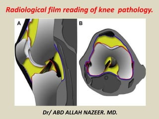
  • 1. Radiological film reading of knee pathology. Dr/ ABD ALLAH NAZEER. MD.
  • 3.
  • 4.
  • 5. Grade 1 myxoid degeneration of the Meniscus.
  • 6.
  • 7. Grade 1 myxoid degeneration of the Meniscus.
  • 8.
  • 9. Grade 11 myxoid degeneration of the Meniscus.
  • 10.
  • 11. Grade 11 myxoid degeneration of the Meniscus.
  • 12.
  • 13. Intrameniscal signal intensity into a medial meniscal tear over 48 months. Images show intrameniscal signal intensity grade 2 (arrowhead) at baseline, having progressed to grade 3 (arrowhead) at 24-month follow-up. At 48 months, the signal is visible on several sections and indicates a horizontal meniscal tear in the posterior horn (arrow).
  • 14. Normal Anatomy Meniscal Tear. Normal meniscal anatomy. (a) Three-dimensional model (left) and cross-sectional diagram (right) of the semi lunar meniscus highlight the concave surface, which conforms to the morphology of the femoral condyles. The result is increased contact area and a tapered central free edge. Circumferentially oriented collagen bundles (blue cylinders) provide hoop strength and course parallel to the long axis of the meniscus, while radial fibers form a lattice and provide additional structural support. (b) Three-dimensional model shows the meniscus subdivided into thirds (the anterior horn [AH], body [B], and posterior horn [PH]) and attached to the tibia via the anterior and posterior root ligaments. Note the proximity of the roots to the tibial attachment sites for the anterior cruciate ligament (ACL) (black *) and posterior cruciate ligament (PCL) (white *). (c) Sagittal PD-weighted MR image shows the striated appearance of the anterior horn of the LM (arrow) due to contributing insertional fibers that originate from the ACL.
  • 15. Normal meniscal anatomy. (a) Three- dimensional model (left) and cross-sectional diagram (right) of the semilunar meniscus highlight the concave surface, which conforms to the morphology of the femoral condyles. The result is increased contact area and a tapered central free edge. Circumferentially oriented collagen bundles (blue cylinders) provide hoop strength and course parallel to the long axis of the meniscus, while radial fibers form a lattice and provide additional structural support. (b) Three-dimensional model shows the meniscus subdivided into thirds (the anterior horn [AH], body [B], and posterior horn [PH]) and attached to the tibia via the anterior and posterior root ligaments. Note the proximity of the roots to the tibial attachment sites for the anterior cruciate ligament (ACL) (black *) and posterior cruciate ligament (PCL) (white *). (c) Sagittal PD- weighted MR image shows the striated appearance of the anterior horn of the LM (arrow) due to contributing insertional fibers that originate from the ACL.
  • 16. Normal MR imaging appearance of the menisci. (a) Sagittal PD-weighted MR images show the typical bow-tie configuration of the meniscal body (left) and the opposing triangles of the horns (center and right). The posterior horn of the MM (arrow) is larger than the anterior horn, whereas the horns of the LM are similar in size and shape. (b) Coronal PD-weighted MR images demonstrate the triangular morphology of the meniscal body (left) and the elongated wedge shape of the posterior horn (right), which is continuous with the crescentic posterior meniscal root.
  • 17. Normal MR imaging appearance of the menisci. (a) Sagittal PD-weighted MR images show the typical bow-tie configuration of the meniscal body (left) and the opposing triangles of the horns (center and right). The posterior horn of the MM (arrow) is larger than the anterior horn, whereas the horns of the LM are similar in size and shape. (b) Coronal PD-weighted MR images demonstrate the triangular morphology of the meniscal body (left) and the elongated wedge shape of the posterior horn (right), which is continuous with the crescentic posterior meniscal root.
  • 18. Surrounding anatomy. (a) Sagittal PD-weighted MR image (left) shows an apparent anterior root tear (black arrow), which, when followed on multiple imaging sections and better depicted on the axial fluid-sensitive reconstructed MR image (right), is shown to be the normal transverse meniscal ligament (white arrow). (b) Sagittal PD-weighted MR image shows the Humphry (white arrow) and Wrisberg (black arrow) ligaments, which are located anterior and posterior to the PCL, respectively. (c) Sagittal T2-weighted MR image shows the posterosuperior (arrow) and anteroinferior (arrowhead) fascicles, which form the roof and floor of the popliteal hiatus, respectively. (d) Sagittal T2-weighted MR image obtained at a more medial level than c depicts the posteroinferior fascicle (arrow). Note the adjacent MFL (arrowhead). (e) Sagittal T2-weighted MR image shows a torn posterosuperior fascicle (arrow), which was found to be associated with an arthroscopically proved peripheral longitudinal LM tear. Note the “kissing” bone contusions from an ACL tear.
  • 19. Anatomic variants. (a) Coronal fat-suppressed PD-weighted MR image through the body of the menisci shows a discoid LM (arrow) that measures 19 mm (normally <15 mm). (b) Sagittal PD-weighted MR image shows the typical rippled appearance of a meniscal flounce (arrowhead). (c) Sagittal PD-weighted MR image (left) shows marrow signal intensity within the MM posterior horn (black arrow), a finding that corresponds to a meniscal ossicle (white arrow) seen on the lateral radiograph (right).
  • 20. Expected MR imaging appearance of a horizontal tear. (a) Three-dimensional model (center) shows a horizontal tear (arrow), and diagrams show the expected appearance of the tear on sagittal (left) and coronal (right) MR images. Dashed lines = orientation of imaging planes. (b) Sagittal T2-weighted MR image shows a tear of the posterior horn (arrow) and an associated multiloculated parameniscal cyst (arrowhead). (c) Coronal PD-weighted MR image shows a horizontal tear of the meniscal body (arrow) that contacts the superior articular surface.
  • 21. Expected MR imaging appearance of a horizontal tear. (a) Three-dimensional model (center) shows a horizontal tear (arrow), and diagrams show the expected appearance of the tear on sagittal (left) and coronal (right) MR images. Dashed lines = orientation of imaging planes. (b) Sagittal T2- weighted MR image shows a tear of the posterior horn (arrow) and an associated multiloculated parameniscal cyst (arrowhead). (c) Coronal PD- weighted MR image shows a horizontal tear of the meniscal body (arrow) that contacts the superior articular surface.
  • 22.
  • 23. Fat-suppressed proton density-weighted (4a) sagittal and (4b) coronal images reveal a horizontal tear of the posterior horn of the medial meniscus (arrows), extending to the tibial surface.
  • 24.
  • 25. Horizontal tear of the posterior horn of the medial meniscus.
  • 26.
  • 27. Horizontal tear of the posterior horn of the medial meniscus, extending to the tibial surface.
  • 28.
  • 29. Horizontal tear of the posterior horn of the medial meniscus, extending to the tibial surface.
  • 30.
  • 31. Horizontal tear of the lateral meniscus.
  • 32.
  • 33. Horizontal flap tear: Graphic demonstrating displaced horizontal tear. Coronal and sagittal PD FS MR show displaced fragments in the posterior aspect of the joint near the PCL.
  • 34. Longitudinal tear. (a) Three-dimensional model (left) and cross-sectional diagram (right) show a longitudinal tear (black arrows) extending to both articular surfaces, running along the long axis of the meniscus and dissecting it between the longitudinal collagen bundles (blue cylinders). A longitudinal tear separates the free edge from the periphery. (b) Arthroscopic image from Movie 1 shows a longitudinal tear parallel to the free edge. Inset shows the arthroscopic field of view.
  • 35. Expected MR imaging appearance of a longitudinal tear. (a) Three-dimensional model (center) shows a typical longitudinal tear (arrow) that divides the meniscus into inner and outer halves, and diagrams show the expected appearance of the tear on sagittal (left) and coronal (right) MR images. Dashed lines = orientation of imaging planes. (b) Axial fluid-sensitive reformatted MR image shows a peripheral longitudinal tear involving the posterior body and posterior horn that extends into the posterior root arrows). (c) Sagittal PD-weighted MR image shows a peripheral longitudinal tear with increased intrasubstance signal intensity that unequivocally contacts the articular surface in a vertical orientation (arrow). This type of tear should not extend to the free edge.
  • 36.
  • 37. Peripheral longitudinal tear extending to the tibial surface within the posterior horn of the medial meniscus.
  • 38.
  • 39. Peripheral longitudinal tear extending to the articular surface within the posterior horn of the medial meniscus.
  • 40.
  • 41. Vertical flap tars (Parrot-beak tears): Graphic and axial PD FS MR show an oblique radial (D) or longitudinal tear entering the free edge of the meniscus with displacement of the edge (A), resulting in a defect shaped like a parrot's beak or the letter "V” (B,C).
  • 42. Radial (Root)tear. (a) Three-dimensional model (left) and cross-sectional diagram (right) show a radial tear (black arrows) that involves the free edge and is perpendicular to the long axis of the meniscus. The circumferential fibers responsible for resisting hoop strength are sequentially torn. Blue cylinders = longitudinal collagen bundles. (b) Arthroscopic image from Movie 2 shows a radial tear that involves the free edge. Inset shows the arthroscopic field of view.
  • 43. MR imaging signs of a radial tear. Sagittal PD-weighted MR images demonstrate the cleft sign (arrowhead in a), the truncated triangle sign of a partial-thickness tear (arrowhead in b), and the ghost meniscus sign associated with a full-thickness tear (arrow in c). Each tear involves the free edge of the meniscus.
  • 44.
  • 45. Marching cleft sign of a radial tear. (a) Sagittal PD-weighted contiguous MR images show a vertically oriented cleft (arrows) “marching” from the free edge into the substance of the meniscus at the junction of the body and anterior horn. (b) Axial fluid-sensitive reconstructed MR image shows the oblique course of a radial tear (arrow) with respect to the sagittal plane, which accounts for the MR imaging appearance of a tear propagating out of plane.
  • 46.
  • 47. Complete root tear. (a, b) Coronal PD-weighted (a) and axial fluid-sensitive reconstructed (b) MR images show a complete posterior root tear (arrow).
  • 48.
  • 50.
  • 51. (12a) A radial tear (arrow) truncates the central attachment/root of the posterior horn of the medial meniscus on this fat suppressed proton density-weighted coronal image. Associated medial subluxation of the meniscal body(arrow)
  • 52.
  • 53. Radial tear with truncation of the posterior root of the medial meniscus. Axial, coronal and sagittal PD FS MR show meniscal extrusion (B) and the "ghost meniscus" sign of absent meniscal tissue (C).
  • 54.
  • 55.
  • 56.
  • 57. Small radial tear of the lateral meniscus.
  • 59. Radial tear of the posterior horn of the medial meniscus (small arrow). Wrisberg ligament
  • 60.
  • 61. (16a) Sagittal and (16b) axial proton density weighted images reveal a very large radial tear (arrows) that extends broadly across the entire width of the anterior body of the lateral meniscus.
  • 62.
  • 63. Complex tear. Sagittal PD-weighted MR image shows both vertical (arrowhead) and horizontal (arrow) components. Complex tears typically appear fragmented.
  • 64.
  • 65. A GRE T2*-weighted sagittal image reveals a complex tear of the posterior horn of the medial meniscus, having horizontal (arrows) and longitudinal (arrowhead) components.
  • 66. Bucket-handle tear. (a) Diagram shows a longitudinal tear (white arrow), with central displacement (red arrows) of the inner segment or “handle.” (b) Axial fluid-sensitive reconstructed MR image shows a rare bucket-handle tear that involves only about one-third of the meniscus (arrow). Typically this type of tear involves the majority of the meniscus.
  • 67.
  • 68. Displaced tear. (a) Coronal T2-weighted MR image shows a flipped fragment within the intercondylar notch (arrow) from a complex tear of the MM. (b) Sagittal PD-weighted MR image shows a large flipped fragment (arrow) in the popliteal recess from a torn LM. (c) Coronal PD-weighted MR image (left) and corresponding anteroposterior radiograph (right) show a laterally displaced meniscal fragment (arrowheads) extending into the superior recess, with central areas of chondrocalcinosis.
  • 69.
  • 70. MR imaging signs of a bucket-handle tear. (a) Sagittal PD-weighted MR image shows the absent bow-tie sign, with non- visualization of the meniscal body (arrows). (b) Sagittal PD-weighted MR image shows the fragment within the intercondylar notch sign, with a centrally displaced meniscal fragment (arrow) eccentric to the PCL. This finding is commonly seen with lateral tears because an intact ACL impedes the fragment from reaching the level of the PCL. (c) Sagittal PD-weighted MR image shows the double PCL sign, with a displaced fragment from the MM anterior and parallel to the PCL (arrow). (d) Sagittal PD-weighted MR image shows the double anterior horn sign, with a meniscal fragment (white arrow) posterior to and displacing the native anterior horn (black arrow) and a markedly diminutive posterior horn (arrowhead).
  • 71.
  • 73.
  • 74. Fat-suppressed coronal images demonstrate before and after images following repair of a bucket handle tear. In (17a), the preoperative study, a large displaced "handle" (arrow) from the body of the lateral meniscus is seen near the intercondylar notch. Only a small peripheral rim of meniscal tissue (arrowhead) is present at the native site of the lateral meniscus.
  • 75.
  • 76. Bucket-handle tear (BHT): Graphic, axial, coronal and sagittal PD FS MR show large intercondylar notch fragments (A-E), creating a "double PCL" sign anterior to the posterior cruciate ligament (D) and a “double delta” sign (D).Broken bucket- handle tear (E).
  • 77.
  • 78. Flipped meniscus: Graphic, axial, coronal and sagittal PD FS MR show a lateral flipped meniscus. In this tear, a large portion of the posterior horn is displaced anteriorly and lies adjacent to the anterior horn. Notice the "double meniscus" sign (D,E). Severely truncated posterior horn (C, D, E).
  • 79.
  • 80. Lateral Displaced Meniscal Bucket Handle Tear: MRI
  • 81.
  • 82. Lateral Displaced Meniscal Bucket Handle Tear.
  • 83.
  • 84. Lateral Displaced Meniscal Bucket Handle Tear.
  • 85.
  • 86. (8a) The curvilinear course of oblique tears often results in abnormal vertical signal (arrows) that progresses towards or away from the free edge of the meniscus on consecutive images, as seen in these sequential images of an oblique tear (arrows) of the posterior horn of the medial meniscus.
  • 87.
  • 89.
  • 91. Normal intrameniscal signal evolution in the site of surgery, with very good final result.
  • 92. Normal healing sequence. Typical worsening in the first scans - intrameniscal signal after surgery is worse then before surgery. No healing disturbance.
  • 93. Normal healing sequence. Typical worsening in the first scans - intrameniscal signal after surgery is worse then before surgery. Any of these scans without sequentional imaging would be difficult to differentiate from disturbed healing.
  • 94. Abnormal healing of degenerated meniscus.
  • 96. Partial tear of the proximal anteromedial band of the anterior cruciate ligament and tear of the medial collateral ligament. Anterior cruciate ligament.
  • 97.
  • 98. A fat-suppressed proton-density sagittal view of an acute partial ACL tear depicts focal increased signal of the ACL (arrowheads) and a wavy contour of posterior fibers, which remain in continuity (arrow).
  • 99.
  • 101.
  • 103.
  • 105.
  • 106. Partial tear of anterior cruciate ligament. Oblique sagittal T2-weighted MR shows diffuse high signal intensity within the ligament itself.
  • 107.
  • 108. Sagittal T2 weighted image of partial anterior cruciate ligament (ACL) tear. The ACL appears lax and concave in appearance.
  • 109.
  • 110.
  • 112.
  • 113. Partial tear of the ACL.
  • 114.
  • 116.
  • 117. Sagittal T2W MRI demonstrates typical appearance of ACL tear at the mid-substance with fibres discontinuity of ACL.
  • 118.
  • 119. Sagittal T2W MRI image demonstrates avulsion of ACL from tibial attachment.
  • 120.
  • 121. LEFT: Acute ACL-tear. ACL fibers too flat compared to condylar roof. RIGHT: Discontinuity of fibers.
  • 122.
  • 123. Primary signs of anterior cruciate ligament tear. Sagittal intermediate-weighted images of three different patients showing different patterns of anterior cruciate ligament (ACL) tear. A: Typical appearance of ACL tear at the mid-substance with fibres discontinuity of ACL (arrowheads). Residual stumps on femoral (asterisk) and tibial sides (white arrow) are lax, thickened and increased in signal intensity; B: Chronic ACL tear with absence of normal ACL fibres compatible with complete resorption of fibres. PCL (Curved black arrow); C: Acute high grade intrasubstance tear as characterized by thickening and edematous change of ACL fibres which show increased signal intensity (white arrows). The fibres are still in continuity suggestive of partial ACL tear.
  • 124.
  • 125. (a) Normal sagittal image of ACL seen on 2 slices. The ligament is intact (green line) and parallels Blumesaat line (white line). (b-e) Sag PD images of different patients with complete ACL tear (red line). (b) ACL fibres are discontinuous, (c-d) different patients with mid ACL complete tear with flattened distal fibres. (e) Complete ACL tear with small bunched distal fibres. There is also complete tear of the proximal PCL (blue line).
  • 126.
  • 127. (a) Sagittal T2-weighted MRI, complete disruption of the ACL fibres. (b) Avulsion of the tibial attachment of the ACL (arrows). (c) Same patient on CT.
  • 128. PCL.
  • 130.
  • 132.
  • 133. Multi-planar images showing partial high grade tear of the mid-distal PCL (red arrow).
  • 134.
  • 135. Complete ACL tear and Partial PCL tear.
  • 136.
  • 137. Sagittal PD & T2 FS MRI image demonstrates complete tear of PCL.
  • 138.
  • 140.
  • 142.
  • 144.
  • 145. Sag PD images of 6 different patients with complete PCL tears (red line). Note is also made of complete ACL tears in all cases.
  • 146.
  • 147. ACL Mucoid degeneration with cyst-formation (intra-osseous ganglion). Mucoid material is squeezed from between the ACL-fibers into the bone.
  • 148.
  • 150.
  • 152. A 3-D cutaway at the intercondylar notch in the sagittal plane reveals normal positioning for a patellar tendon ACL autograft. The tibial tunnel should lie posterior to the line drawn parallel to the intercondylar notch (red) and the femoral attachment should lie posterior to a line drawn parallel to the cortex of the distal femoral diaphysis (blue).
  • 153.
  • 154. The sagittal image reveals diffuse edema and abnormal laxity (arrows) along the course of the ACL graft, compatible with graft rupture. A displaced femoral fixation pin (arrowhead) is also apparent.
  • 155.
  • 156. Femoral tunnel is normally positioned at the junction of the physeal scar and posterior intercondylar roof (asterisk). Abnormal placement of the femoral tunnel (asterisk), which lies significantly anterior to a line drawn along the posterior cortex of the femoral diaphysis with graft degeneration.
  • 157.
  • 158. Tibial tunnel cyst after ACL graft reconstruction.
  • 159.
  • 160. Cyclops lesion (arrowheads) attached to the ACL (arrow) with a head-like appearance, showing a focal area of discoloration resembling an eye (curved arrow)
  • 161.
  • 162. Hypointense to isointense nodule (arrowhead) attached to the anterior surface of the anterior cruciate ligament (ACL) graft (arrow). Sagittal T1W MRI image (C) shows a hypointense nodule (arrowhead) in the anterior intercondylar notch, related to Cyclops.
  • 163.
  • 164. ACL reconstruction are provided. Diffuse abnormal fluid signal intensity is seen along the course of the graft (arrows) on the sagittal image, compatible with extensive graft ganglion formation.
  • 165.
  • 166. ACL Surgery Failure with stem cells injection.
  • 168. Grade 1 MCL Sprain. Collateral ligaments.
  • 169.
  • 170. Grade II MCL Sprain
  • 171.
  • 172. Cor PD images of 3 patients showing thickened MCL with some disruption of fibres proximally in keeping with partial tears.
  • 173.
  • 174. Cor PD images of different patients with complete distal MCL tear. Top left image shows complete disruption of distal fibres (yellow arrow). Top right and bottom left images show complete MCL tear with retracted and coiled up MCL (red arrow), tear of the medial meniscus is also seen (black arrow). Bottom right image shows complete discontinuity of MCL with hemorrhage (orange arrow) with muscle tears and hematoma in vastus lateralis (yellow star).
  • 175.
  • 176. Superficial MCL-tear (yellow arrow) and deep MCL tear (orange arrow).
  • 177.
  • 178.
  • 179.
  • 180. LEFT: distal rupture of fibular collateral ligament. RIGHT: biceps femoris tendon and collateral ligament do not attach.
  • 181.
  • 182. Posterolateral corner injury with complete tear of the lateral collateral ligament (blue arrow) and biceps tendon (white arrow). There is also avulsion of the popliteal tendon (not shown) with muscle edema (red star). There is fluid decompressed out of the lateral side of the knee joint into the subcutaneous tissues and interfacial plane (yellow star).
  • 184. Complete tear of the medial patellar retinaculum with defect (white arrow) with retracted fibres in the patellofemoral compartment (yellow arrow). There is also a partial tear of the lateral patellar retinaculum (orange arrow) with lateral femoral condyle contusion (red star) and fascial edema (blue arrow). Patellar retinaculum
  • 185.
  • 186. Ax PD FS images (a) with complete medial patellar retinaculum tear (red arrow) and capsular disruption (orange star). (b) Shows complete lateral patellar retinaculum and iliotibial band tear (orange arrow) with capsular disruption (orange star). Bony contusions are seen on the contralateral femoral condyle (yellow star).
  • 187.
  • 188. Iliotibial Band Friction syndrome: no fat between iliotibial band (yellow arrow) and the lateral condyle.
  • 189.
  • 190. Cor PD images from 3 different patients showing complete iliotibial band tear (red arrow) with capsular disruption (orange star)
  • 191.
  • 193.
  • 194. A sagittal T2 fat sat image depicts a high grade partial tear of the quadriceps tendon. The distal edge (red arrow) of the tendon is well seen, as is the large hematoma (green arrow). The patella (pink arrow) is positioned more inferiorly than normal, and the patellar tendon (white arrow) is lax, suggesting that this is functionally a complete tear. The deep layer of the quadriceps tendon, composed of the vastus intermedius (yellow arrow), remains intact.
  • 195.
  • 196. Partial quadriceps tendon tear: T2W-images.LEFT: Abnormal attachment of tendon. RIGHT: Most of tendon is retracted (red arrow) deep part (vastus intermedius) is still intact.
  • 197.
  • 198. LEFT: Torn tendon with pre-existing tendinopathy (red arrow). RIGHT: Intact vastus intermedius tendon.
  • 199.
  • 200. LEFT: Partial quadriceps tear. Only rectus femoris tendon is torn (blue arrow). RIGHT: Pre-existing tendinopathy (yellow circle) on axial image.
  • 201.
  • 202. The quadriceps tendon attaches to the superior aspect of the patella and, together with the quadriceps muscles of the thigh and the patellar ligament, allows knee extension. Strains and overuse injuries to the quadriceps tendon are common and result in microscopic tears and inflammation. Following a quadriceps tendon injury, patients may or may not be able to ambulate. Tendon rupture can be manifested as abnormal thickening of the tendon (left image) or as complete, revealing retraction of the quadriceps tendon (right image). Individuals with a complete tear will be unable to extend the knee and often have a palpable defect at the site of tendon rupture. Initial management consists of immobilization, crutches, rest, ice, and elevation. Complete tendon rupture requires surgical repair.
  • 203.
  • 204. A, B, Sagittal MRI scans demonstrating a quadriceps tendon tear at the osteotendinous junction.
  • 205.
  • 206. Simultaneous Bilateral Quadriceps Tendon Rupture in Patient with Chronic Renal Failure.
  • 207.
  • 209.
  • 210. Complete quadriceps tear. Sag T2W-images. No continuity. Hematoma in between.
  • 211.
  • 212. Patellar tendinopathy and Partial patellar tendon tear.
  • 213.
  • 214. Complete Patellar tendon tear. Image on the right shows hemorrhagic bursitis ( low signal in bursa).
  • 215.
  • 216. Patellar sleeve avulsion. Fracture line only visible on the coronal images.
  • 217.
  • 218. LEFT: Bone bruise lateral condyle (yellow circle). Normal MCL (green arrow) but missing patellar femoral ligament anterior to it. RIGHT: Medial patellar femoral ligament thorn from femoral attachment.
  • 219.
  • 220. LEFT: Bone bruise medial patella (green arrow).RIGHT: Cartilage fracture.
  • 221.
  • 223.
  • 224.
  • 225.
  • 227.
  • 229.
  • 231.
  • 232.
  • 233.
  • 235.
  • 236.
  • 237.
  • 238.
  • 239.
  • 241.
  • 242. Osgood-Schlutter disease. A, Lateral radiograph. B, Proton density sagittal image of the knee demonstrates tibial tuberosity fragmentation. C, A T2-weighted fat suppression sagittal image demonstrates edema of the tibial fragmentation (arrow) and adjacent Hoffa’s fat pad.
  • 243.
  • 244.
  • 246.
  • 248.
  • 249. Stable Osteochondritis Dissecans. Stable Osteochondritis Dissecans with arthrogram.
  • 250.
  • 251.
  • 252.
  • 254.
  • 256.
  • 258.
  • 259.
  • 260. Superficial infrapatellar bursitis. A 42-year-old female presented with an anterior knee swelling. Sagittal proton-density fat saturated (A) and sagittal gradient-echo T2W (B) images show a distended superficial infrapatellar bursa (arrows)
  • 261.
  • 263.
  • 264. Suprapatellar bursitis. A 29-year-old male presented with internal derangement of the knee: sagittal proton density fat saturated (A) and axial T2W gradient-echo (B) images show a distended suprapatellar bursa (arrows) and in addition, a partial tear of the anterior cruciate ligament (thick arrow in A)
  • 265.
  • 266. Medial collateral ligament bursitis located between the deep MCL (yellow arrow) and the superficial MCL (green arrow).
  • 267.
  • 268. Medial collateral ligament bursitis. A 54-year-old female presented with knee swelling: sagittal true inversion recovery magnitude (TIRM) (A) and axial T2W (B) images show a distended medial collateral ligament burse (arrows)
  • 269.
  • 270. Pes anserine bursitis. A 32-year-old female presented with pain along the posteromedial aspect of the knee: Coronal proton density (A) and axial T2W (B) images show a distended pes anserine bursa (arrows). The pes anserinus tendons (arrowhead in B) are seen inferior to the bursa
  • 271.
  • 272. Ilio-tibial bursitis with distended ilio-tibial bursa.
  • 273.
  • 274. Baker's cyst. A 48-year-old male presented with swelling in the popliteal fossa: sagittal (A) and axial (B) T2W gradient-echo images show a distended gastrocnemius--semimembranosus bursa (arrows).
  • 275.
  • 276.
  • 277.
  • 278.
  • 279.
  • 280.
  • 281.
  • 282.
  • 283.
  • 284. Infrapatellar (Hoffa’s fat pad) ganglion cyst arising from the anterior cruciate ligament.
  • 285.
  • 286. Large infrapatellar ganglionic cyst of the knee fat pad.
  • 287.
  • 288. T1-weighted sagittal and (9b) proton density-weighted axial images demonstrate a large, fluid signal intensity mass (arrows) which fills the infrapatellar fat pad in this patient who presented with palpable anterior knee swelling. The well-defined, homogeneous appearance of the lesion is typical for a benign ganglion, but in light of lesion size, intravenous contrast was administered
  • 289.
  • 290. Ganglion cyst of the ACL.
  • 291.
  • 292. Ganglion cyst of the PCL.
  • 293.
  • 295.
  • 296. Patella Alta. Measurement of the Insall-Salvatti Index on Sagittal 2D GE MRI images of the left (6a) and right (6b) knees of the patient in the unknown case demonstrates ISI values which are diagnostic of patella alta.
  • 297.
  • 298. Patella Baja. Demonstration of an abnormal MRI Insall-Salvatti Index measurement on a patient with patella baja (7a). The patient in 7b and 7c is post operative tibial tubercle transfer and demonstrates thickening of the patellar tendon insertion (arrows) and scarring in the infrapatellar fat pad (asterisk) (7b) and an abnormal MRI Insall-Salvatti Index of 0.51 (7c) which is diagnostic of patella baja.
  • 299.