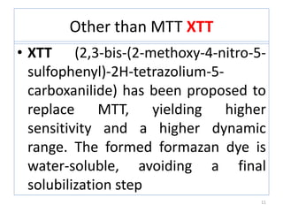
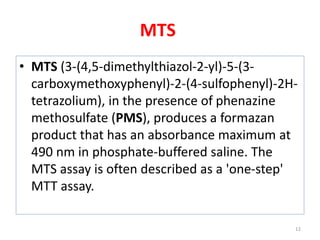
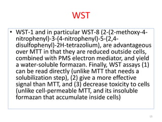
The MTT assay is a colorimetric assay that uses the yellow tetrazolium dye MTT to measure cellular metabolic activity as a proxy for cell viability. In the assay, viable cells containing NAD(P)H-dependent oxidoreductase enzymes reduce MTT to purple formazan crystals, which are then dissolved and quantified by spectrophotometry. The degree of color formation correlates with the number of viable cells present. The MTT assay is inexpensive, does not require cell transfer, and can be used to assess cell proliferation, cytotoxicity, and apoptosis for a variety of cell types.
The MTT assay and the MTS assay are colorimetric assays for measuring the activity of enzymes that reduce MTT or close dyes (XTT, MTS, WSTs) to formazan dyes, giving a purple color The main application allows to assess the viability (cell counting) and the proliferation of cells (cell culture assays)
It can also be used to determine cytotoxicity of potential medicinal agents and toxic materials, since those agents would stimulate or inhibit cell viability and growth
Equipments used , types of culture and media, subculturing, secondary culture, finite & continuous cell lines, cryopreservation and applications of cell culture
Principles of cell viability assays by surendra.pptxSurendra Chowdary
1.DYE EXCLUSION ASSAYS:
Dye exclusion assays are the simplest methods that are based on utilization of different dyes such as trypan blue, eosin, congo red, and erythrosine B, which are excluded by the living cells, but not by dead cells.
For these assays, although staining procedure is quite straightforward, experimental procedure may be time-consuming in case of large sample sizes.
a. Trypan blue stain assay:
Trypan blue stain assay has initially been developed in 1975 to measure viable cell count and is still used as a confirmatory test for measuring changes in viable cell number caused by a drug or toxin.
Trypan blue stain, a large negatively charged molecule, is one of the simplest assays that are used to determine the number of viable cells in a cell suspension.
Principle:
The principle of this assay is that living cells have intact cell membranes that exclude the trypan blue stain, whereas dead cells do not.
Cell suspension is mixed with the trypan blue stain and examined visually under light microscopy to determine whether cells include or exclude the stain.
A viable cell will have a clear cytoplasm, whereas a nonviable cell will have a blue cytoplasm.
Reagent preparation:
To perform the trypan blue stain assay, 0.4% trypan blue stain and phosphate- buffered saline (PBS) or serum-free medium are obtained.
Trypan blue stain should be stored in dark and filtered after prolonged storage.
As trypan blue stain binds to serum proteins and causing misleading results, serum-free medium should be used to obtain reliable results.
Assay Protocol:
The cell suspension to be tested is centrifuged at 100 g for 5 min.
The supernatant is discarded and the pellet is resuspended in 1-ml PBS solution or serum-free medium.
Then, one portion of this cell suspension is mixed with one portion of trypan blue stain.
The mixture is allowed to stay at room temperature for 3 min. It is important to note that the cells should be counted within 3–5 min of mixing with trypan blue, as longer incubation periods will lead to cell death and hence reduced viability counts.
Following the incubation, a drop of the mixture is applied to a hemocytometer, which is placed on the stage of a binocular microscope.
Viable cells will remain unstained, and nonviable cells will stain, in the hemocytometer and these cells are counted separately.
.
Calculation:
After counting viable and nonviable cells, the total number of viable cells per milliliter of aliquot is determined by multiplying the total number of viable cells by 2, which is the dilution factor for trypan blue.
Similarly, total number of cells per milliliter of aliquot is determined by addition of number of viable and nonviable cells and multiplying it by 2.
Then, the percentage of viable cells is calculated using the following equation.
% Viable cells = Total number of viable cells per milliliter of aliquot × 100.
Total number of cells per milliliter of aliquot
2.COLORIMETRIC ASSAYS:
Colorimetric assays
In vitro methods of screening of anticancer agentsNikitaSavita
Cancer- It is the leading cause of mortality. Cancer is the disease which is categorized by abnormal cell growth with the dormant to spread to other parts of body.
Comparison of different methods to measure cell viabilitycreativebioarray22
In vitro assessment of cellular viability has become a generic approach in addressing a vast range of biological questions in many areas of biomedical research. Cell viability assays are often used to screen collections of compounds to determine if the test molecules have effects on cell proliferation or show direct cytotoxic effects that eventually lead to cell death. According to the number and type of cells used, choosing a proper assay method to decided whether the expected outcome is valid or not.
This work is done in IIT-M (Indian Institute of Technology- Madras) with help of Indian Academy of Science during June 2011-Oct 2011 under Dr Karunagaran Devarajan sir
Embracing GenAI - A Strategic ImperativePeter Windle
Artificial Intelligence (AI) technologies such as Generative AI, Image Generators and Large Language Models have had a dramatic impact on teaching, learning and assessment over the past 18 months. The most immediate threat AI posed was to Academic Integrity with Higher Education Institutes (HEIs) focusing their efforts on combating the use of GenAI in assessment. Guidelines were developed for staff and students, policies put in place too. Innovative educators have forged paths in the use of Generative AI for teaching, learning and assessments leading to pockets of transformation springing up across HEIs, often with little or no top-down guidance, support or direction.
This Gasta posits a strategic approach to integrating AI into HEIs to prepare staff, students and the curriculum for an evolving world and workplace. We will highlight the advantages of working with these technologies beyond the realm of teaching, learning and assessment by considering prompt engineering skills, industry impact, curriculum changes, and the need for staff upskilling. In contrast, not engaging strategically with Generative AI poses risks, including falling behind peers, missed opportunities and failing to ensure our graduates remain employable. The rapid evolution of AI technologies necessitates a proactive and strategic approach if we are to remain relevant.
Read| The latest issue of The Challenger is here! We are thrilled to announce that our school paper has qualified for the NATIONAL SCHOOLS PRESS CONFERENCE (NSPC) 2024. Thank you for your unwavering support and trust. Dive into the stories that made us stand out!
Macroeconomics- Movie Location
This will be used as part of your Personal Professional Portfolio once graded.
Objective:
Prepare a presentation or a paper using research, basic comparative analysis, data organization and application of economic information. You will make an informed assessment of an economic climate outside of the United States to accomplish an entertainment industry objective.
Unit 8 - Information and Communication Technology (Paper I).pdfThiyagu K
This slides describes the basic concepts of ICT, basics of Email, Emerging Technology and Digital Initiatives in Education. This presentations aligns with the UGC Paper I syllabus.
2024.06.01 Introducing a competency framework for languag learning materials ...Sandy Millin
http://sandymillin.wordpress.com/iateflwebinar2024
Published classroom materials form the basis of syllabuses, drive teacher professional development, and have a potentially huge influence on learners, teachers and education systems. All teachers also create their own materials, whether a few sentences on a blackboard, a highly-structured fully-realised online course, or anything in between. Despite this, the knowledge and skills needed to create effective language learning materials are rarely part of teacher training, and are mostly learnt by trial and error.
Knowledge and skills frameworks, generally called competency frameworks, for ELT teachers, trainers and managers have existed for a few years now. However, until I created one for my MA dissertation, there wasn’t one drawing together what we need to know and do to be able to effectively produce language learning materials.
This webinar will introduce you to my framework, highlighting the key competencies I identified from my research. It will also show how anybody involved in language teaching (any language, not just English!), teacher training, managing schools or developing language learning materials can benefit from using the framework.
Palestine last event orientationfvgnh .pptxRaedMohamed3
An EFL lesson about the current events in Palestine. It is intended to be for intermediate students who wish to increase their listening skills through a short lesson in power point.
Instructions for Submissions thorugh G- Classroom.pptxJheel Barad
This presentation provides a briefing on how to upload submissions and documents in Google Classroom. It was prepared as part of an orientation for new Sainik School in-service teacher trainees. As a training officer, my goal is to ensure that you are comfortable and proficient with this essential tool for managing assignments and fostering student engagement.
4. • MTT, a yellow tetrazole is reduced
to purpleformazan in living cells.
• A solubilization solution
• dimethyl sulfoxide
• an acidified ethanol solution
• Isopropanol
• detergent sodium dodecyl sulfate in
diluted hydrochloric acid) is added to dissolve the
insoluble purple formazan product into a colored
solution.
4
6. PRINCIPLE
• The MTT assay is a colorimetric assay for
assessing cell metabolic activity.
• NAD(P)H-dependent cellular oxido reductase
enzymes may, under defined conditions,
reflect the number of viable cells present.
• These enzymes are capable of reducing the
tetrazolium dye MTT 3-(4,5-dimethylthiazol-2-
yl)-2,5-diphenyltetrazolium bromide to its
insoluble formazan, which has a purple color.
6
7. MTT ASSAY:Principle:
• This colorimetric assay uses reduction of a
yellow tetrazolium salt (3-(4,5-
dimethylthiazol-2-yl)-2,diphenyltetrazolium
bromide, or MTT) to measure cellular
metabolic activity as a proxy for cell viability.
7
8. MTT assays are usually done in the dark since
the MTT reagent is sensitive to light.
8
9. Principle
• Viable cells contain NAD(P)H-dependent
oxidoreductase enzymes which reduce the
MTT reagent to formazan, an insoluble
crystalline product with a deep purple color.
• Formazan crystals are then dissolved using a
solubilizing solution ( organic solvent s-eg.
Isopropanol/DMSO) and absorbance is
measured at 500-600 nanometers using a plate-
reader.
9
10. • The darker the solution, the greater the
number of viable, metabolically active
cells
• The absorbance of this colored solution
can be quantified by measuring at a
certain wavelength (usually between 500
and 600 nm) by a spectrophotometer.
• The degree of light absorption depends
on the solvent.
10
11. Other than MTT XTT
• XTT (2,3-bis-(2-methoxy-4-nitro-5-
sulfophenyl)-2H-tetrazolium-5-
carboxanilide) has been proposed to
replace MTT, yielding higher
sensitivity and a higher dynamic
range. The formed formazan dye is
water-soluble, avoiding a final
solubilization step
11
13. WST
• WST-1 and in particular WST-8 (2-(2-methoxy-4-
nitrophenyl)-3-(4-nitrophenyl)-5-(2,4-
disulfophenyl)-2H-tetrazolium), are advantageous
over MTT in that they are reduced outside cells,
combined with PMS electron mediator, and yield
a water-soluble formazan. Finally, WST assays (1)
can be read directly (unlike MTT that needs a
solubilization step), (2) give a more effective
signal than MTT, and (3) decrease toxicity to cells
(unlike cell-permeable MTT, and its insoluble
formazan that accumulate inside cells)
13
14. Tetrazolium dye assays can also be
used to measure
• Cytotoxicity (loss of viable cells)
• Cytostatic activity (shift from
proliferation to quiescence [ it is a state
of quietness or inactivity) of potential
medicinal agents
• Cytostatic activity of toxic materials.
• Number of viable cells present
14
16. Features of viable cell
They are based on various cell functions such as
– enzyme activity,
– cell membrane permeability,.
- cell adherence,
– ATP production,
– co-enzyme production,
– nucleotide uptake activity
16
17. Used to measure a
markers that indicate the number of
– dead cells (cytotoxicity assay),
– the number of live cells(viability
assay),
• the total number of cells or
• the mechanism of cell death (e.g.,
apoptosis).
17
18. Application of viability assay
• To detect cytotoxic or growth inhibitory
lymphokines
• To detect mammalian cell survival and
proliferations
• To diagnose disease
• To diagnose male infertility
• To screen drugs
18
21. MTT ASSAY PROTOCOL:
Trypsinization or scrapping the cells
Resuspend or splitting cells at 96 well plate
Check the cell confluency (if 90%-100% of the cell
then it is ready for using)
100~ 200 µl of desired drugs or compound in
each well (usually 1 compound /drugs should
have triplicates)
Incubate the cells (12~24 hours)
Dissolve 1mg MTT in 1ml PBS solution then the
molar concentration will be 2.41m M
21
22. In each well add 20 µl MTT reagent and rest 160
µl culture media and incubate it for 3~4 hours
check the cells to see the purple precipitation
and the purple color is clearly visible then discard
the media carefully as it does not drain the cells.
after discarding media add 100 µl DMSO/
isopropanol
cover it with aluminum foil and mildly shake it for
10 ~15 minutes
Read plate in fluorescent Reader – measure OD
in 570nm (background wavelength is 630nm).
22
23. MASS = CONCENTRATION X VOLUME X MOLECULAR WEIGHT
Interpreting results
• The absorbance reading of the blank must be
subtracted from all samples.
• Absorbance readings from test samples must
then be divided by those of the control and
multiplied by 100 to give percentage cell viability
or proliferation (see formula below).
• Absorbance values greater than the control
indicate cell proliferation, while lower values
suggest cell death or inhibition of proliferation.
23
24. % Viable cells = abs (sam)- abs (blank)
abs (control) – abs (blank)
TROUBLE SHOOTING:REAGENTS:
• MTT is light-sensitive, and should be kept in the dark as much as
possible. It is always a good idea to cover MTT bottles with foil.
• Make sure you do not contaminate your MTT stock. If it is green
or blue (it should be yellow), you can assume it has either been
contaminated by cells or bacteria, or exposed to light. If properly
looked after and stored at 4°C, your MTT stock can last up to 18
months.
• Always handle MTT using the appropriate personal protection
equipment, as it is can cause skin and eye irritation.
• To dissolve formazan crystals, you can use DMSO, acidified
isopropanol, or SDS. Test to find out which works best for your
experiment.
24
25. Absorbance values
• Your absorbance readings should fall between 0.75 and 1.25. If your
readings are too low, try increasing the number of cells plated or the
incubation time, and make sure your cell culture conditions are
appropriate.
• On the other hand, plating too many cells per well will yield very high
absorbance readings, as will contamination by yeast or bacteria.
• To determine the optimal cell number per well, prepare and plate serial
dilutions of cells in medium from 106 to 10³ cells/mL. This will help
you decide which number of cells yields appropriate absorbance values.
• Always include a positive control (untreated cells) and a blank (well
containing medium only), and plate these and samples in triplicate.
• Always wash cells with PBS (step 2) before adding MTT in order to
remove dead cells and cellular debris, which could give inaccurate
results.
25
26. ADVANTAGES:
• No transfer of the cells, the entire assay is
performed in a single microplate
• MTT is metabolised by all cells , the assay can be
used with all cell types.
• In expensive.
DISADVANTAGES:
• Cannot take multiple time points in a single assay.
• Cells with low metabolic activity (eg:
lymphocytes) must be used in high numbers.
26
27. APPLICATIONS:
• Cell proliferation assays
• Cytotoxicity analysis
• Apoptosis screening
• Anticancer drug
• Predictive drug testing for tumors
• Genotoxicity
27