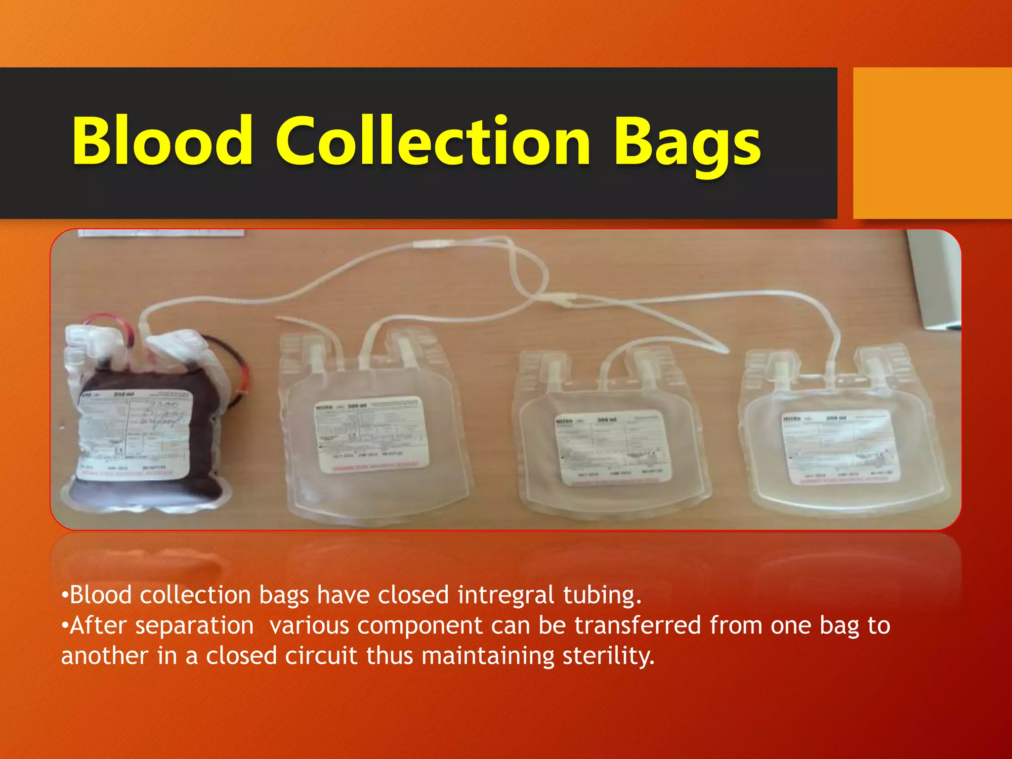
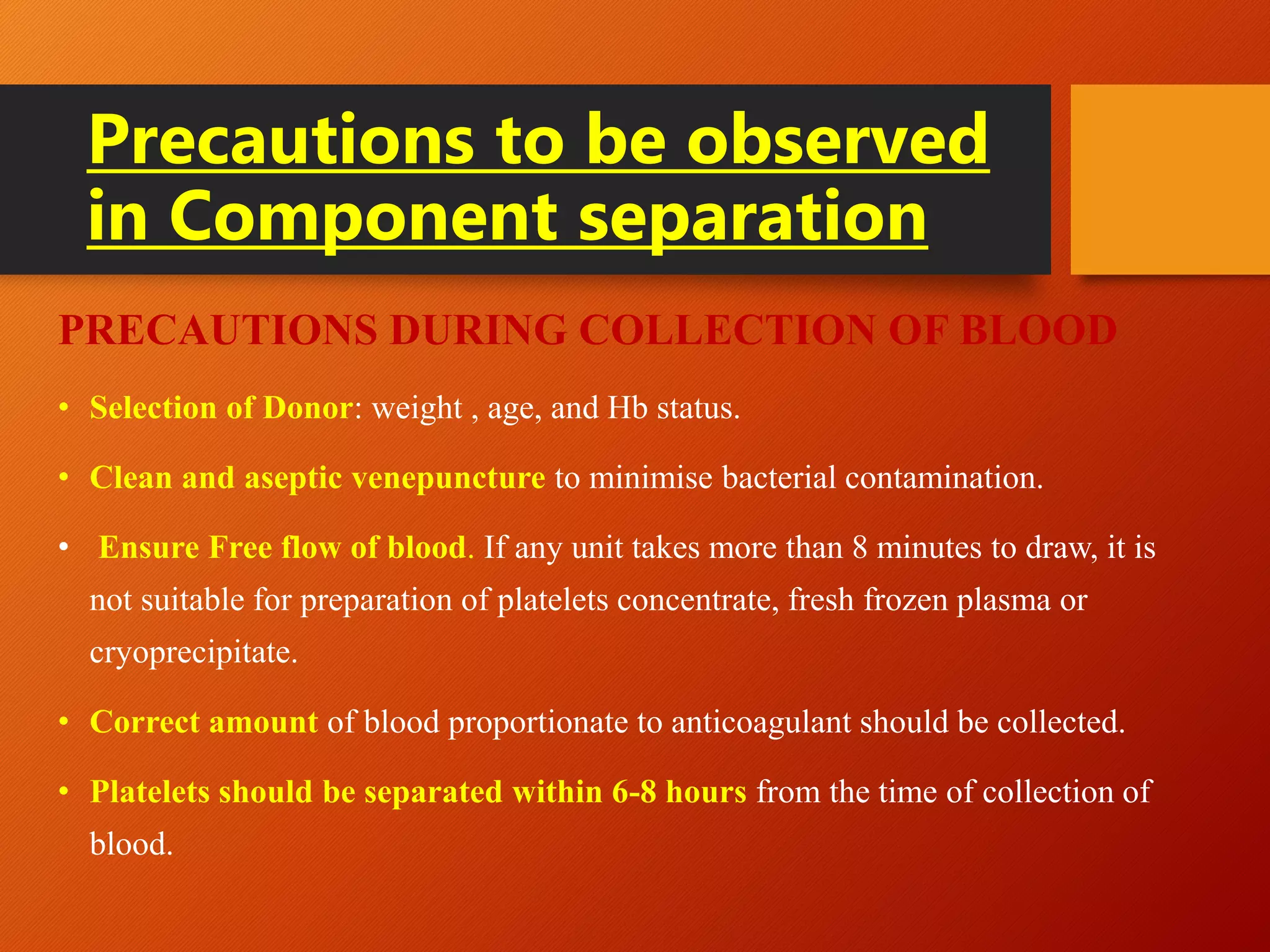
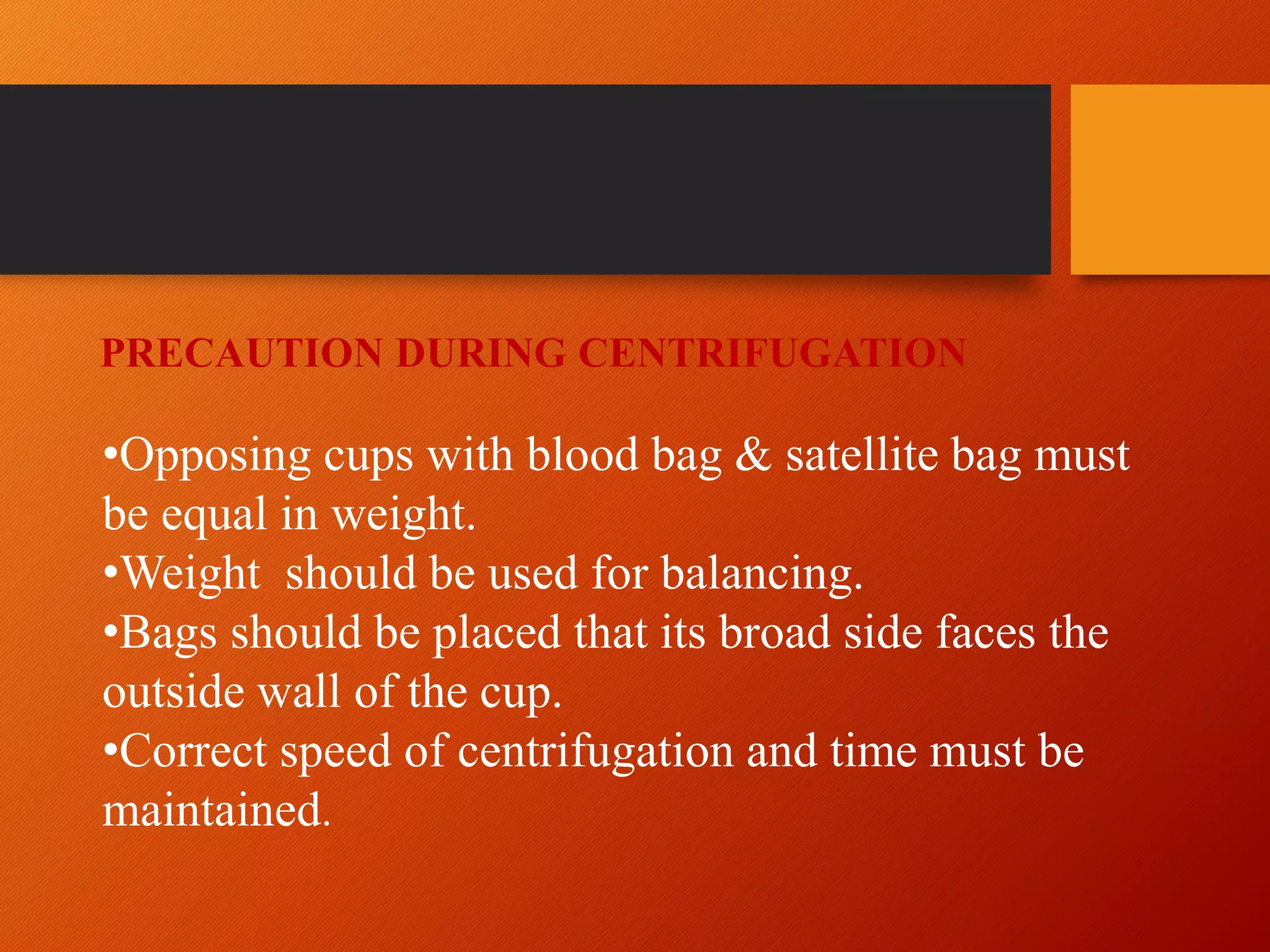
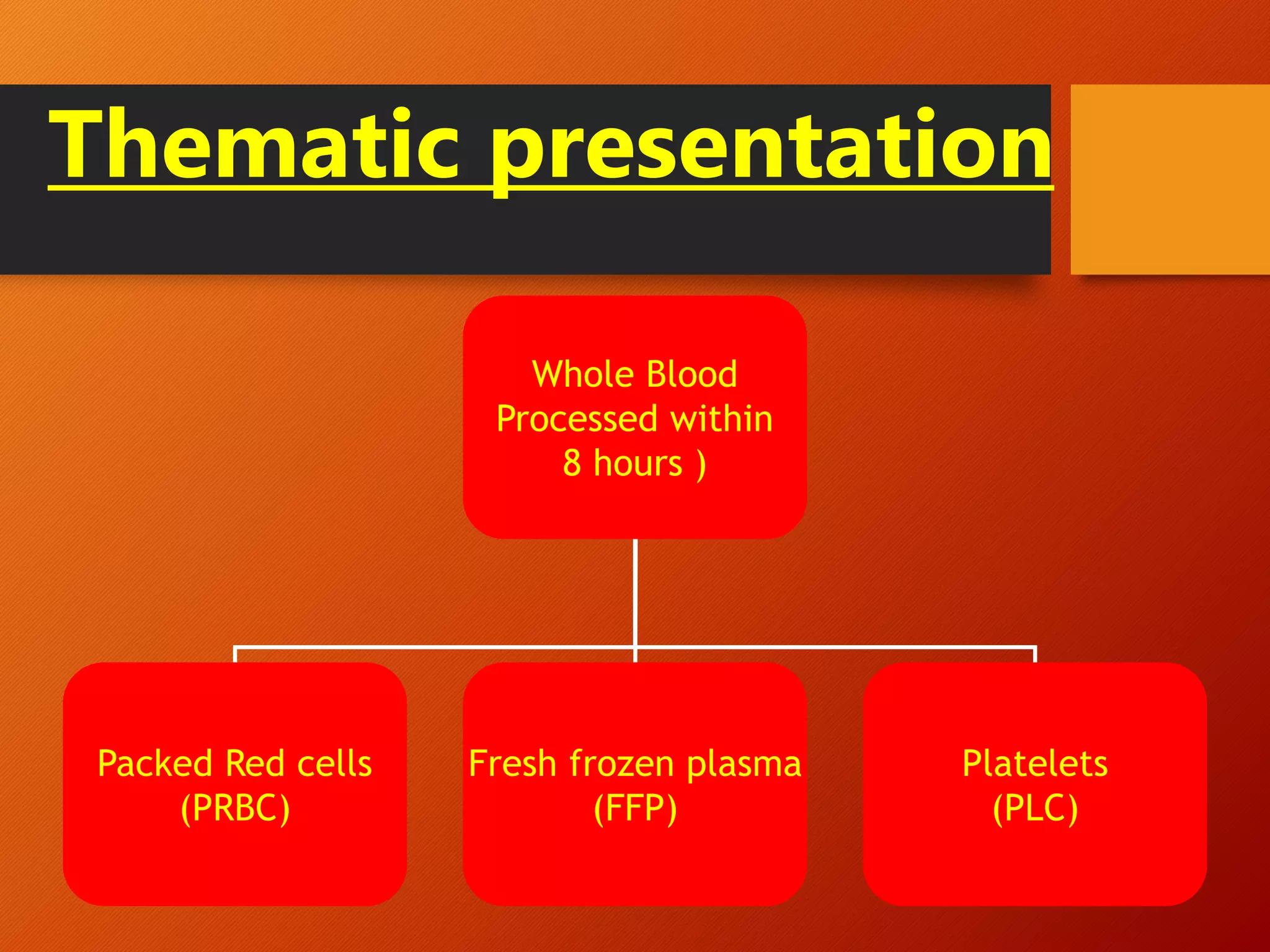
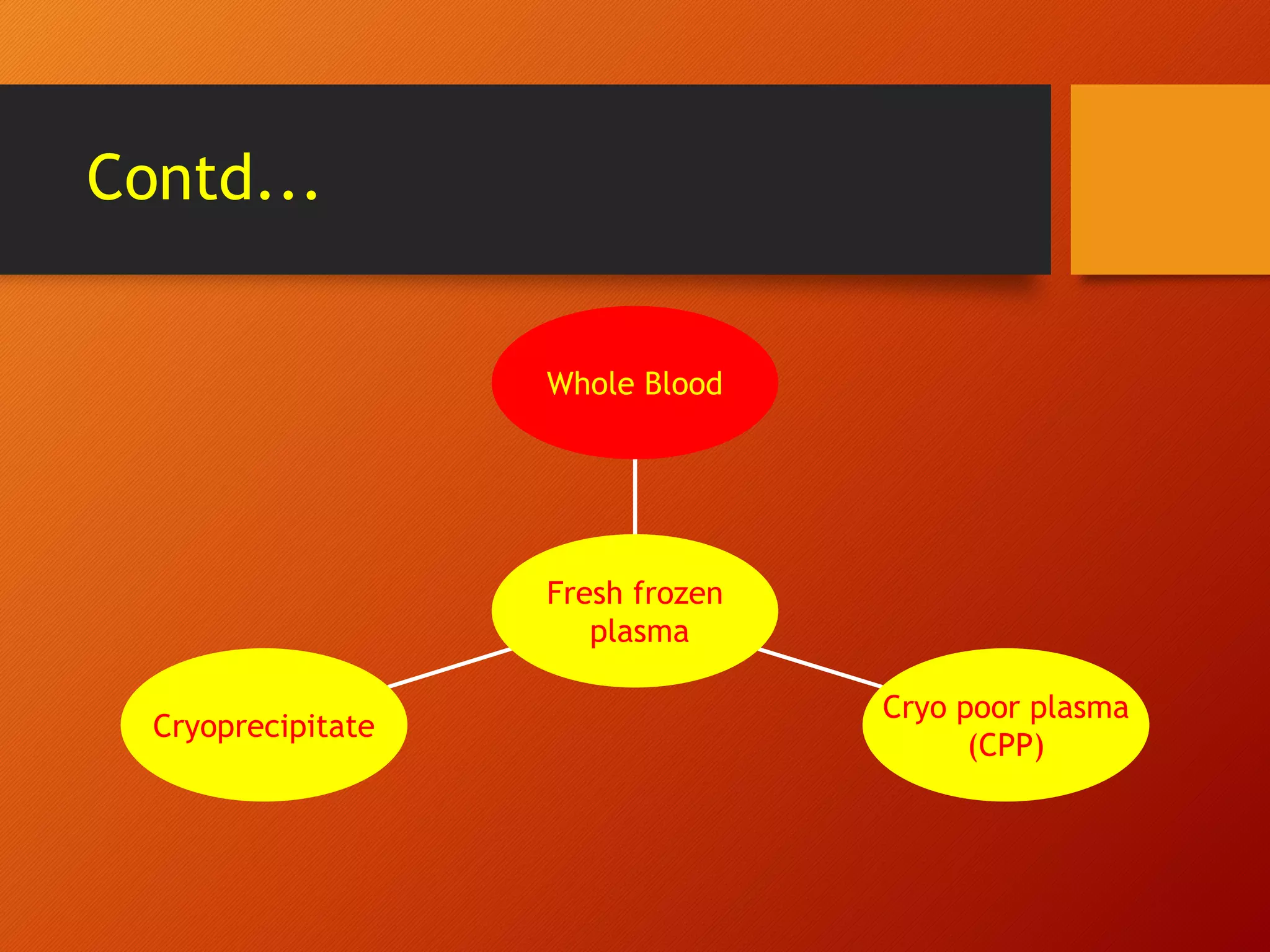
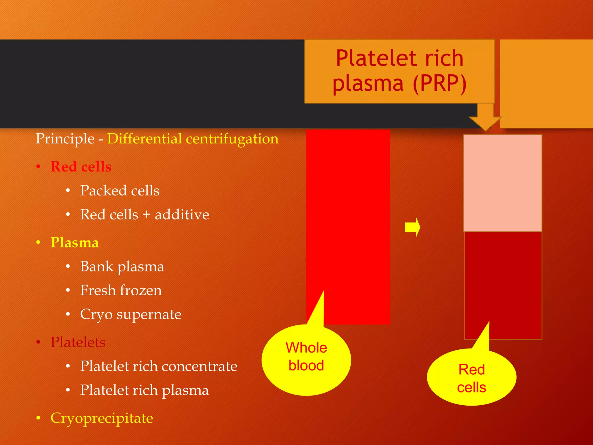
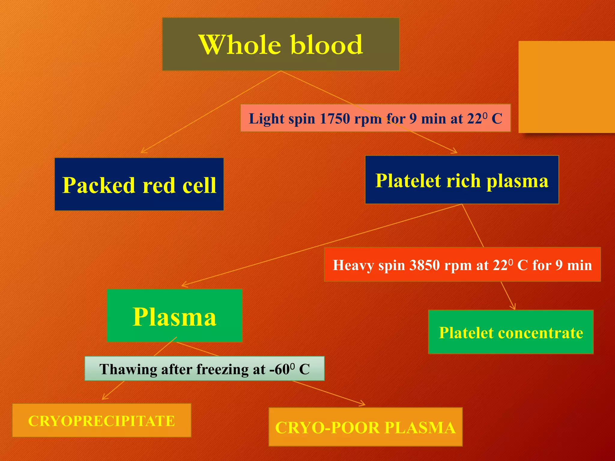
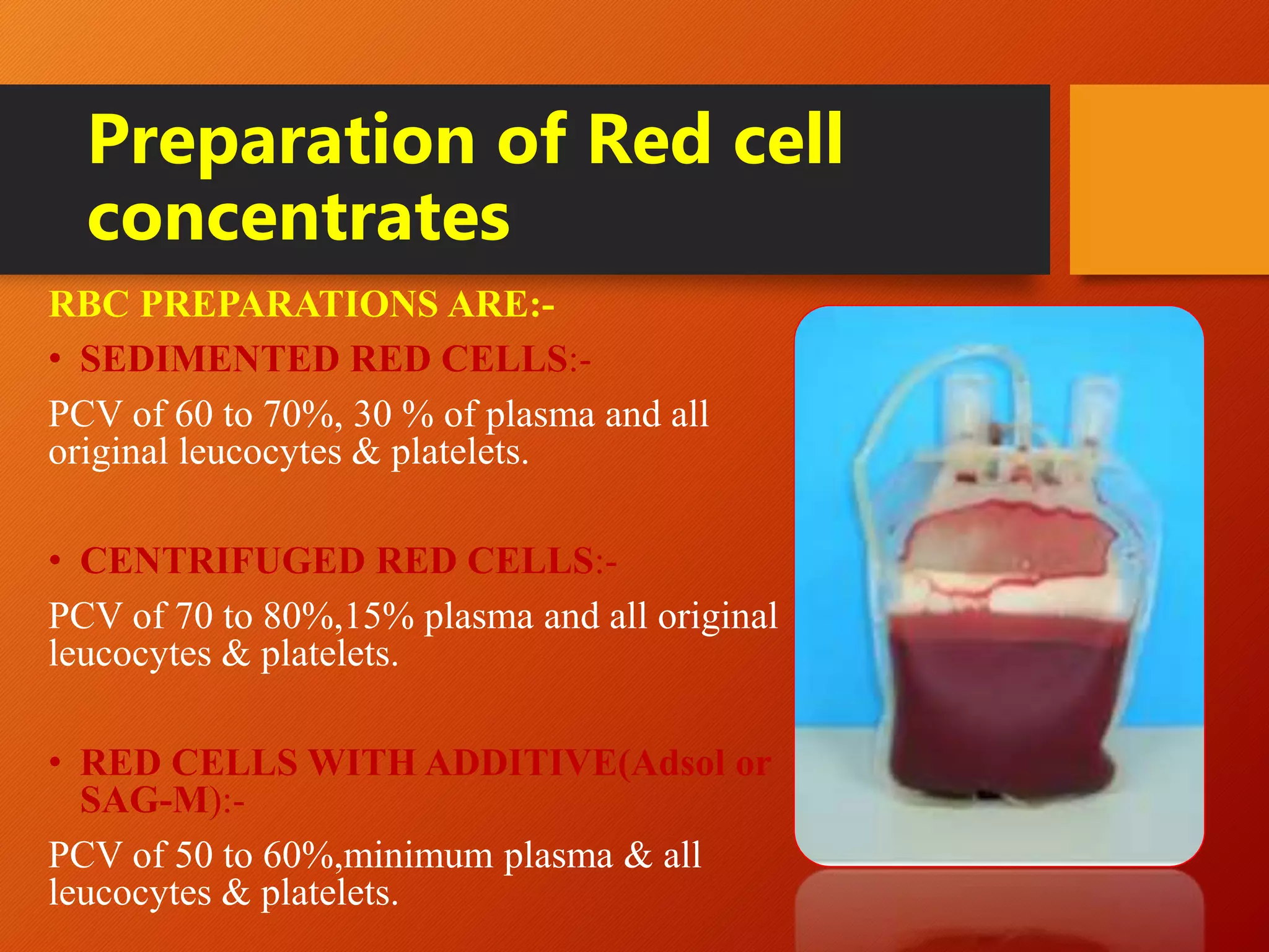
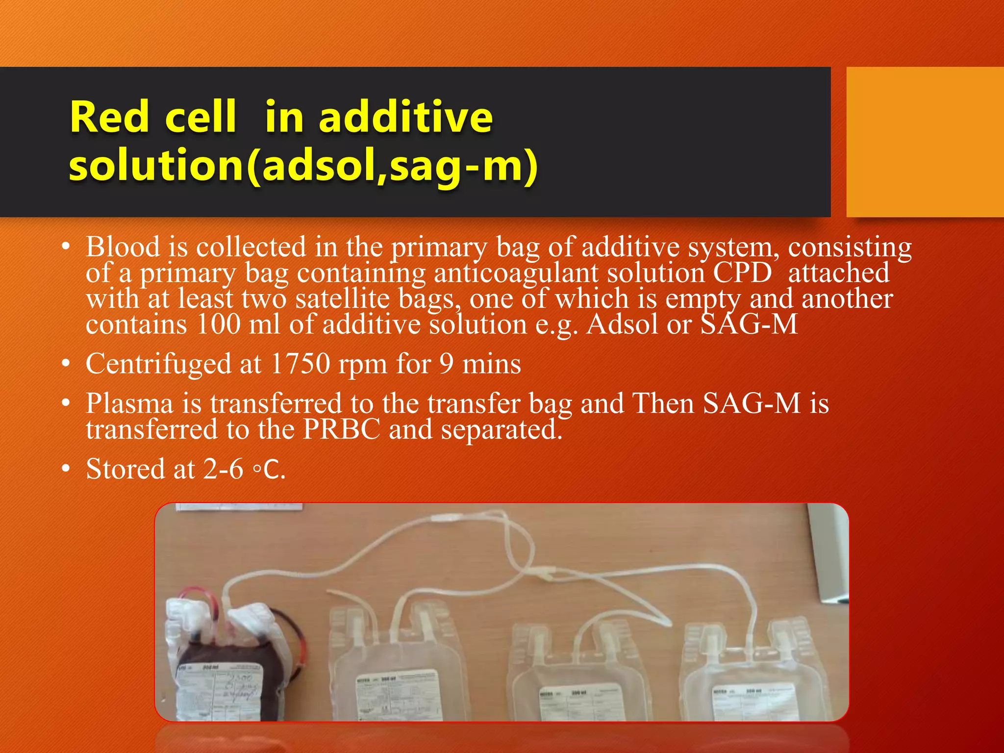
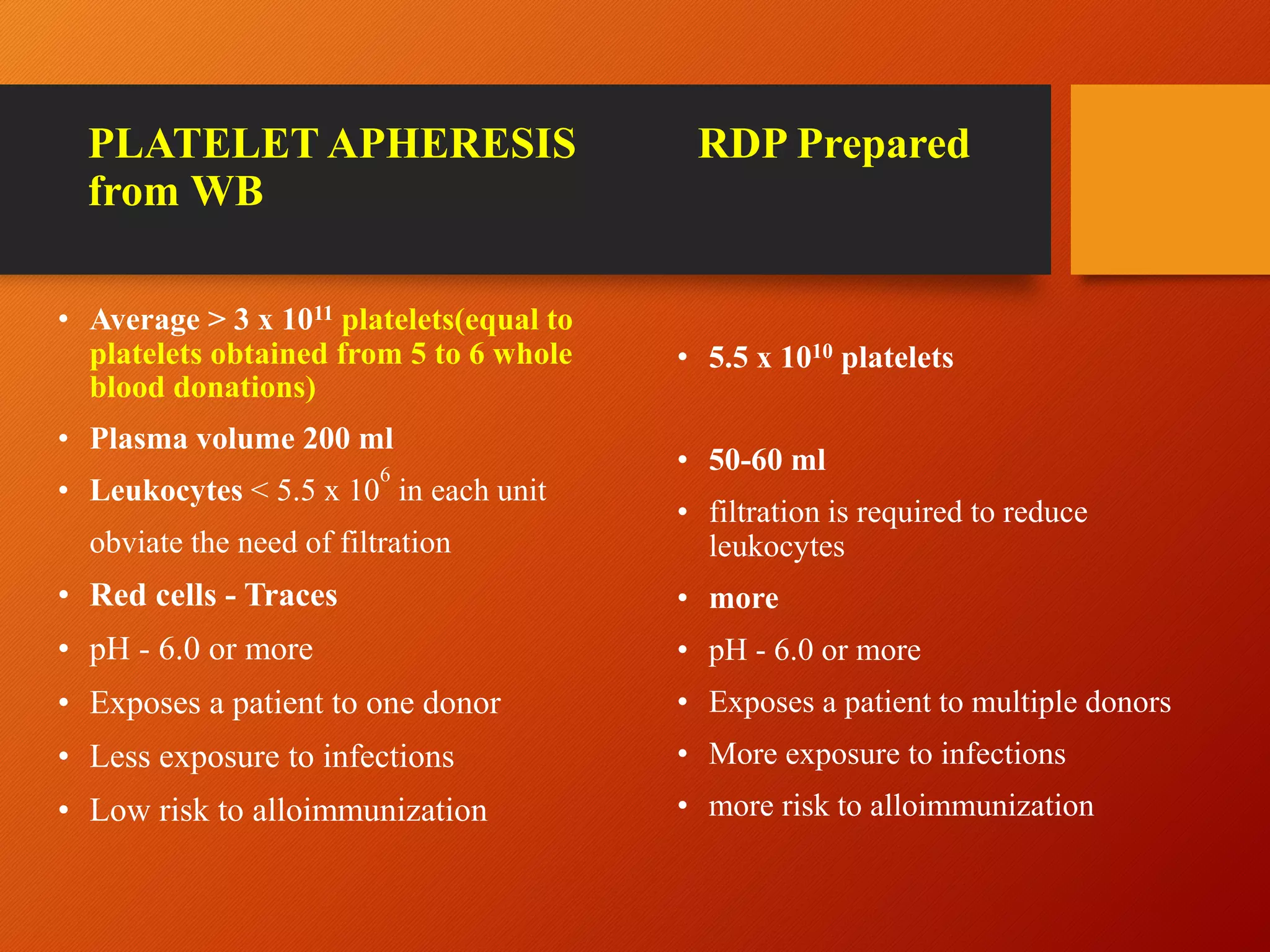
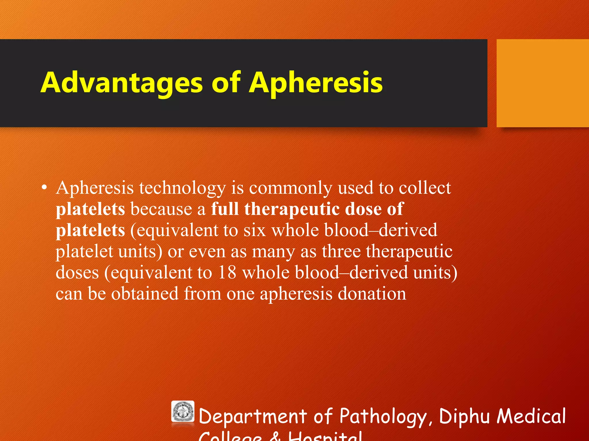
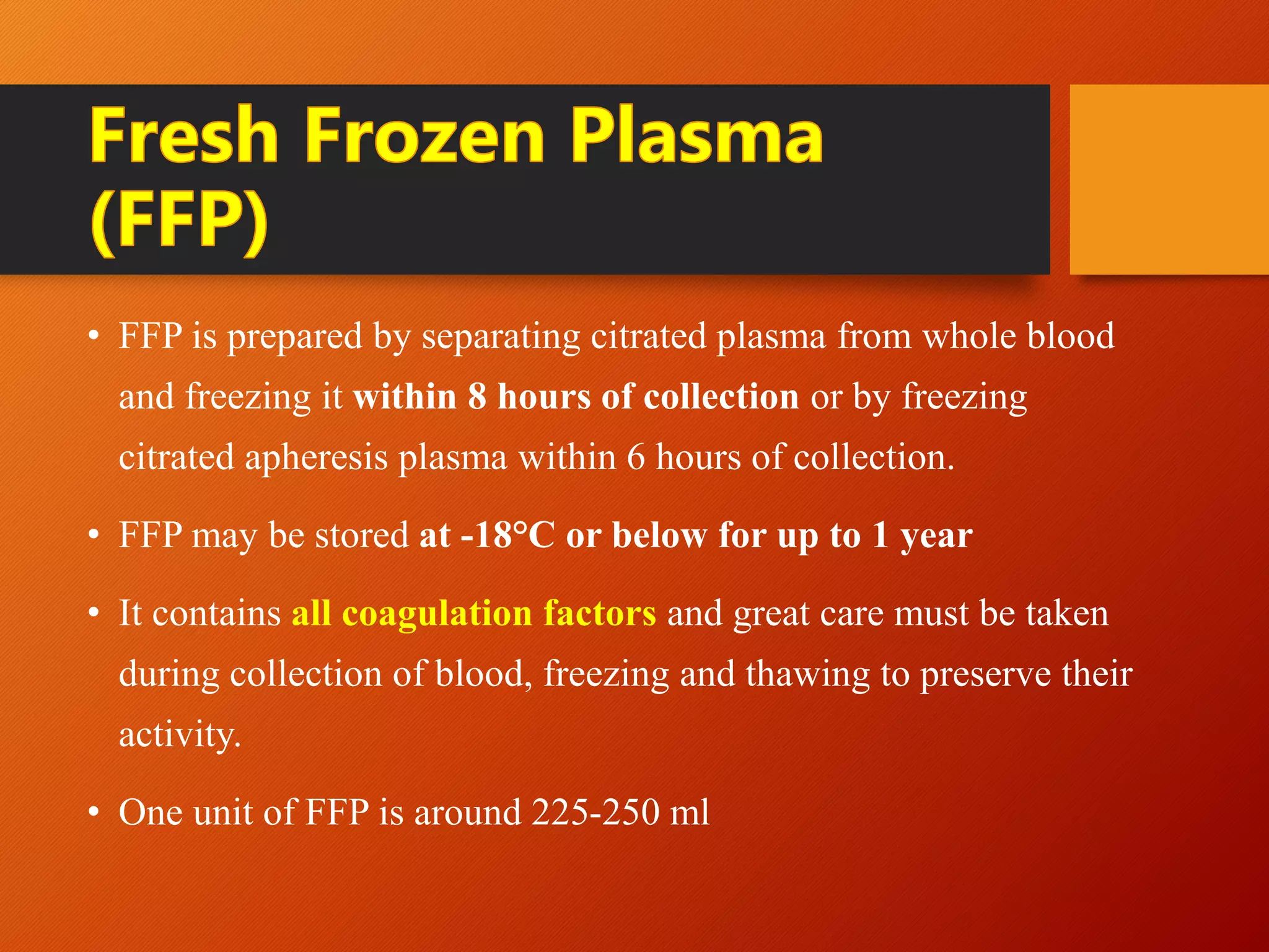
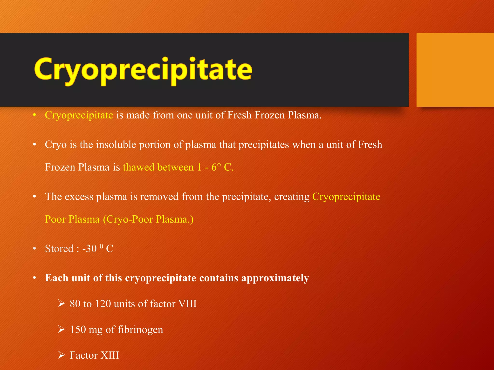
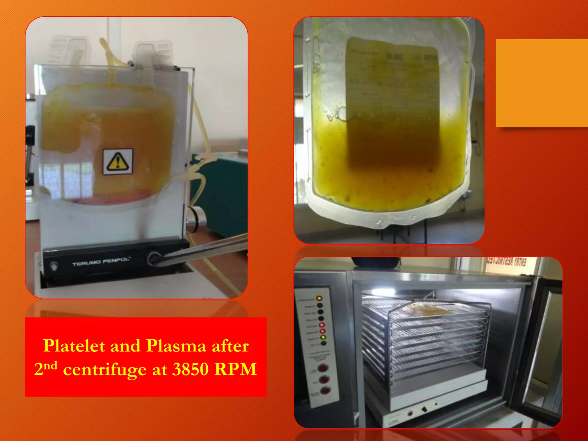
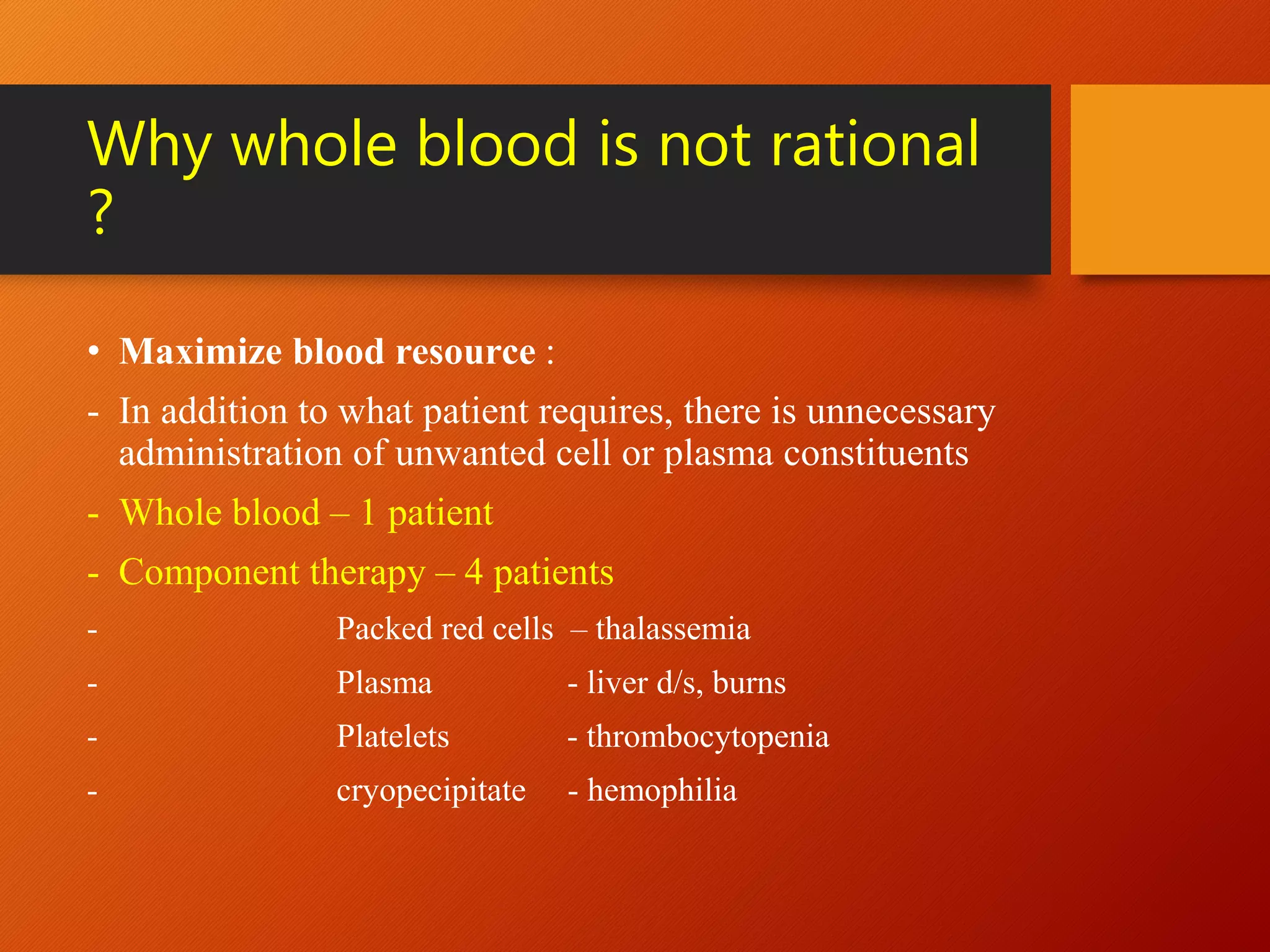
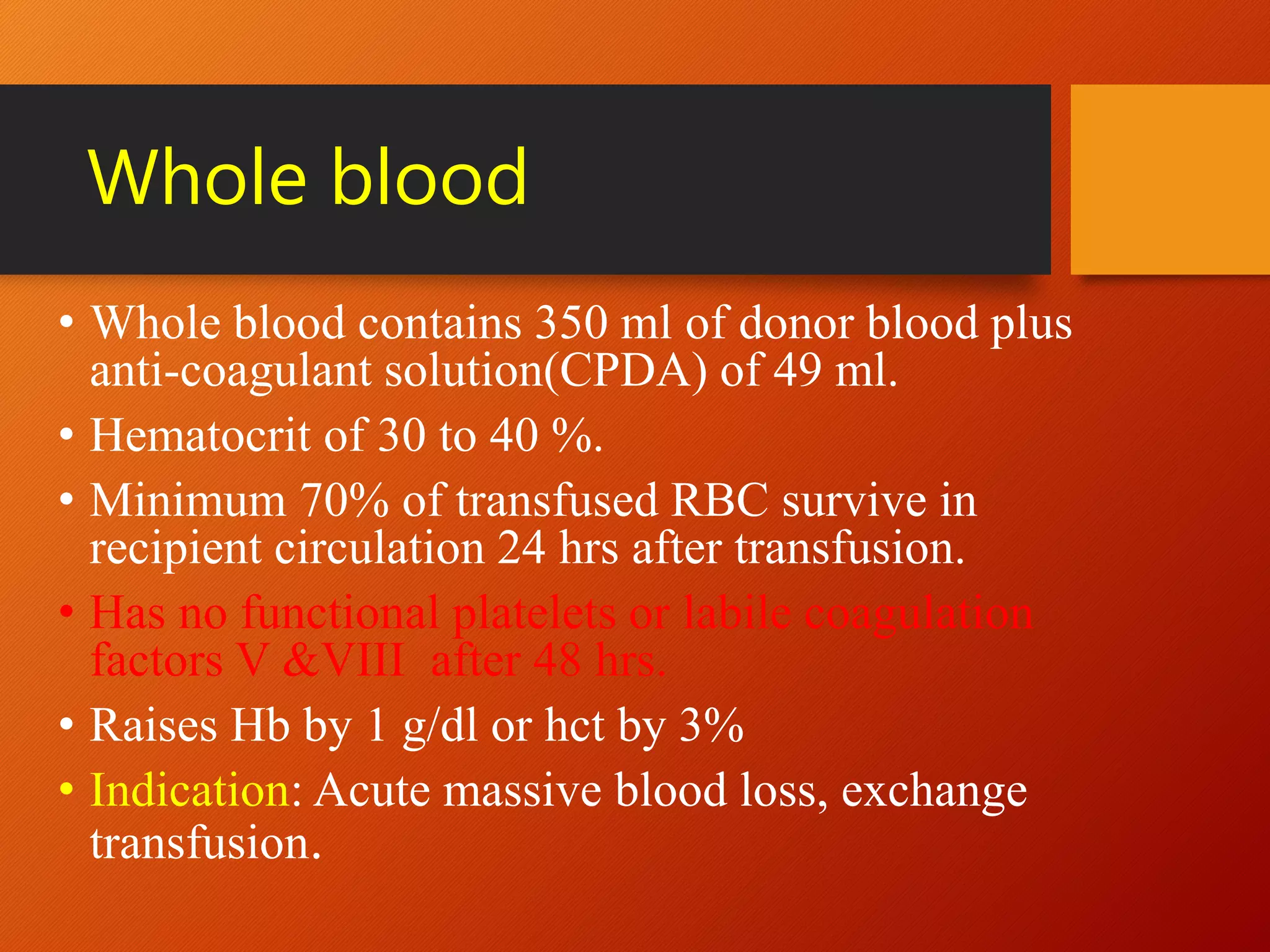
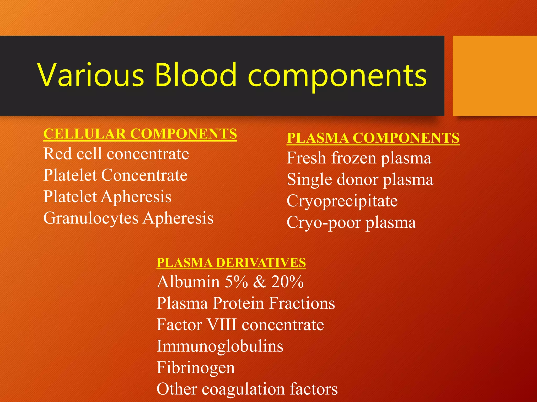
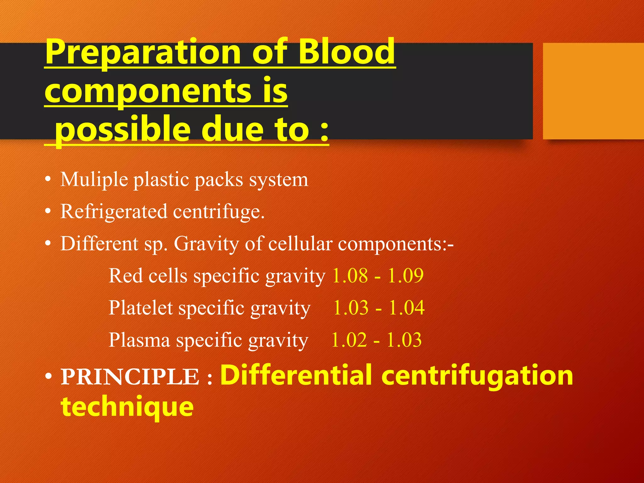
Blood components -
Principles of separation &
indications
Dr. Topon Narzary, MD (Path)
Assistant Professor, Pathology
Department
Diphu Medical College & Hospital,
Diphu (Assam)
Department of Pathology, Diphu Medical
Introduction
“Blood is the best substitute of lost blood”
Effective blood transfusion therapy depends on
availability of different blood components
Components when used separately or in
combination can meet most patients transfusion
needs and keep the risk of transfusion to minimum.
History
• 1628 : William Harvey introduces the controversial concept of circulation.
• 1667: Jean Baptist Denis in France and Richard Lower in England
separately report giving the first human blood transfusion with blood from
lambs.
• 1818: James Blundell performs the first successful transfusion of human
blood to a patient.
• 1914 : Sodium citrate is found to prevent blood from clotting, allowing
blood to be stored between collection and transfusion.
• 1936 : Chicago’s Cook County Hospital establishes the first true “blood
bank” in the United States.
• 1950-1960 : Component therapy started.
Why
Component separation
is
justified ?
1. Separation of blood components allows optimal survival for each
component.
2. Allows transfusing specific blood components according to the
need of the patient
3. Avoids use of unnecessary component which may be
contraindicated in a patient.
4. Several patients can be treated from one unit of donated blood.
5. Use of blood components supplements blood supply and adds to the
blood inventory.
Why whole blood is not rational
?
• Maximize blood resource :
- In addition to what patient requires, there is unnecessary
administration of unwanted cell or plasma constituents
- Whole blood – 1 patient
- Component therapy – 4 patients
- Packed red cells – thalassemia
- Plasma - liver d/s, burns
- Platelets - thrombocytopenia
- cryopecipitate - hemophilia
Whole blood
• Whole blood contains 350 ml of donor blood plus
anti-coagulant solution(CPDA) of 49 ml.
• Hematocrit of 30 to 40 %.
• Minimum 70% of transfused RBC survive in
recipient circulation 24 hrs after transfusion.
• Has no functional platelets or labile coagulation
factors V &VIII after 48 hrs.
• Raises Hb by 1 g/dl or hct by 3%
• Indication: Acute massive blood loss, exchange
transfusion.
Various Blood components
CELLULAR COMPONENTS
Red cell concentrate
Platelet Concentrate
Platelet Apheresis
Granulocytes Apheresis
PLASMA COMPONENTS
Fresh frozen plasma
Single donor plasma
Cryoprecipitate
Cryo-poor plasma
PLASMA DERIVATIVES
Albumin 5% & 20%
Plasma Protein Fractions
Factor VIII concentrate
Immunoglobulins
Fibrinogen
Other coagulation factors
Preparation of Blood
components is
possible due to :
• Muliple plastic packs system
• Refrigerated centrifuge.
• Different sp. Gravity of cellular components:-
Red cells specific gravity 1.08 - 1.09
Platelet specific gravity 1.03 - 1.04
Plasma specific gravity 1.02 - 1.03
• PRINCIPLE : Differential centrifugation
technique
Mathematical formulae
RPM Formula:
RPM = √[RCF/(r x 1.118)] x 1 x 10⁵
RCF (g Force) = Relative Centrifugal Force
r = radius of centrifuge rotor in cm
(rotational radius)
G Force (RCF) Formula:
Relative Centrifugal Force (RCF) in g = (RPM)2 x 1.118 x 10-5 x r
r = radius of centrifuge rotor in cm
(rotational radius)
Convers
ion of
centrifu
gal
speed
from g
to rpm
Guidelines
• Transfusion Medicine Technical Manual (WHO)
• NACO (National AIDS Control Organization)
• American Association of Blood Banks (AABB)
Department of Pathology, Diphu Medical
Blood Collection Bags
•Blood collection bags have closed intregral tubing.
•After separation various component can be transferred from one bag to
another in a closed circuit thus maintaining sterility.
Interconnected Bag System
Double packs system: Red cells, and Plasma only.
Triple packs system: Red cells, Platelet concentrate and Fresh frozen
plasma.
Quad packs system:
Red cells
Platelet concentrate
Cryoprecipitate (factor VIII) and
Cryo- poor plasma.
Precautions to be observed
in Component separation
PRECAUTIONS DURING COLLECTION OF BLOOD
• Selection of Donor: weight , age, and Hb status.
• Clean and aseptic venepuncture to minimise bacterial contamination.
• Ensure Free flow of blood. If any unit takes more than 8 minutes to draw, it is
not suitable for preparation of platelets concentrate, fresh frozen plasma or
cryoprecipitate.
• Correct amount of blood proportionate to anticoagulant should be collected.
• Platelets should be separated within 6-8 hours from the time of collection of
blood.
PRECAUTION DURING CENTRIFUGATION
•Opposing cups with blood bag & satellite bag must
be equal in weight.
•Weight should be used for balancing.
•Bags should be placed that its broad side faces the
outside wall of the cup.
•Correct speed of centrifugation and time must be
maintained.
Thematic presentation
Whole Blood
Processed within
8 hours )
Packed Red cells
(PRBC)
Fresh frozen plasma
(FFP)
Platelets
(PLC)
Contd...
Cryoprecipitate
Cryo poor plasma
(CPP)
Whole Blood
Fresh frozen
plasma
Platelet rich
plasma (PRP)
Principle - Differential centrifugation
• Red cells
• Packed cells
• Red cells + additive
• Plasma
• Bank plasma
• Fresh frozen
• Cryo supernate
• Platelets
• Platelet rich concentrate
• Platelet rich plasma
• Cryoprecipitate
Whole
blood Red
cells
Whole blood
Light spin 1750 rpm for 9 min at 220 C
Packed red cell Platelet rich plasma
Heavy spin 3850 rpm at 220 C for 9 min
Plasma Platelet concentrate
Thawing after freezing at -600 C
CRYOPRECIPITATE CRYO-POOR PLASMA
Preparation of Red cell
concentrates
RBC PREPARATIONS ARE:-
• SEDIMENTED RED CELLS:-
PCV of 60 to 70%, 30 % of plasma and all
original leucocytes & platelets.
• CENTRIFUGED RED CELLS:-
PCV of 70 to 80%,15% plasma and all original
leucocytes & platelets.
• RED CELLS WITH ADDITIVE(Adsol or
SAG-M):-
PCV of 50 to 60%,minimum plasma & all
leucocytes & platelets.
Red cell in additive
solution(adsol,sag-m)
• Blood is collected in the primary bag of additive system, consisting
of a primary bag containing anticoagulant solution CPD attached
with at least two satellite bags, one of which is empty and another
contains 100 ml of additive solution e.g. Adsol or SAG-M
• Centrifuged at 1750 rpm for 9 mins
• Plasma is transferred to the transfer bag and Then SAG-M is
transferred to the PRBC and separated.
• Stored at 2-6 ◦C.
ADVANTAGES
1. Maximum amount of plasma can be removed for
preparation of plasma components.
2. Shelf life increases from 35 days to 42 days.
3. Flow of infusion is increased due to reduced viscosity.
Department of Pathology, Diphu Medical
Preparation of Platelet Rich
Plasma(PRP)& Platelet
concentrate(PLC)
Platelet concentrate can be prepared from:
1. Random donor platelet (prepared from
450 ml whole blood)
2. Single donor platelet prepared by
apheresis
Department of Pathology, Diphu Medical
Random Donor Platelet
Procedure:
1. Collect 350 ml blood by a clean, single venepuncture into CPDA
or Adsol / SAGM triple bags system.
2. Keep the blood bag at room temperature (20-22°C) before
preparing platelets, for not more than 6 hours.
3. Centrifuge the blood bags at 20-24°C at light spin for appropriate
time (1750 rpm for 9minutes).
4. Separate 4/5 of the Platelet rich plasma (PRP) into one satellite bag.
Double seal the tubing between the primary bag and the satellite bags.
Separate the primary bag with red cells. One of the satellite bag
contain PRP. PRP may be used as such or processed further to prepare
Platelet Concentrate (PC).
Schematic
diagram
5. Centrifuge the bag with PRP and another satellite bag at 20-
24°C at ‘heavy spin’ for appropriate time e.g. 3850 rpm for 9
minutes.
6. Express supernatant platelet - poor plasma into another empty
satellite bag.
7. Leave approximately 50 ml of plasma with the platelets and
label it.
8. Leave platelet concentrate (PC) undisturbed at 20-22°C for 1
hour, then re-suspend the platelet in plasma by gently mixing for
l0 min.
9. Store platelet at 20-22°C under constant agitation in platelet
incubator with agitator till used. The shelf life is 3-5 days.
Apheresis
• Apheresis (or hemapheresis) is a Greek word that means
to separate or remove.
• In apheresis blood is withdrawn from a donor or patient
in anticoagulant solution and separated into components.
• One (or more) component is retained and the remaining
constituents are returned to the individual
Department of Pathology, Diphu Medical
Automatic Apheresis machine
PLATELET APHERESIS RDP Prepared
from WB
• Average > 3 x 10¹¹ platelets(equal to
platelets obtained from 5 to 6 whole
blood donations)
• Plasma volume 200 ml
• Leukocytes < 5.5 x 10
6
in each unit
obviate the need of filtration
• Red cells - Traces
• pH - 6.0 or more
• Exposes a patient to one donor
• Less exposure to infections
• Low risk to alloimmunization
• 5.5 x 10¹⁰ platelets
• 50-60 ml
• filtration is required to reduce
leukocytes
• more
• pH - 6.0 or more
• Exposes a patient to multiple donors
• More exposure to infections
• more risk to alloimmunization
• HLA - or platelet- matched
donor product can be
prepared for the patients
who have become refractory
to platelets
• Decreased risk of bacterial
contamination and easy
handling as platelets are
pooled
• Donation by apheresis
requires great commitment
• Sophisticated equipment
required
• Highly trained personnel
required
• Not possible
• More risk of bacterial
contamination
• requires Routine donation can be
made from
• Whole blood
• Not required
• Not required
Advantages of Apheresis
• Apheresis technology is commonly used to collect
platelets because a full therapeutic dose of
platelets (equivalent to six whole blood–derived
platelet units) or even as many as three therapeutic
doses (equivalent to 18 whole blood–derived units)
can be obtained from one apheresis donation
Department of Pathology, Diphu Medical
• FFP is prepared by separating citrated plasma from whole blood
and freezing it within 8 hours of collection or by freezing
citrated apheresis plasma within 6 hours of collection.
• FFP may be stored at -18°C or below for up to 1 year
• It contains all coagulation factors and great care must be taken
during collection of blood, freezing and thawing to preserve their
activity.
• One unit of FFP is around 225-250 ml
• Cryoprecipitate is made from one unit of Fresh Frozen Plasma.
• Cryo is the insoluble portion of plasma that precipitates when a unit of Fresh
Frozen Plasma is thawed between 1 - 6° C.
• The excess plasma is removed from the precipitate, creating Cryoprecipitate
Poor Plasma (Cryo-Poor Plasma.)
• Stored : -30 0 C
• Each unit of this cryoprecipitate contains approximately
 80 to 120 units of factor VIII
 150 mg of fibrinogen
 Factor XIII
Blood Component
Modification
Leukocytes in blood components can cause:
 Non-hemolytic febrile transfusion reaction (NHFTR)
 Human leukocyte antigen (HLA) alloimmunizaion
 Transmission of leukotropic viruses
 Cytomegalovirus(CMV),
 Epstein- Barr virus (EBV)
 Human T-cell lymphotropic virus type 1 (HTLV-1)
 Transfusion related Graft versus host disease
 Transfusion related acute lung injury (TRALI)
 Transfusion related immunosuppression
Methods of the
preparation of
Leucocyte-Reduced Red
cells
Centrifugation and removing
of buffy coat
Filtration
Washing of red cells with
saline
Freezing and thawing of red
cells
Washed Products
• Packed red cell can be washed with normal saline to remove
plasma proteins,white cells,and platelets.
• Washing may also be used to reduce the concentration of
potassium in RBC supernatants, which may be required prior to
massive or rapid infusion of stored RBC to neonates.
• Washing is used primarily to prevent severe allergic reactions,
which are thought to be triggered by donor plasma proteins.(IgA
deficient individual who developed anti-IgAANTIBODIES
Irradiation of Blood
Products
• Gamma-irradiation(25 Gy) of cellular blood
components is used to prevent transfusion-related
GVHD by inhibiting replication of donor
lymphocytes in the blood component.
• USED in immunodeficient individual and patient
receiving blood from first degree relative.
Blood component
preparation in our
department
Plasma and RBC after first
centrifuge
Department of Pathology, Diphu Medical
Platelet and Plasma after
2nd centrifuge at 3850 RPM
Department of Pathology, Diphu Medical
Uses of Blood
components
• Because each whole blood unit constituted approximately
10% of a donor’s blood volume, each component can be
considered roughly 10% replacement therapy for an adult
patient.
Considering one unit Whole blood = 450 ml
Department of Pathology, Diphu Medical
INDICATIONS OF RED CELLS
TRANSFUSION ARE:
In decreased bone marrow production conditions
LEUKEMIA
APLASTIC ANEMIA
In decreased red cells survival conditions
HEMOLYIC ANEMIA
THALASSAEMIA
In bleeding patients
SURGICAL BLEEDING
TRAUMATIC BLEEDING
Leukocytes reduced Red
blood cells
• Multitransfused patients like thalassaemic.
• Leukemia
• Aplastic anemia
• Immunosupressed & Immunodeficient.
• Multiparous women.
• Prevention of recurrent FNHTRs
• Prevention or delay of primary alloimmunization to HLA
antigen
• Prevention of CMV transmission in at risk individual
Washed Red cells
Washing of red cells removes
 70 - 95 % of leukocytes
 15 - 20 ml of red blood cells
 Plasma proteins and microaggregates.
Indications
Currently it is mainly used to prevent allergic reactions .
1. Patients having recurrent attacks FNHTRs
2. IgA deficient patient who has developed anti-IgA .
3. Paraoxymal noctural hemglobiuria (PNH), sensitive to complement.
4. Patients who have developed antibodies to plasma proteins.
Frozen / Deglycerolized
Red cells
• Polge et al. In 1949 observed that If glycerol (cryoprotective
agent) is added to the cells they can be frozen and thawed
without damage for a very long time.
• Two concentrations are used to glycerolize red cells,
• high concentration glycerol 40%
• low concentration glycerol 20%
• Frozen cells are deglycerolized before transfusion. Removal
of glycerol is achieved by washing the RBC with decreasing
conc. of saline.
Use
• Frozen red cells are primarily used for autologous
transfusion
• The storage of rare group blood
Department of Pathology, Diphu Medical
Platelet concentrate
Indications of platelet transfusion when
• Platelet count is < 5000 / μl regardless of clinical condition
• Platelet count is 5000-10000/μL, if there is increased risk of bleeding due to haematological
malignancies.
• Platelet count is 10000-20000/ μL , if thrombocytopenic bleeding is present .
• Chemotherapy for malignancy, if platelet count is 20000/μL
• DIC
• Massive transfusion.
• In major surgery , if platelet count is < 70-80000/ μL
Granulocyte
• According to AABB standards, each leukapheresis should
yield at least 1 × 10¹⁰ granulocytes
Indication :
Severe neutrophil depletion
Antibiotic resistance
• Granulocytes should be administered on a daily basis
until the patient’s endogenous neutrophil count rises to
0.5 × 10⁹/L or until the infection clears (AABB guideline)
Fresh Frozen Plasma :
Contents of 1 unit of FFP prepared from 450 ml of
whole blood
• Plasma 225 - 250 ml
• All coagulation Factors 1 i.u. / ml of each factor
(including Factors V & VIII)
• Fibrinogen 200 - 400 mg
Department of Pathology, Diphu Medical
1. Actively bleeding and multiple coagulation factors deficiencies in
 Liver disease
 Disseminated intravascular coagulation (D1C)
 Coagulopathy in massive transfusion
 TTP
 When specific disorder cannot be or has not yet been identified
2. Familial Factor V deficiency
3. Congenital or acquired coagulation factor deficiency
4. Antithrombin III deficiency
Each bag has approximately:
• Plasma 10- 15 ml
• Factor VIII 80 - 100 i.u.
• Fibrinogen 150 - 250 mg
• von-Willebrand Factor 40 - 70%
• Fibronectin 55 mgm
• Factor XIII 20 - 30% of the
original
Department of Pathology, Diphu Medical
Indications
• Hemophilia A
• von Willebrand’s disease
• Congenital or acquired fibrinogen deficiency
• Acquired Factor VIII deficiency (e.g. DIC, massive transfusion)
• Factor XIII deficiency
Fibrin glue :
• Cryoprecipitate has also been used topically, along with
thrombin and calcium, as a “fibrin glue.”
Albumin: Prepared by ethanol fractionation.
Found in 5 % or 25% preparation
Solvent/treated – Treated plasma: Prepared from many FFP units treated with organic
solvents to make then free from lipid enveloped viruses ( HBV HIV etc) but it
doesn’t have any effect on non lipid enveloped viruses (HAV, Parvo virus )
Coagulation factor concentrate: Factor VIII concentrate
Intramuscular immunoglobulins: Contains 95% IgG and small amount of IgA and IgM
Intravenous immune globulins: IV Rh immune globulin.
Conclusion
• Fully automated machines have revolutionized the component
preparation.
• Collaboration between transfusion medicine professional and
clinicians is important.
• Proper care must be taken to prevent transfusion of infection to
the recipient.
• Proper quality control should be ensured each and every time.
Department of Pathology, Diphu Medical
Tube sealer CRYOFUGE (CENTRIFUGE)
Department of Pathology, Diphu Medical
Thank
you
Department of Pathology, Diphu Medical

Blood component – Principles of separation & indication.pptx

  • 1.
    Blood components - Principlesof separation & indications Dr. Topon Narzary, MD (Path) Assistant Professor, Pathology Department Diphu Medical College & Hospital, Diphu (Assam) Department of Pathology, Diphu Medical
  • 2.
    Introduction “Blood is thebest substitute of lost blood” Effective blood transfusion therapy depends on availability of different blood components Components when used separately or in combination can meet most patients transfusion needs and keep the risk of transfusion to minimum.
  • 3.
    History • 1628 :William Harvey introduces the controversial concept of circulation. • 1667: Jean Baptist Denis in France and Richard Lower in England separately report giving the first human blood transfusion with blood from lambs. • 1818: James Blundell performs the first successful transfusion of human blood to a patient. • 1914 : Sodium citrate is found to prevent blood from clotting, allowing blood to be stored between collection and transfusion. • 1936 : Chicago’s Cook County Hospital establishes the first true “blood bank” in the United States. • 1950-1960 : Component therapy started.
  • 4.
  • 5.
    1. Separation ofblood components allows optimal survival for each component. 2. Allows transfusing specific blood components according to the need of the patient 3. Avoids use of unnecessary component which may be contraindicated in a patient. 4. Several patients can be treated from one unit of donated blood. 5. Use of blood components supplements blood supply and adds to the blood inventory.
  • 6.
    Why whole bloodis not rational ? • Maximize blood resource : - In addition to what patient requires, there is unnecessary administration of unwanted cell or plasma constituents - Whole blood – 1 patient - Component therapy – 4 patients - Packed red cells – thalassemia - Plasma - liver d/s, burns - Platelets - thrombocytopenia - cryopecipitate - hemophilia
  • 7.
    Whole blood • Wholeblood contains 350 ml of donor blood plus anti-coagulant solution(CPDA) of 49 ml. • Hematocrit of 30 to 40 %. • Minimum 70% of transfused RBC survive in recipient circulation 24 hrs after transfusion. • Has no functional platelets or labile coagulation factors V &VIII after 48 hrs. • Raises Hb by 1 g/dl or hct by 3% • Indication: Acute massive blood loss, exchange transfusion.
  • 8.
    Various Blood components CELLULARCOMPONENTS Red cell concentrate Platelet Concentrate Platelet Apheresis Granulocytes Apheresis PLASMA COMPONENTS Fresh frozen plasma Single donor plasma Cryoprecipitate Cryo-poor plasma PLASMA DERIVATIVES Albumin 5% & 20% Plasma Protein Fractions Factor VIII concentrate Immunoglobulins Fibrinogen Other coagulation factors
  • 9.
    Preparation of Blood componentsis possible due to : • Muliple plastic packs system • Refrigerated centrifuge. • Different sp. Gravity of cellular components:- Red cells specific gravity 1.08 - 1.09 Platelet specific gravity 1.03 - 1.04 Plasma specific gravity 1.02 - 1.03 • PRINCIPLE : Differential centrifugation technique
  • 10.
    Mathematical formulae RPM Formula: RPM= √[RCF/(r x 1.118)] x 1 x 10⁵ RCF (g Force) = Relative Centrifugal Force r = radius of centrifuge rotor in cm (rotational radius) G Force (RCF) Formula: Relative Centrifugal Force (RCF) in g = (RPM)2 x 1.118 x 10-5 x r r = radius of centrifuge rotor in cm (rotational radius)
  • 11.
  • 12.
    Guidelines • Transfusion MedicineTechnical Manual (WHO) • NACO (National AIDS Control Organization) • American Association of Blood Banks (AABB) Department of Pathology, Diphu Medical
  • 13.
    Blood Collection Bags •Bloodcollection bags have closed intregral tubing. •After separation various component can be transferred from one bag to another in a closed circuit thus maintaining sterility.
  • 14.
    Interconnected Bag System Doublepacks system: Red cells, and Plasma only. Triple packs system: Red cells, Platelet concentrate and Fresh frozen plasma. Quad packs system: Red cells Platelet concentrate Cryoprecipitate (factor VIII) and Cryo- poor plasma.
  • 15.
    Precautions to beobserved in Component separation PRECAUTIONS DURING COLLECTION OF BLOOD • Selection of Donor: weight , age, and Hb status. • Clean and aseptic venepuncture to minimise bacterial contamination. • Ensure Free flow of blood. If any unit takes more than 8 minutes to draw, it is not suitable for preparation of platelets concentrate, fresh frozen plasma or cryoprecipitate. • Correct amount of blood proportionate to anticoagulant should be collected. • Platelets should be separated within 6-8 hours from the time of collection of blood.
  • 16.
    PRECAUTION DURING CENTRIFUGATION •Opposingcups with blood bag & satellite bag must be equal in weight. •Weight should be used for balancing. •Bags should be placed that its broad side faces the outside wall of the cup. •Correct speed of centrifugation and time must be maintained.
  • 17.
    Thematic presentation Whole Blood Processedwithin 8 hours ) Packed Red cells (PRBC) Fresh frozen plasma (FFP) Platelets (PLC)
  • 18.
  • 19.
    Platelet rich plasma (PRP) Principle- Differential centrifugation • Red cells • Packed cells • Red cells + additive • Plasma • Bank plasma • Fresh frozen • Cryo supernate • Platelets • Platelet rich concentrate • Platelet rich plasma • Cryoprecipitate Whole blood Red cells
  • 20.
    Whole blood Light spin1750 rpm for 9 min at 220 C Packed red cell Platelet rich plasma Heavy spin 3850 rpm at 220 C for 9 min Plasma Platelet concentrate Thawing after freezing at -600 C CRYOPRECIPITATE CRYO-POOR PLASMA
  • 21.
    Preparation of Redcell concentrates RBC PREPARATIONS ARE:- • SEDIMENTED RED CELLS:- PCV of 60 to 70%, 30 % of plasma and all original leucocytes & platelets. • CENTRIFUGED RED CELLS:- PCV of 70 to 80%,15% plasma and all original leucocytes & platelets. • RED CELLS WITH ADDITIVE(Adsol or SAG-M):- PCV of 50 to 60%,minimum plasma & all leucocytes & platelets.
  • 22.
    Red cell inadditive solution(adsol,sag-m) • Blood is collected in the primary bag of additive system, consisting of a primary bag containing anticoagulant solution CPD attached with at least two satellite bags, one of which is empty and another contains 100 ml of additive solution e.g. Adsol or SAG-M • Centrifuged at 1750 rpm for 9 mins • Plasma is transferred to the transfer bag and Then SAG-M is transferred to the PRBC and separated. • Stored at 2-6 ◦C.
  • 23.
    ADVANTAGES 1. Maximum amountof plasma can be removed for preparation of plasma components. 2. Shelf life increases from 35 days to 42 days. 3. Flow of infusion is increased due to reduced viscosity. Department of Pathology, Diphu Medical
  • 24.
    Preparation of PlateletRich Plasma(PRP)& Platelet concentrate(PLC) Platelet concentrate can be prepared from: 1. Random donor platelet (prepared from 450 ml whole blood) 2. Single donor platelet prepared by apheresis Department of Pathology, Diphu Medical
  • 25.
    Random Donor Platelet Procedure: 1.Collect 350 ml blood by a clean, single venepuncture into CPDA or Adsol / SAGM triple bags system. 2. Keep the blood bag at room temperature (20-22°C) before preparing platelets, for not more than 6 hours. 3. Centrifuge the blood bags at 20-24°C at light spin for appropriate time (1750 rpm for 9minutes). 4. Separate 4/5 of the Platelet rich plasma (PRP) into one satellite bag. Double seal the tubing between the primary bag and the satellite bags. Separate the primary bag with red cells. One of the satellite bag contain PRP. PRP may be used as such or processed further to prepare Platelet Concentrate (PC).
  • 26.
  • 27.
    5. Centrifuge thebag with PRP and another satellite bag at 20- 24°C at ‘heavy spin’ for appropriate time e.g. 3850 rpm for 9 minutes. 6. Express supernatant platelet - poor plasma into another empty satellite bag. 7. Leave approximately 50 ml of plasma with the platelets and label it. 8. Leave platelet concentrate (PC) undisturbed at 20-22°C for 1 hour, then re-suspend the platelet in plasma by gently mixing for l0 min. 9. Store platelet at 20-22°C under constant agitation in platelet incubator with agitator till used. The shelf life is 3-5 days.
  • 28.
    Apheresis • Apheresis (orhemapheresis) is a Greek word that means to separate or remove. • In apheresis blood is withdrawn from a donor or patient in anticoagulant solution and separated into components. • One (or more) component is retained and the remaining constituents are returned to the individual Department of Pathology, Diphu Medical
  • 29.
  • 30.
    PLATELET APHERESIS RDPPrepared from WB • Average > 3 x 10¹¹ platelets(equal to platelets obtained from 5 to 6 whole blood donations) • Plasma volume 200 ml • Leukocytes < 5.5 x 10 6 in each unit obviate the need of filtration • Red cells - Traces • pH - 6.0 or more • Exposes a patient to one donor • Less exposure to infections • Low risk to alloimmunization • 5.5 x 10¹⁰ platelets • 50-60 ml • filtration is required to reduce leukocytes • more • pH - 6.0 or more • Exposes a patient to multiple donors • More exposure to infections • more risk to alloimmunization
  • 31.
    • HLA -or platelet- matched donor product can be prepared for the patients who have become refractory to platelets • Decreased risk of bacterial contamination and easy handling as platelets are pooled • Donation by apheresis requires great commitment • Sophisticated equipment required • Highly trained personnel required • Not possible • More risk of bacterial contamination • requires Routine donation can be made from • Whole blood • Not required • Not required
  • 32.
    Advantages of Apheresis •Apheresis technology is commonly used to collect platelets because a full therapeutic dose of platelets (equivalent to six whole blood–derived platelet units) or even as many as three therapeutic doses (equivalent to 18 whole blood–derived units) can be obtained from one apheresis donation Department of Pathology, Diphu Medical
  • 33.
    • FFP isprepared by separating citrated plasma from whole blood and freezing it within 8 hours of collection or by freezing citrated apheresis plasma within 6 hours of collection. • FFP may be stored at -18°C or below for up to 1 year • It contains all coagulation factors and great care must be taken during collection of blood, freezing and thawing to preserve their activity. • One unit of FFP is around 225-250 ml
  • 34.
    • Cryoprecipitate ismade from one unit of Fresh Frozen Plasma. • Cryo is the insoluble portion of plasma that precipitates when a unit of Fresh Frozen Plasma is thawed between 1 - 6° C. • The excess plasma is removed from the precipitate, creating Cryoprecipitate Poor Plasma (Cryo-Poor Plasma.) • Stored : -30 0 C • Each unit of this cryoprecipitate contains approximately  80 to 120 units of factor VIII  150 mg of fibrinogen  Factor XIII
  • 35.
  • 36.
    Leukocytes in bloodcomponents can cause:  Non-hemolytic febrile transfusion reaction (NHFTR)  Human leukocyte antigen (HLA) alloimmunizaion  Transmission of leukotropic viruses  Cytomegalovirus(CMV),  Epstein- Barr virus (EBV)  Human T-cell lymphotropic virus type 1 (HTLV-1)  Transfusion related Graft versus host disease  Transfusion related acute lung injury (TRALI)  Transfusion related immunosuppression
  • 37.
    Methods of the preparationof Leucocyte-Reduced Red cells Centrifugation and removing of buffy coat Filtration Washing of red cells with saline Freezing and thawing of red cells
  • 38.
    Washed Products • Packedred cell can be washed with normal saline to remove plasma proteins,white cells,and platelets. • Washing may also be used to reduce the concentration of potassium in RBC supernatants, which may be required prior to massive or rapid infusion of stored RBC to neonates. • Washing is used primarily to prevent severe allergic reactions, which are thought to be triggered by donor plasma proteins.(IgA deficient individual who developed anti-IgAANTIBODIES
  • 39.
    Irradiation of Blood Products •Gamma-irradiation(25 Gy) of cellular blood components is used to prevent transfusion-related GVHD by inhibiting replication of donor lymphocytes in the blood component. • USED in immunodeficient individual and patient receiving blood from first degree relative.
  • 40.
  • 42.
    Plasma and RBCafter first centrifuge
  • 43.
  • 44.
    Platelet and Plasmaafter 2nd centrifuge at 3850 RPM
  • 45.
  • 46.
    Uses of Blood components •Because each whole blood unit constituted approximately 10% of a donor’s blood volume, each component can be considered roughly 10% replacement therapy for an adult patient. Considering one unit Whole blood = 450 ml Department of Pathology, Diphu Medical
  • 47.
    INDICATIONS OF REDCELLS TRANSFUSION ARE: In decreased bone marrow production conditions LEUKEMIA APLASTIC ANEMIA In decreased red cells survival conditions HEMOLYIC ANEMIA THALASSAEMIA In bleeding patients SURGICAL BLEEDING TRAUMATIC BLEEDING
  • 48.
    Leukocytes reduced Red bloodcells • Multitransfused patients like thalassaemic. • Leukemia • Aplastic anemia • Immunosupressed & Immunodeficient. • Multiparous women. • Prevention of recurrent FNHTRs • Prevention or delay of primary alloimmunization to HLA antigen • Prevention of CMV transmission in at risk individual
  • 49.
    Washed Red cells Washingof red cells removes  70 - 95 % of leukocytes  15 - 20 ml of red blood cells  Plasma proteins and microaggregates. Indications Currently it is mainly used to prevent allergic reactions . 1. Patients having recurrent attacks FNHTRs 2. IgA deficient patient who has developed anti-IgA . 3. Paraoxymal noctural hemglobiuria (PNH), sensitive to complement. 4. Patients who have developed antibodies to plasma proteins.
  • 50.
    Frozen / Deglycerolized Redcells • Polge et al. In 1949 observed that If glycerol (cryoprotective agent) is added to the cells they can be frozen and thawed without damage for a very long time. • Two concentrations are used to glycerolize red cells, • high concentration glycerol 40% • low concentration glycerol 20% • Frozen cells are deglycerolized before transfusion. Removal of glycerol is achieved by washing the RBC with decreasing conc. of saline.
  • 51.
    Use • Frozen redcells are primarily used for autologous transfusion • The storage of rare group blood Department of Pathology, Diphu Medical
  • 52.
    Platelet concentrate Indications ofplatelet transfusion when • Platelet count is < 5000 / μl regardless of clinical condition • Platelet count is 5000-10000/μL, if there is increased risk of bleeding due to haematological malignancies. • Platelet count is 10000-20000/ μL , if thrombocytopenic bleeding is present . • Chemotherapy for malignancy, if platelet count is 20000/μL • DIC • Massive transfusion. • In major surgery , if platelet count is < 70-80000/ μL
  • 53.
    Granulocyte • According toAABB standards, each leukapheresis should yield at least 1 × 10¹⁰ granulocytes Indication : Severe neutrophil depletion Antibiotic resistance • Granulocytes should be administered on a daily basis until the patient’s endogenous neutrophil count rises to 0.5 × 10⁹/L or until the infection clears (AABB guideline)
  • 54.
    Fresh Frozen Plasma: Contents of 1 unit of FFP prepared from 450 ml of whole blood • Plasma 225 - 250 ml • All coagulation Factors 1 i.u. / ml of each factor (including Factors V & VIII) • Fibrinogen 200 - 400 mg Department of Pathology, Diphu Medical
  • 55.
    1. Actively bleedingand multiple coagulation factors deficiencies in  Liver disease  Disseminated intravascular coagulation (D1C)  Coagulopathy in massive transfusion  TTP  When specific disorder cannot be or has not yet been identified 2. Familial Factor V deficiency 3. Congenital or acquired coagulation factor deficiency 4. Antithrombin III deficiency
  • 56.
    Each bag hasapproximately: • Plasma 10- 15 ml • Factor VIII 80 - 100 i.u. • Fibrinogen 150 - 250 mg • von-Willebrand Factor 40 - 70% • Fibronectin 55 mgm • Factor XIII 20 - 30% of the original Department of Pathology, Diphu Medical
  • 57.
    Indications • Hemophilia A •von Willebrand’s disease • Congenital or acquired fibrinogen deficiency • Acquired Factor VIII deficiency (e.g. DIC, massive transfusion) • Factor XIII deficiency Fibrin glue : • Cryoprecipitate has also been used topically, along with thrombin and calcium, as a “fibrin glue.”
  • 58.
    Albumin: Prepared byethanol fractionation. Found in 5 % or 25% preparation Solvent/treated – Treated plasma: Prepared from many FFP units treated with organic solvents to make then free from lipid enveloped viruses ( HBV HIV etc) but it doesn’t have any effect on non lipid enveloped viruses (HAV, Parvo virus ) Coagulation factor concentrate: Factor VIII concentrate Intramuscular immunoglobulins: Contains 95% IgG and small amount of IgA and IgM Intravenous immune globulins: IV Rh immune globulin.
  • 59.
    Conclusion • Fully automatedmachines have revolutionized the component preparation. • Collaboration between transfusion medicine professional and clinicians is important. • Proper care must be taken to prevent transfusion of infection to the recipient. • Proper quality control should be ensured each and every time. Department of Pathology, Diphu Medical
  • 60.
  • 61.
  • 62.

Editor's Notes