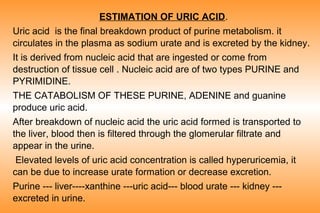
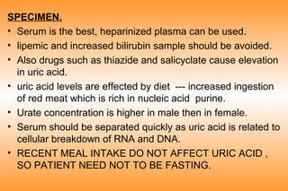
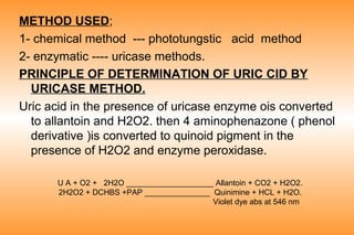
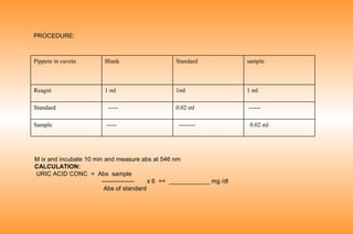
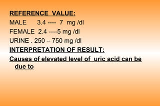
ESTIMATION OF URIC ACID.
Uric acid is the final breakdown product of purine metabolism. it
circulates in the plasma as sodium urate and is excreted by the kidney.
It is derived from nucleic acid that are ingested or come from
destruction of tissue cell . Nucleic acid are of two types PURINE and
PYRIMIDINE.
THE CATABOLISM OF THESE PURINE, ADENINE and guanine
produce uric acid.
After breakdown of nucleic acid the uric acid formed is transported to
the liver, blood then is filtered through the glomerular filtrate and
appear in the urine.
Elevated levels of uric acid concentration is called hyperuricemia, it
can be due to increase urate formation or decrease excretion.
Purine --- liver----xanthine ---uric acid--- blood urate --- kidney --excreted in urine.
OBJECTIVE.
• To know the uric acid level in the body.
• To diagnose a case of hyperuricemia.
SPECIMEN.
• Serum is the best, heparinized plasma can be used.
• lipemic and increased bilirubin sample should be avoided.
• Also drugs such as thiazide and salicyclate cause elevation
in uric acid.
• uric acid levels are effected by diet --- increased ingestion
of red meat which is rich in nucleic acid purine.
• Urate concentration is higher in male then in female.
• Serum should be separated quickly as uric acid is related to
cellular breakdown of RNA and DNA.
• RECENT MEAL INTAKE DO NOT AFFECT URIC ACID ,
SO PATIENT NEED NOT TO BE FASTING.
METHOD USED;
1- chemical method --- phototungstic acid method
2- enzymatic ---- uricase methods.
PRINCIPLE OF DETERMINATION OF URIC CID BY
URICASE METHOD.
Uric acid in the presence of uricase enzyme ois converted
to allantoin and H2O2. then 4 aminophenazone ( phenol
derivative )is converted to quinoid pigment in the
presence of H2O2 and enzyme peroxidase.
U A + O2 + 2H2O ____________________ Allantoin + CO2 + H2O2.
2H2O2 + DCHBS +PAP _______________ Quinimine + HCL + H2O.
Violet dye abs at 546 nm
PROCEDURE:

Pippete in cuvete

Blank

Standard

sample

Reagnt

1 ml

1ml

1 ml
------

Standard

-----

0.02 ml

Sample

-----

--------

M ix and incubate 10 min and measure abs at 546 nm
CALCULATION:
URIC ACID CONC = Abs sample
---------------x 6 == ____________ mg /dl
Abs of standard

0.02 ml
REFERENCE VALUE:
MALE
3.4 ---- 7 mg /dl
FEMALE 2.4 ----5 mg /dl
URINE . 250 – 750 mg /dl
INTERPRETATION OF RESULT:
Causes of elevated level of uric acid can be
due to
INREASED PRODUCTION.
PRIMARY CAUSE.

2 SECONDARY

-LEISH NYHAN SYNDROME.
Increase dietary inta
-GLUCOSE 6 PHOSPHATE DEFFICIENCY.
Intake
increase tissue cell
breakdown due to
LUKEMIA.
Cytotoxic drugs
DECREASE EXCRETION;
Renal insufficiency due to CRF , drugs.
HYPERURICEMIA can lead to disease condition
called GOUT.
G out is a clinical syndrome characterized by
hyperuricemia nad acute arthritis . acute gout tat
by deposition of sodium urate crystals which
cause inflammation
s/s pain and inflammation of joints.

Estimation of uric acid

  • 1.
    ESTIMATION OF URICACID. Uric acid is the final breakdown product of purine metabolism. it circulates in the plasma as sodium urate and is excreted by the kidney. It is derived from nucleic acid that are ingested or come from destruction of tissue cell . Nucleic acid are of two types PURINE and PYRIMIDINE. THE CATABOLISM OF THESE PURINE, ADENINE and guanine produce uric acid. After breakdown of nucleic acid the uric acid formed is transported to the liver, blood then is filtered through the glomerular filtrate and appear in the urine. Elevated levels of uric acid concentration is called hyperuricemia, it can be due to increase urate formation or decrease excretion. Purine --- liver----xanthine ---uric acid--- blood urate --- kidney --excreted in urine.
  • 2.
    OBJECTIVE. • To knowthe uric acid level in the body. • To diagnose a case of hyperuricemia.
  • 3.
    SPECIMEN. • Serum isthe best, heparinized plasma can be used. • lipemic and increased bilirubin sample should be avoided. • Also drugs such as thiazide and salicyclate cause elevation in uric acid. • uric acid levels are effected by diet --- increased ingestion of red meat which is rich in nucleic acid purine. • Urate concentration is higher in male then in female. • Serum should be separated quickly as uric acid is related to cellular breakdown of RNA and DNA. • RECENT MEAL INTAKE DO NOT AFFECT URIC ACID , SO PATIENT NEED NOT TO BE FASTING.
  • 4.
    METHOD USED; 1- chemicalmethod --- phototungstic acid method 2- enzymatic ---- uricase methods. PRINCIPLE OF DETERMINATION OF URIC CID BY URICASE METHOD. Uric acid in the presence of uricase enzyme ois converted to allantoin and H2O2. then 4 aminophenazone ( phenol derivative )is converted to quinoid pigment in the presence of H2O2 and enzyme peroxidase. U A + O2 + 2H2O ____________________ Allantoin + CO2 + H2O2. 2H2O2 + DCHBS +PAP _______________ Quinimine + HCL + H2O. Violet dye abs at 546 nm
  • 5.
    PROCEDURE: Pippete in cuvete Blank Standard sample Reagnt 1ml 1ml 1 ml ------ Standard ----- 0.02 ml Sample ----- -------- M ix and incubate 10 min and measure abs at 546 nm CALCULATION: URIC ACID CONC = Abs sample ---------------x 6 == ____________ mg /dl Abs of standard 0.02 ml
  • 6.
    REFERENCE VALUE: MALE 3.4 ----7 mg /dl FEMALE 2.4 ----5 mg /dl URINE . 250 – 750 mg /dl INTERPRETATION OF RESULT: Causes of elevated level of uric acid can be due to
  • 7.
    INREASED PRODUCTION. PRIMARY CAUSE. 2SECONDARY -LEISH NYHAN SYNDROME. Increase dietary inta -GLUCOSE 6 PHOSPHATE DEFFICIENCY. Intake increase tissue cell breakdown due to LUKEMIA. Cytotoxic drugs
  • 8.
    DECREASE EXCRETION; Renal insufficiencydue to CRF , drugs. HYPERURICEMIA can lead to disease condition called GOUT. G out is a clinical syndrome characterized by hyperuricemia nad acute arthritis . acute gout tat by deposition of sodium urate crystals which cause inflammation s/s pain and inflammation of joints.