Downloaded 320 times


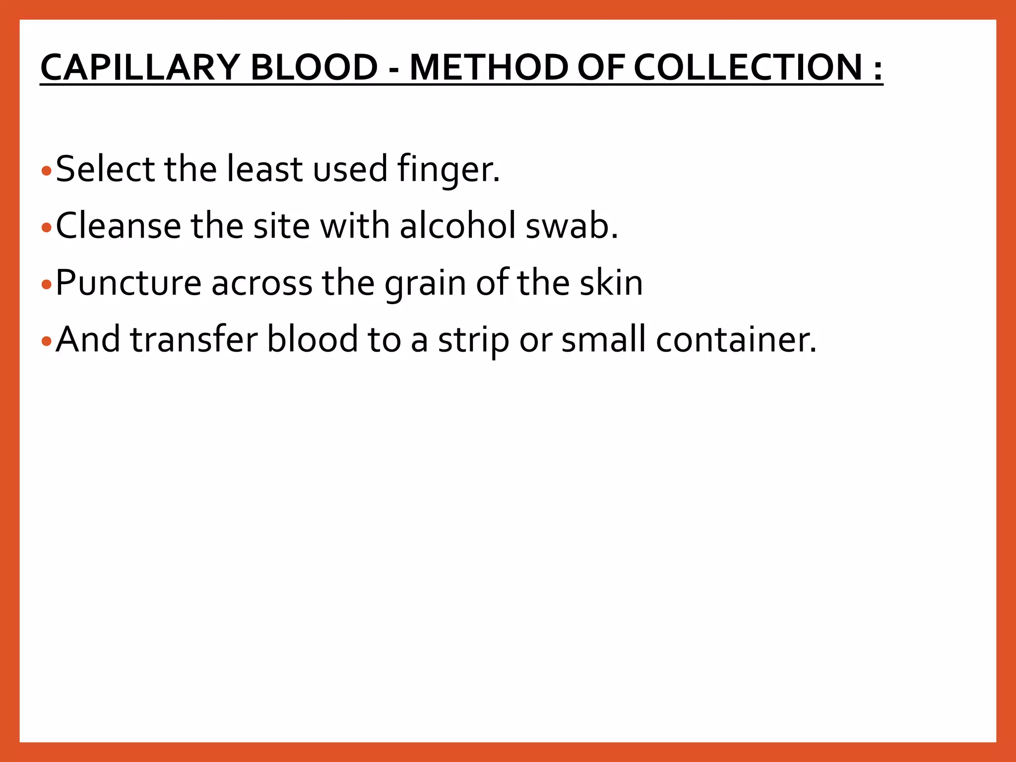


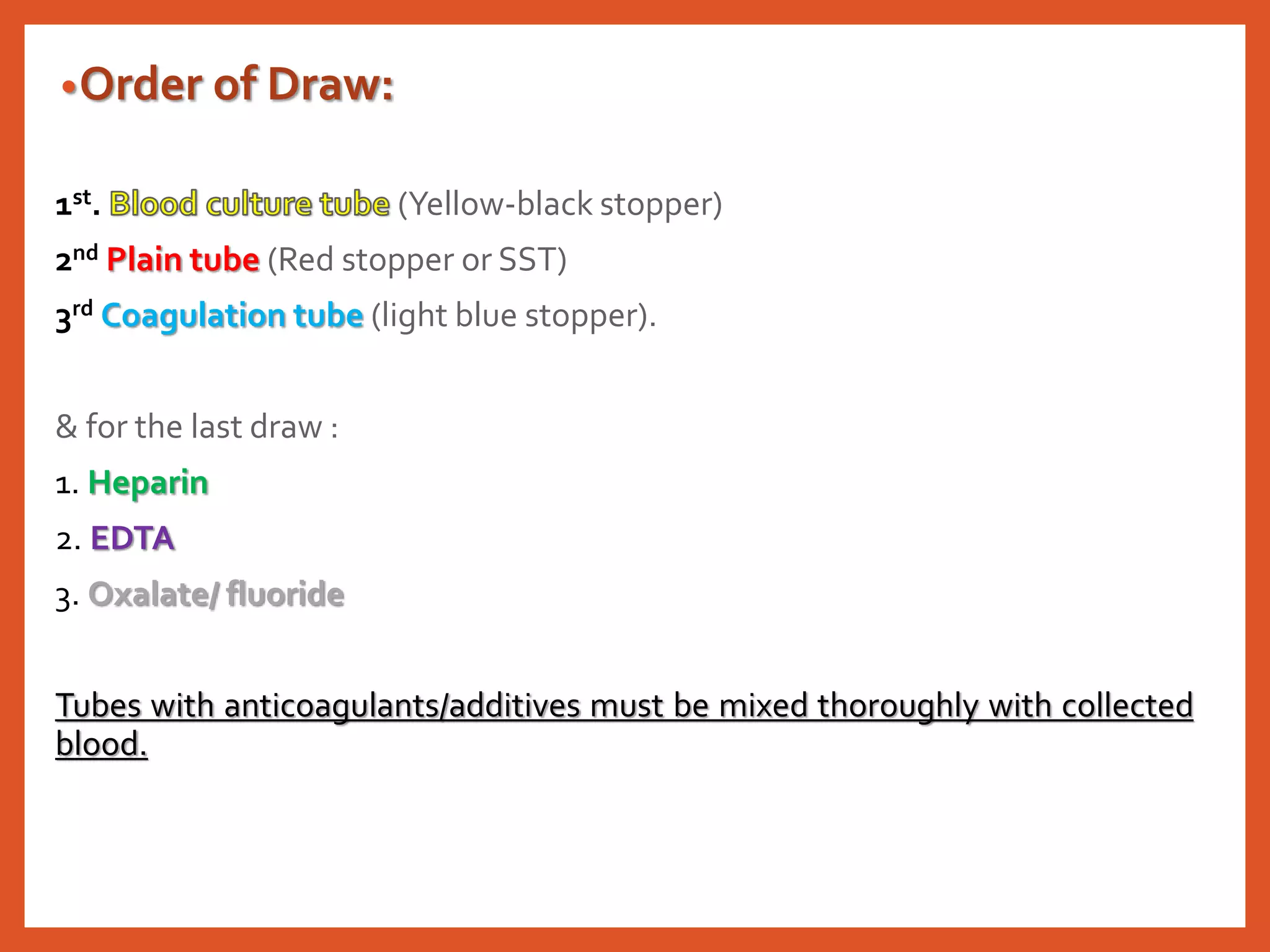


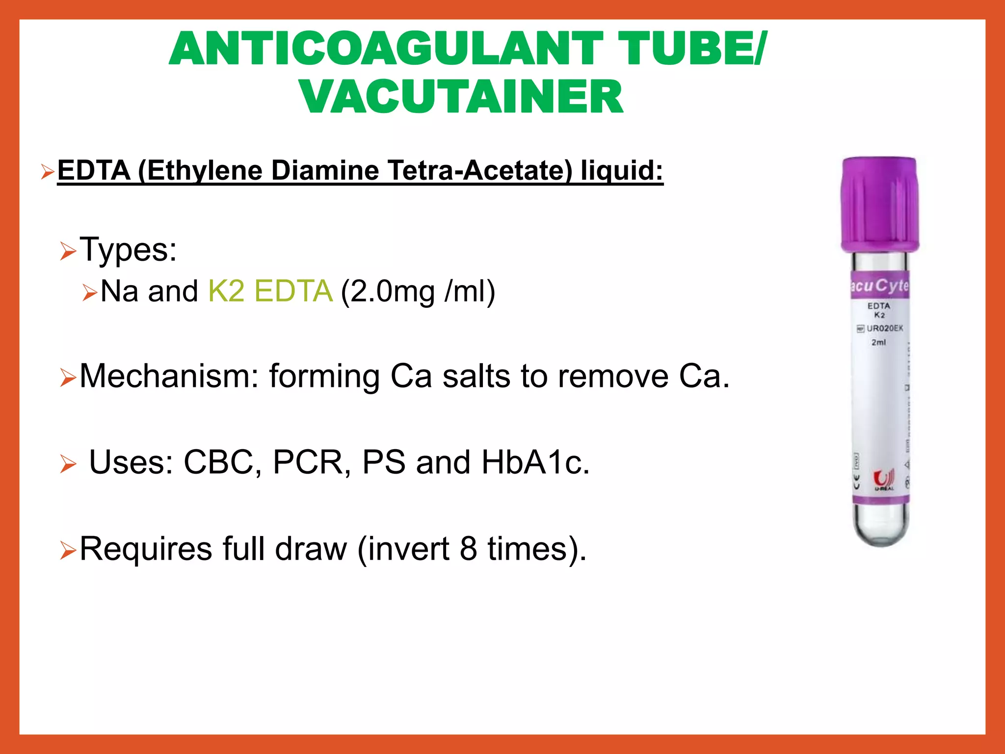


The document discusses different methods of blood collection including capillary, venous, and arterial blood. It describes various anticoagulants used in blood collection tubes and their purposes, such as EDTA for cell counts and citrate for coagulation studies. The document also outlines the proper order for drawing blood into collection tubes and the use of blood banks for storing blood components.


