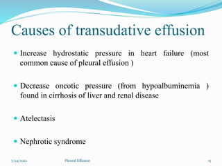
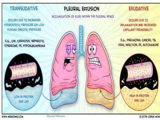
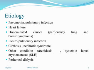
7/24/2022 1
Pleural Effusion
Prepared By:
Pranita Aryal
Roll No. 07
BNS 3rd Year
TU, IOM
7/24/2022 2
Pleural Effusion
7/24/2022 3
Pleural Effusion
7/24/2022 4
Pleural Effusion
7/24/2022 5
Pleural Effusion
7/24/2022 6
Pleural Effusion
Introduction
 Pleural effusion a collection of fluid in the pleural space ,
is rarely a primary disease process but is usually
secondary to other disease.
 Normally, the pleural space contains a small amount of
fluid (5 to 15ml), which acts as a lubricant that allows the
pleural surface to move without friction.
7/24/2022 7
Pleural Effusion
Cont….
 Pleural fluid normally seeps continually into the pleural
space from the capillaries lining the parietal pleura and is
reabsorbed by the visceral pleural capillaries and
lymphatic system.
 Any condition that interferes with either secretion or
drainage of this fluid leads to pleural effusion.
7/24/2022 8
Pleural Effusion
Definition
Pleural effusion is an abnormal, excessive collection of
fluid in the pleural spaces.
Too much fluid impairs the ability of the lung to expand and
move.
It may be a complication of heart failure, TB, pneumonia,
pulmonary infection, nephrotic syndrome
7/24/2022 9
Pleural Effusion
7/24/2022 10
Pleural Effusion
7/24/2022 11
Pleural Effusion
Epidemiology
 The incidence in U.S is estimated to be atleast 1.5 million
cases annually. Most cases are caused by CHF, bacterial
pneumonia.
 In industralized countries the prevalence rate is 320 cases
per 1lakh people.
 The incidence is equal between the sexes.Nearly two
thirds of malignant pleural effusion occur in women
associated with breast malignancies.
7/24/2022 12
Pleural Effusion
Classification
 Transudative effusion
 Exudatives effusion
7/24/2022 13
Pleural Effusion
Transudative Effusion
Transudative effusion is also known as hydrothorax, occur
primarily in non inflammatory conditions; is an
accumulation of low protein , low cell count fluid.It is
caused by fluid leaking into the pleural space.
It has a clear fluid similar to blood serum.
7/24/2022 14
Pleural Effusion
Causes of transudative effusion
 Increase hydrostatic pressure in heart failure (most
common cause of pleural effusion )
 Decrease oncotic pressure (from hypoalbuminemia )
found in cirrhosis of liver and renal disease
 Atelectasis
 Nephrotic syndrome
7/24/2022 15
Pleural Effusion
Characteristics of Tranudative
effusion
 Occurs primarily in non-inflammatory conditions.
 Low protein, low-cell-count fluid.
 Clear to faint yellow tinge, no odor.
 pH 7.40-7.55
 Specific gravity <1.015.
 Protein content<3g100ml.
7/24/2022 16
Pleural Effusion
Exudative effusion
 Exudative effusion contain a fluid rich in protein and
cellular elements that oozes out of blood vessels due to
inflammation.
 An exudates effusion result from increased capillary
permeability characteristic of inflammatory reaction.
 A fluid is cloudy containing cells and much protein.
7/24/2022 17
Pleural Effusion
Cont…
 This types of effusion occurs secondary to condition such
as:
 Tuberculosis
 malignancies , pulmonary infection
 pulmonary embolism
 Empyema
7/24/2022 18
Pleural Effusion
Characteristics of Exudate effusion
 Often turbid, bloody or purulrnt.
 pH<7.30
 Specific gravity <1.016.
 Protein content<3g100ml
 High protein fluid
7/24/2022 19
Pleural Effusion
7/24/2022 20
Pleural Effusion
Etiology
 Pneumonia, pulmonary infection
 Heart failure
 Disseminated cancer (particularly lung and
breast,lymphoma)
 Pleuro-pulmonary infection
 Cirrhosis , nephrotic syndrome
 Other condition sarcoidosis , systemic lupus
erythematosus (SLE)
 Peritoneal dialysis
7/24/2022 21
Pleural Effusion
Etiology contd..
 Viral infection : Other condition sarcoidosis , systemic
lupus erythematosus (SLE)
 Peritoneal dialysis
(Excessive fluid may accumulate because the body does not
handle fluid properly (such as in congestive heart failure, or
kidney and liver disease). The fluid in pleural effusions also may
result from inflammation, such as in pneumonia, autoimmune
disease, and many other conditions).
7/24/2022 22
Pleural Effusion
Pathophysiology
 Transudative effusion
 Due to different etiological factors.
 Increase hydrostatic pressure ,decrease oncotic pressure
 Unable to remain the fluid with in a intravascular space
 Fluid shift interstitial space (effusion)
7/24/2022 23
Pleural Effusion
exudative effusion
(pathophysiology)
 Invasion of microbes
 Initiation of inflammatory reaction
 Vasodilation increase capillary permeability
 Leak of plasma protein decrease oncotic pressure
7/24/2022 24
Pleural Effusion
Cont…..
Fluid shift into interstitial space
effusion
7/24/2022 25
Pleural Effusion
Mechanism
 Altered permeability (e.g.inflammation.
 Reduction in intravascular oncotic pressure
(e.g.hypoalbuminemia)
 Increased capillary hydrostatic pressure
(e.g.congestive heart failure)
 Decreased lymphatic drainage.
 Increased fluid in peritoneal cavity, with migration
across the diaphragm via the lymphatics (e.g. hepatic
cirrhosis, peritoneal dialysis).
7/24/2022 26
Pleural Effusion
Clinical Manifestation
 The clinical presentation of pleural effusion depends on
the amount of fluid present and the underlying causes. If
the effusion is small (250ml) ,it’s presence may be
discovered only on chest x-ray
 Many patient have no symptoms at the time a pleural
effusion is discovered .
7/24/2022 27
Pleural Effusion
Clinical Manifestation contd..
 Fever with chills
 Cough
 Dyspnea
 Dullness and flatness on chest percussion
 Decreased or absent breathe sound
 Shortness of breathe.
7/24/2022 28
Pleural Effusion
Cont …
 Pleuritic chest pain
 Dyspnea
- the chest pain is usually sharp and is exacerbated by
movement of the pleural surfaces ,as with deep inspiration ,
coughing and sneezing .
7/24/2022 29
Pleural Effusion
Diagnostic evaluation
 History taking
7/24/2022 30
Pleural Effusion
Physical examination
7/24/2022 31
Pleural Effusion
CT-Scan
7/24/2022 32
Pleural Effusion
Chest x-ray
7/24/2022 33
Pleural Effusion
Thoracentesis
7/24/2022 34
Pleural Effusion
Pleural biopsy
7/24/2022 35
Pleural Effusion
Analysis of pleural fluid
7/24/2022 36
Pleural Effusion
Complication
 Large effusion can lead to respiratory failure.
 Disseminated cancer.
 Pleuropulmonary infection.
7/24/2022 37
Pleural Effusion
Management
General management:
 Treatment is aim at underlying cause eg heart disease ,
tuberculosis infection ,cancer
 Thoracocentesis is done to remove fluids. Collect a
specimen and relieve dyspnea.
 Intrapleural instillation of medicine: tetrascycline,
doxycycline
7/24/2022 38
Pleural Effusion
For malignant effusion
 Chest tube drainage, radiation and chemotherapy ,surgical
pleumonectomy, pleuroperitoneal shunt or pleurodesis
 thoracocentesis may be provided only transient benefits in
malignant because effusion may reaccumulate within few
days
7/24/2022 39
Pleural Effusion
Surgical management
 Surgical pleurectomy:
Pleurectomy is a type of surgery in which part of the
pleura is removed. This procedure helps to prevent
fluid from collecting in the affected area and is used
for the treatment of mesothelioma, a pleural
mesothelial cancer.
7/24/2022 40
Pleural Effusion
Nursing Management
Assessment:
 Obtain history of previous pulmonary condition
 Assess patient for dyspnea and tachypnea
 Auscultation and percussion of the lungs of abnormalities
7/24/2022 41
Pleural Effusion
Nursing diagnosis
 Impaired gas exchange related to decreased function of
lung tissue
 Ineffective breathing pattern related to compromised lung
expansion
 Acute pain related to inflammatory process
 Anxiety related to inability to take deep breaths
 Risk for infection r/t pooling of fluid in lung space
7/24/2022 42
Pleural Effusion
Nursing intervention:
To improve gas exchange:
 Assess respiration; quality, rate ,rhythm, depth.
 Observe colour of skin, mucous membrane and nail beds
for presence of cyanosis.
 Advice patient for compelte bed rest.
 Encourage use of relaxation technique such as deep
breathing.
 Administer 02 by appropriate means: nasal prongs , mask ,
venture mask.
7/24/2022 43
Pleural Effusion
Maintain effective pattern:
 Check ou respiratory function, resiratory distress and
changes in vital function.
 Maintain a position of comfort, with a head of bed slightly
elevated.
 Turn to affected site.
 Administer oxygen therapy as prescribed to maintain
oxygen level.
 Maintain calm environment.
 Check drainage if inserted.
7/24/2022 44
Pleural Effusion
To reduce pain:
 Assess the patient by using pain rating scale for intensity,
characteristics and location of pain.
 Assist patient on deep breathing exercise and relaxation
technique.
 Assist the patient to change position.
 Maintain calm environment.
 Administer analgesic for pain as prescribed.
7/24/2022 45
Pleural Effusion
To reduce risk of infection:
 Demonstrate and encourage good hand washing
technique.
 Limit visitors.
 Promote adequate nutrition intake which facilitate healing
process.
 Encourage adequate rest with moderate activity.
7/24/2022 46
Pleural Effusion
Cont..
Patient education and health maintenance:
 Provide care after pleurodesis
 Monitor for excessive pain and give analgesic
 Assist patient undergoing instillation of intrapleural
lignocaine if pain is not relief
 Administer oxygen as indicated dyspnea and hypoxemia
7/24/2022 47
Pleural Effusion
Cont….
 Observed patient’s breathing pattern , oxygen saturation
and vital sign for assessment weather improvement or
deterioration
7/24/2022 48
Pleural Effusion
Complication:
 A lung that is surrounded by excess fluid for a long
time may be damaged.
 Pleural fluid that becomes infected may turn into an
abscess, called an empyema, which will need to be
drained with a chest tube.
 Pneumothorax (air in the chest cavity) can be a
complication of the thoracentesis procedure.
7/24/2022 49
Pleural Effusion
Care of Chest Tube Drainage
Never lift drain above chest level
 The unit and all tubing should be below patient ’s chest
level to facilitate drainage
 Tubing should have no kinks or obstructions that may
inhibit drainage
 Ensure all connections between chest tubes and drainage
unit are tight and secure
 Connections should have cable ties in place
7/24/2022 50
Pleural Effusion
Care of Chest Tube Drainage
contd
 Tubing should be anchored to the patient ’s skin to prevent
pulling of the drain prevent accidental removal.
 In PICU and NICU tubing should also be secured to patient bed
prevent accidental removal .
 Ensure the unit is securely positioned on its stand or hanging on
the bed.
 Ensure the water seal is maintained at 2cm at all times.
 Measure the output of drainage.
7/24/2022 51
Pleural Effusion
7/24/2022 52
Pleural Effusion

pleural effusion

  • 1.
  • 2.
    Prepared By: Pranita Aryal RollNo. 07 BNS 3rd Year TU, IOM 7/24/2022 2 Pleural Effusion
  • 3.
  • 4.
  • 5.
  • 6.
  • 7.
    Introduction  Pleural effusiona collection of fluid in the pleural space , is rarely a primary disease process but is usually secondary to other disease.  Normally, the pleural space contains a small amount of fluid (5 to 15ml), which acts as a lubricant that allows the pleural surface to move without friction. 7/24/2022 7 Pleural Effusion
  • 8.
    Cont….  Pleural fluidnormally seeps continually into the pleural space from the capillaries lining the parietal pleura and is reabsorbed by the visceral pleural capillaries and lymphatic system.  Any condition that interferes with either secretion or drainage of this fluid leads to pleural effusion. 7/24/2022 8 Pleural Effusion
  • 9.
    Definition Pleural effusion isan abnormal, excessive collection of fluid in the pleural spaces. Too much fluid impairs the ability of the lung to expand and move. It may be a complication of heart failure, TB, pneumonia, pulmonary infection, nephrotic syndrome 7/24/2022 9 Pleural Effusion
  • 10.
  • 11.
  • 12.
    Epidemiology  The incidencein U.S is estimated to be atleast 1.5 million cases annually. Most cases are caused by CHF, bacterial pneumonia.  In industralized countries the prevalence rate is 320 cases per 1lakh people.  The incidence is equal between the sexes.Nearly two thirds of malignant pleural effusion occur in women associated with breast malignancies. 7/24/2022 12 Pleural Effusion
  • 13.
    Classification  Transudative effusion Exudatives effusion 7/24/2022 13 Pleural Effusion
  • 14.
    Transudative Effusion Transudative effusionis also known as hydrothorax, occur primarily in non inflammatory conditions; is an accumulation of low protein , low cell count fluid.It is caused by fluid leaking into the pleural space. It has a clear fluid similar to blood serum. 7/24/2022 14 Pleural Effusion
  • 15.
    Causes of transudativeeffusion  Increase hydrostatic pressure in heart failure (most common cause of pleural effusion )  Decrease oncotic pressure (from hypoalbuminemia ) found in cirrhosis of liver and renal disease  Atelectasis  Nephrotic syndrome 7/24/2022 15 Pleural Effusion
  • 16.
    Characteristics of Tranudative effusion Occurs primarily in non-inflammatory conditions.  Low protein, low-cell-count fluid.  Clear to faint yellow tinge, no odor.  pH 7.40-7.55  Specific gravity <1.015.  Protein content<3g100ml. 7/24/2022 16 Pleural Effusion
  • 17.
    Exudative effusion  Exudativeeffusion contain a fluid rich in protein and cellular elements that oozes out of blood vessels due to inflammation.  An exudates effusion result from increased capillary permeability characteristic of inflammatory reaction.  A fluid is cloudy containing cells and much protein. 7/24/2022 17 Pleural Effusion
  • 18.
    Cont…  This typesof effusion occurs secondary to condition such as:  Tuberculosis  malignancies , pulmonary infection  pulmonary embolism  Empyema 7/24/2022 18 Pleural Effusion
  • 19.
    Characteristics of Exudateeffusion  Often turbid, bloody or purulrnt.  pH<7.30  Specific gravity <1.016.  Protein content<3g100ml  High protein fluid 7/24/2022 19 Pleural Effusion
  • 20.
  • 21.
    Etiology  Pneumonia, pulmonaryinfection  Heart failure  Disseminated cancer (particularly lung and breast,lymphoma)  Pleuro-pulmonary infection  Cirrhosis , nephrotic syndrome  Other condition sarcoidosis , systemic lupus erythematosus (SLE)  Peritoneal dialysis 7/24/2022 21 Pleural Effusion
  • 22.
    Etiology contd..  Viralinfection : Other condition sarcoidosis , systemic lupus erythematosus (SLE)  Peritoneal dialysis (Excessive fluid may accumulate because the body does not handle fluid properly (such as in congestive heart failure, or kidney and liver disease). The fluid in pleural effusions also may result from inflammation, such as in pneumonia, autoimmune disease, and many other conditions). 7/24/2022 22 Pleural Effusion
  • 23.
    Pathophysiology  Transudative effusion Due to different etiological factors.  Increase hydrostatic pressure ,decrease oncotic pressure  Unable to remain the fluid with in a intravascular space  Fluid shift interstitial space (effusion) 7/24/2022 23 Pleural Effusion
  • 24.
    exudative effusion (pathophysiology)  Invasionof microbes  Initiation of inflammatory reaction  Vasodilation increase capillary permeability  Leak of plasma protein decrease oncotic pressure 7/24/2022 24 Pleural Effusion
  • 25.
    Cont….. Fluid shift intointerstitial space effusion 7/24/2022 25 Pleural Effusion
  • 26.
    Mechanism  Altered permeability(e.g.inflammation.  Reduction in intravascular oncotic pressure (e.g.hypoalbuminemia)  Increased capillary hydrostatic pressure (e.g.congestive heart failure)  Decreased lymphatic drainage.  Increased fluid in peritoneal cavity, with migration across the diaphragm via the lymphatics (e.g. hepatic cirrhosis, peritoneal dialysis). 7/24/2022 26 Pleural Effusion
  • 27.
    Clinical Manifestation  Theclinical presentation of pleural effusion depends on the amount of fluid present and the underlying causes. If the effusion is small (250ml) ,it’s presence may be discovered only on chest x-ray  Many patient have no symptoms at the time a pleural effusion is discovered . 7/24/2022 27 Pleural Effusion
  • 28.
    Clinical Manifestation contd.. Fever with chills  Cough  Dyspnea  Dullness and flatness on chest percussion  Decreased or absent breathe sound  Shortness of breathe. 7/24/2022 28 Pleural Effusion
  • 29.
    Cont …  Pleuriticchest pain  Dyspnea - the chest pain is usually sharp and is exacerbated by movement of the pleural surfaces ,as with deep inspiration , coughing and sneezing . 7/24/2022 29 Pleural Effusion
  • 30.
    Diagnostic evaluation  Historytaking 7/24/2022 30 Pleural Effusion
  • 31.
  • 32.
  • 33.
  • 34.
  • 35.
  • 36.
    Analysis of pleuralfluid 7/24/2022 36 Pleural Effusion
  • 37.
    Complication  Large effusioncan lead to respiratory failure.  Disseminated cancer.  Pleuropulmonary infection. 7/24/2022 37 Pleural Effusion
  • 38.
    Management General management:  Treatmentis aim at underlying cause eg heart disease , tuberculosis infection ,cancer  Thoracocentesis is done to remove fluids. Collect a specimen and relieve dyspnea.  Intrapleural instillation of medicine: tetrascycline, doxycycline 7/24/2022 38 Pleural Effusion
  • 39.
    For malignant effusion Chest tube drainage, radiation and chemotherapy ,surgical pleumonectomy, pleuroperitoneal shunt or pleurodesis  thoracocentesis may be provided only transient benefits in malignant because effusion may reaccumulate within few days 7/24/2022 39 Pleural Effusion
  • 40.
    Surgical management  Surgicalpleurectomy: Pleurectomy is a type of surgery in which part of the pleura is removed. This procedure helps to prevent fluid from collecting in the affected area and is used for the treatment of mesothelioma, a pleural mesothelial cancer. 7/24/2022 40 Pleural Effusion
  • 41.
    Nursing Management Assessment:  Obtainhistory of previous pulmonary condition  Assess patient for dyspnea and tachypnea  Auscultation and percussion of the lungs of abnormalities 7/24/2022 41 Pleural Effusion
  • 42.
    Nursing diagnosis  Impairedgas exchange related to decreased function of lung tissue  Ineffective breathing pattern related to compromised lung expansion  Acute pain related to inflammatory process  Anxiety related to inability to take deep breaths  Risk for infection r/t pooling of fluid in lung space 7/24/2022 42 Pleural Effusion
  • 43.
    Nursing intervention: To improvegas exchange:  Assess respiration; quality, rate ,rhythm, depth.  Observe colour of skin, mucous membrane and nail beds for presence of cyanosis.  Advice patient for compelte bed rest.  Encourage use of relaxation technique such as deep breathing.  Administer 02 by appropriate means: nasal prongs , mask , venture mask. 7/24/2022 43 Pleural Effusion
  • 44.
    Maintain effective pattern: Check ou respiratory function, resiratory distress and changes in vital function.  Maintain a position of comfort, with a head of bed slightly elevated.  Turn to affected site.  Administer oxygen therapy as prescribed to maintain oxygen level.  Maintain calm environment.  Check drainage if inserted. 7/24/2022 44 Pleural Effusion
  • 45.
    To reduce pain: Assess the patient by using pain rating scale for intensity, characteristics and location of pain.  Assist patient on deep breathing exercise and relaxation technique.  Assist the patient to change position.  Maintain calm environment.  Administer analgesic for pain as prescribed. 7/24/2022 45 Pleural Effusion
  • 46.
    To reduce riskof infection:  Demonstrate and encourage good hand washing technique.  Limit visitors.  Promote adequate nutrition intake which facilitate healing process.  Encourage adequate rest with moderate activity. 7/24/2022 46 Pleural Effusion
  • 47.
    Cont.. Patient education andhealth maintenance:  Provide care after pleurodesis  Monitor for excessive pain and give analgesic  Assist patient undergoing instillation of intrapleural lignocaine if pain is not relief  Administer oxygen as indicated dyspnea and hypoxemia 7/24/2022 47 Pleural Effusion
  • 48.
    Cont….  Observed patient’sbreathing pattern , oxygen saturation and vital sign for assessment weather improvement or deterioration 7/24/2022 48 Pleural Effusion
  • 49.
    Complication:  A lungthat is surrounded by excess fluid for a long time may be damaged.  Pleural fluid that becomes infected may turn into an abscess, called an empyema, which will need to be drained with a chest tube.  Pneumothorax (air in the chest cavity) can be a complication of the thoracentesis procedure. 7/24/2022 49 Pleural Effusion
  • 50.
    Care of ChestTube Drainage Never lift drain above chest level  The unit and all tubing should be below patient ’s chest level to facilitate drainage  Tubing should have no kinks or obstructions that may inhibit drainage  Ensure all connections between chest tubes and drainage unit are tight and secure  Connections should have cable ties in place 7/24/2022 50 Pleural Effusion
  • 51.
    Care of ChestTube Drainage contd  Tubing should be anchored to the patient ’s skin to prevent pulling of the drain prevent accidental removal.  In PICU and NICU tubing should also be secured to patient bed prevent accidental removal .  Ensure the unit is securely positioned on its stand or hanging on the bed.  Ensure the water seal is maintained at 2cm at all times.  Measure the output of drainage. 7/24/2022 51 Pleural Effusion
  • 52.