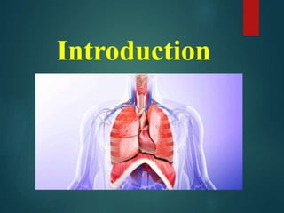
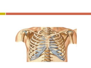
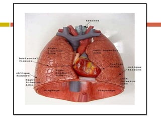
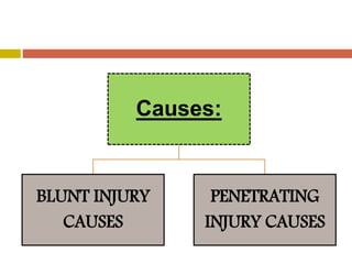
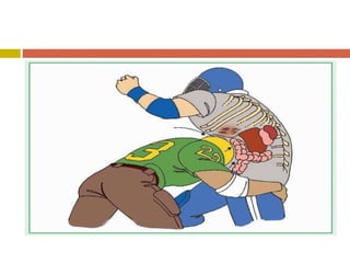
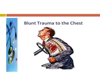
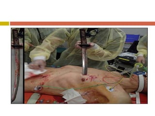
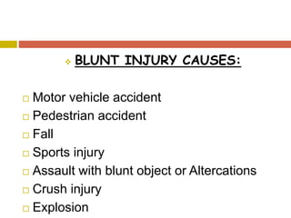
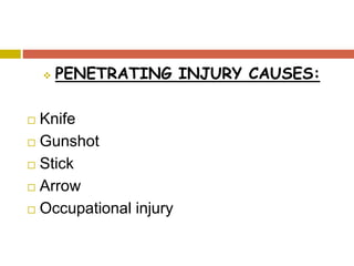
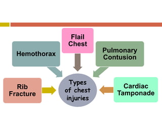
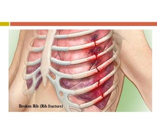
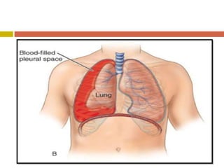
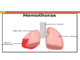
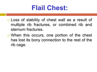
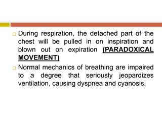
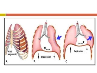
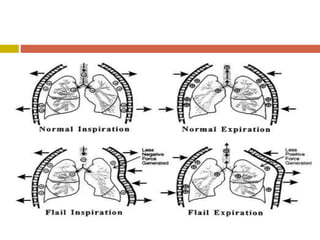
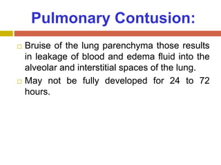
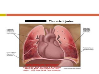
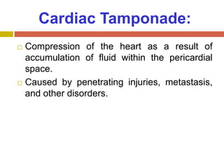
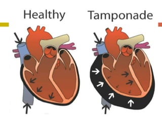
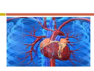
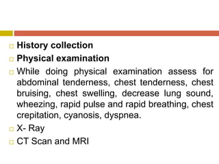
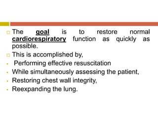
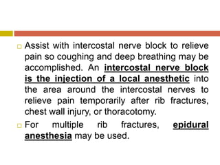
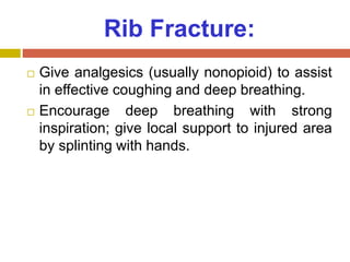
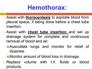
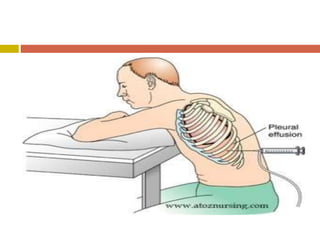
CHEST INJURY
Presented By,
Ms. Ekta. S.
Patel,
1st Year M.Sc
Nursing,
Introduction
Introduction:
 Traumatic injuries to the chest contribute to
75% of all traumatic deaths.
 Thoracic injuries range from simple rib
fractures to complex life-threatening rupture
of organs.
 The mechanisms of injuries causing chest
trauma are separated into two categories:
blunt trauma and penetrating trauma.
 Chest injuries are potentially life-threatening
because of immediate disturbances of
cardiorespiratory physiology and
haemorrhage and later developments of
infection, damaged lung and thoracic cage.
Definition
Definition:
 A chest injury is define as, “it is a form of
injury to the chest including the ribs, heart
and lungs, great vessels, trachea and
esophagus.”
Incidence
Incidence:
 25% of all death form traumatic injury.
Causes
Causes:
BLUNT INJURY
CAUSES
PENETRATING
INJURY CAUSES
 BLUNT INJURY CAUSES:
 Motor vehicle accident
 Pedestrian accident
 Fall
 Sports injury
 Assault with blunt object or Altercations
 Crush injury
 Explosion
 PENETRATING INJURY CAUSES:
 Knife
 Gunshot
 Stick
 Arrow
 Occupational injury
Types of chest
injuries
Types
of chest
injuries
Rib
Fracture
Hemothorax
Flail
Chest
Pulmonary
Contusion
Cardiac
Tamponade
Rib Fracture:
 Most common chest injury.
 May interfere with ventilation and may
lacerate underlying lung.
Hemothorax:
 Blood in pleural space as a result of
penetrating or blunt chest trauma.
 Accompanies a high percentage of chest
injuries.
 Can result in hidden blood loss.
Flail Chest:
 Loss of stability of chest wall as a result of
multiple rib fractures, or combined rib and
sternum fractures.
 When this occurs, one portion of the chest
has lost its bony connection to the rest of the
rib cage.
 During respiration, the detached part of the
chest will be pulled in on inspiration and
blown out on expiration (PARADOXICAL
MOVEMENT)
 Normal mechanics of breathing are impaired
to a degree that seriously jeopardizes
ventilation, causing dyspnea and cyanosis.
Pulmonary Contusion:
 Bruise of the lung parenchyma those results
in leakage of blood and edema fluid into the
alveolar and interstitial spaces of the lung.
 May not be fully developed for 24 to 72
hours.
Cardiac Tamponade:
 Compression of the heart as a result of
accumulation of fluid within the pericardial
space.
 Caused by penetrating injuries, metastasis,
and other disorders.
Clinical
manifestation:
Respiratory
 Dyspnea, respiratory distress
 Cough with or without haemoptysis
 Cyanosis of mouth, face, nail beds, mucous
membranes
 Tracheal deviation
 Audible air escaping from chest wound
 Decreased breath sounds on side of injury
 Decreased O2 saturation
 Frothy secretions
Cardiovascular
 Rapid, thready pulse
 Decreased BP
 Narrowed pulse pressure
 Asymmetric BP values in arms
 Distended neck veins
 Muffled heart sounds
 Chest pain
 Crunching sound synchronous with heart
sounds
 Dysrhythmias
Surface Findings
 Bruising
 Abrasions
 Open chest wound
 Asymmetric chest movement
 Subcutaneous emphysema
Diagnostic
evaluation
 History collection
 Physical examination
 While doing physical examination assess for
abdominal tenderness, chest tenderness, chest
bruising, chest swelling, decrease lung sound,
wheezing, rapid pulse and rapid breathing, chest
crepitation, cyanosis, dyspnea.
 X- Ray
 CT Scan and MRI
Management
 The goal is to restore normal
cardiorespiratory function as quickly as
possible.
 This is accomplished by,
 Performing effective resuscitation
 While simultaneously assessing the patient,
 Restoring chest wall integrity,
 Reexpanding the lung.
 Assist with intercostal nerve block to relieve
pain so coughing and deep breathing may be
accomplished. An intercostal nerve block
is the injection of a local anesthetic into
the area around the intercostal nerves to
relieve pain temporarily after rib fractures,
chest wall injury, or thoracotomy.
 For multiple rib fractures, epidural
anesthesia may be used.
Rib Fracture:
 Give analgesics (usually nonopioid) to assist
in effective coughing and deep breathing.
 Encourage deep breathing with strong
inspiration; give local support to injured area
by splinting with hands.
Hemothorax:
 Assist with thoracentesis to aspirate blood from
pleural space, if being done before a chest tube
insertion.
 Assist with chest tube insertion and set up
drainage system for complete and continuous
removal of blood and air.
Auscultate lungs and monitor for relief of
dyspnea.
Monitor amount of blood loss in drainage.
 Replace volume with I.V. fluids or blood
products.
Flail Chest:
 Stabilize the flail portion of the chest with
hands; apply a pressure dressing and turn
the patient on injured side, or place 10-lb
sandbag at site of flail.
 Thoracic epidural analgesia may be used
for some patients to relieve pain and
improve ventilation.
Pulmonary Contusion:
 Employ mechanical ventilation to keep lungs
inflated.
 Administer diuretics to reduce edema.
 Correct metabolic acidosis with I.V. sodium
bicarbonate.
 Use PAP monitoring.
 Monitor for development of pneumonia.
 If respiratory failure is present, prepare for
immediate ET intubation and mechanical
ventilation treats underlying pulmonary
contusion and serves to stabilize the thoracic
cage for healing of fractures, improves alveolar
ventilation, and restores thoracic cage stability
and intrathoracic volume by decreasing work of
breathing.
 Prepare for operative stabilization of chest
wall in select patients.
Cardiac Tamponade:
 Assist with pericardiocentesis to provide
emergency relief and improve hemodynamic
function until surgery can be undertaken.
 Prepare for emergency thoracotomy to
control bleeding and to repair cardiac injury.
ADDITIONAL
RESPONSIBILITIES:
 Secure and support the airway as indicated.
 Prepare for tracheostomy if indicated.
Tracheostomy helps to clear
tracheobronchial tree, helps the patient
breathe with less effort, decreases the
amount of dead airspace in the respiratory
tree, and helps reduce paradoxical motion.
When used with mechanical ventilation,
provides a closed system and stabilizes the
chest.
 Secure one or more I.V. lines for fluid
replacement, and obtain blood for
baseline studies, such as hemoglobin
level and hematocrit.
 Monitor serial CVP readings to prevent
hypovolemia and circulatory overload.
 Monitor ABG/Spo2 results to determine
need for supplemental oxygen,
mechanical ventilation.
 Obtain urinary output hourly to evaluate
tissue perfusion.
 Continue to monitor thoracic drainage to
provide information about rate of blood loss,
whether bleeding has stopped, whether
surgical intervention is necessary.
 Institute ECG monitoring for early
detection and treatment of cardiac
dysrhythmias (dysrhythmias are a
frequent cause of death in chest trauma).
 Maintain ongoing surveillance for
complications:
Aspiration
Atelectasis
Pneumonia
Mediastinal/subcutaneous emphysema
Respiratory failure
Patient education and
health maintenance:
 Instruct patient in splinting techniques.
 Make sure patient is aware of importance of
automobile seat belt use to reduce serious
chest injuries caused by automobile
accidents.
 Teach patient to report signs of complications
increasing dyspnea, fever, and cough.
Summary
Introduction
Definition
Incidence
Causes
Types
Clinical manifestation
Diagnostic evaluation
Management
› What is the definition of chest injury?
› What are the causes of chest injury?
› What are the types of chest injury?
› What are the diagnostic evaluation for
chest injury?
Feedback questions:
Thank You!!!

Chest injury

  • 1.
    CHEST INJURY Presented By, Ms.Ekta. S. Patel, 1st Year M.Sc Nursing,
  • 2.
  • 3.
    Introduction:  Traumatic injuriesto the chest contribute to 75% of all traumatic deaths.  Thoracic injuries range from simple rib fractures to complex life-threatening rupture of organs.  The mechanisms of injuries causing chest trauma are separated into two categories: blunt trauma and penetrating trauma.
  • 4.
     Chest injuriesare potentially life-threatening because of immediate disturbances of cardiorespiratory physiology and haemorrhage and later developments of infection, damaged lung and thoracic cage.
  • 5.
  • 6.
    Definition:  A chestinjury is define as, “it is a form of injury to the chest including the ribs, heart and lungs, great vessels, trachea and esophagus.”
  • 9.
  • 10.
    Incidence:  25% ofall death form traumatic injury.
  • 11.
  • 12.
  • 16.
     BLUNT INJURYCAUSES:  Motor vehicle accident  Pedestrian accident  Fall  Sports injury  Assault with blunt object or Altercations  Crush injury  Explosion
  • 17.
     PENETRATING INJURYCAUSES:  Knife  Gunshot  Stick  Arrow  Occupational injury
  • 18.
  • 19.
  • 20.
    Rib Fracture:  Mostcommon chest injury.  May interfere with ventilation and may lacerate underlying lung.
  • 22.
    Hemothorax:  Blood inpleural space as a result of penetrating or blunt chest trauma.  Accompanies a high percentage of chest injuries.  Can result in hidden blood loss.
  • 25.
    Flail Chest:  Lossof stability of chest wall as a result of multiple rib fractures, or combined rib and sternum fractures.  When this occurs, one portion of the chest has lost its bony connection to the rest of the rib cage.
  • 26.
     During respiration,the detached part of the chest will be pulled in on inspiration and blown out on expiration (PARADOXICAL MOVEMENT)  Normal mechanics of breathing are impaired to a degree that seriously jeopardizes ventilation, causing dyspnea and cyanosis.
  • 29.
    Pulmonary Contusion:  Bruiseof the lung parenchyma those results in leakage of blood and edema fluid into the alveolar and interstitial spaces of the lung.  May not be fully developed for 24 to 72 hours.
  • 32.
    Cardiac Tamponade:  Compressionof the heart as a result of accumulation of fluid within the pericardial space.  Caused by penetrating injuries, metastasis, and other disorders.
  • 34.
  • 36.
    Respiratory  Dyspnea, respiratorydistress  Cough with or without haemoptysis  Cyanosis of mouth, face, nail beds, mucous membranes  Tracheal deviation  Audible air escaping from chest wound  Decreased breath sounds on side of injury  Decreased O2 saturation  Frothy secretions
  • 38.
    Cardiovascular  Rapid, threadypulse  Decreased BP  Narrowed pulse pressure  Asymmetric BP values in arms  Distended neck veins  Muffled heart sounds
  • 39.
     Chest pain Crunching sound synchronous with heart sounds  Dysrhythmias
  • 41.
    Surface Findings  Bruising Abrasions  Open chest wound  Asymmetric chest movement  Subcutaneous emphysema
  • 42.
  • 43.
     History collection Physical examination  While doing physical examination assess for abdominal tenderness, chest tenderness, chest bruising, chest swelling, decrease lung sound, wheezing, rapid pulse and rapid breathing, chest crepitation, cyanosis, dyspnea.  X- Ray  CT Scan and MRI
  • 44.
  • 45.
     The goalis to restore normal cardiorespiratory function as quickly as possible.  This is accomplished by,  Performing effective resuscitation  While simultaneously assessing the patient,  Restoring chest wall integrity,  Reexpanding the lung.
  • 46.
     Assist withintercostal nerve block to relieve pain so coughing and deep breathing may be accomplished. An intercostal nerve block is the injection of a local anesthetic into the area around the intercostal nerves to relieve pain temporarily after rib fractures, chest wall injury, or thoracotomy.  For multiple rib fractures, epidural anesthesia may be used.
  • 47.
    Rib Fracture:  Giveanalgesics (usually nonopioid) to assist in effective coughing and deep breathing.  Encourage deep breathing with strong inspiration; give local support to injured area by splinting with hands.
  • 48.
    Hemothorax:  Assist withthoracentesis to aspirate blood from pleural space, if being done before a chest tube insertion.  Assist with chest tube insertion and set up drainage system for complete and continuous removal of blood and air. Auscultate lungs and monitor for relief of dyspnea. Monitor amount of blood loss in drainage.  Replace volume with I.V. fluids or blood products.
  • 50.
    Flail Chest:  Stabilizethe flail portion of the chest with hands; apply a pressure dressing and turn the patient on injured side, or place 10-lb sandbag at site of flail.  Thoracic epidural analgesia may be used for some patients to relieve pain and improve ventilation.
  • 51.
    Pulmonary Contusion:  Employmechanical ventilation to keep lungs inflated.  Administer diuretics to reduce edema.  Correct metabolic acidosis with I.V. sodium bicarbonate.  Use PAP monitoring.  Monitor for development of pneumonia.
  • 52.
     If respiratoryfailure is present, prepare for immediate ET intubation and mechanical ventilation treats underlying pulmonary contusion and serves to stabilize the thoracic cage for healing of fractures, improves alveolar ventilation, and restores thoracic cage stability and intrathoracic volume by decreasing work of breathing.  Prepare for operative stabilization of chest wall in select patients.
  • 53.
    Cardiac Tamponade:  Assistwith pericardiocentesis to provide emergency relief and improve hemodynamic function until surgery can be undertaken.  Prepare for emergency thoracotomy to control bleeding and to repair cardiac injury.
  • 55.
    ADDITIONAL RESPONSIBILITIES:  Secure andsupport the airway as indicated.
  • 56.
     Prepare fortracheostomy if indicated. Tracheostomy helps to clear tracheobronchial tree, helps the patient breathe with less effort, decreases the amount of dead airspace in the respiratory tree, and helps reduce paradoxical motion. When used with mechanical ventilation, provides a closed system and stabilizes the chest.
  • 57.
     Secure oneor more I.V. lines for fluid replacement, and obtain blood for baseline studies, such as hemoglobin level and hematocrit.  Monitor serial CVP readings to prevent hypovolemia and circulatory overload.  Monitor ABG/Spo2 results to determine need for supplemental oxygen, mechanical ventilation.
  • 58.
     Obtain urinaryoutput hourly to evaluate tissue perfusion.  Continue to monitor thoracic drainage to provide information about rate of blood loss, whether bleeding has stopped, whether surgical intervention is necessary.
  • 59.
     Institute ECGmonitoring for early detection and treatment of cardiac dysrhythmias (dysrhythmias are a frequent cause of death in chest trauma).
  • 60.
     Maintain ongoingsurveillance for complications: Aspiration Atelectasis Pneumonia Mediastinal/subcutaneous emphysema Respiratory failure
  • 61.
    Patient education and healthmaintenance:  Instruct patient in splinting techniques.  Make sure patient is aware of importance of automobile seat belt use to reduce serious chest injuries caused by automobile accidents.  Teach patient to report signs of complications increasing dyspnea, fever, and cough.
  • 62.
  • 63.
    › What isthe definition of chest injury? › What are the causes of chest injury? › What are the types of chest injury? › What are the diagnostic evaluation for chest injury? Feedback questions:
  • 65.