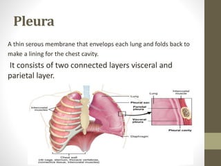
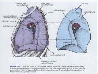
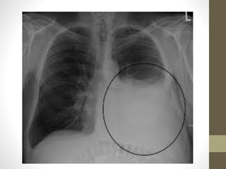
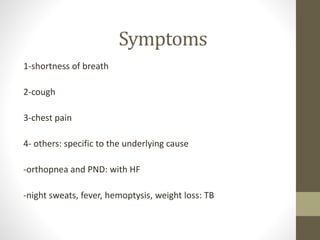
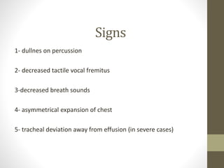
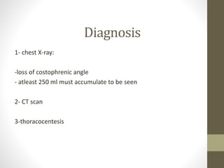
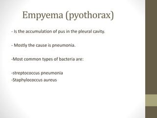
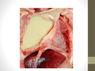
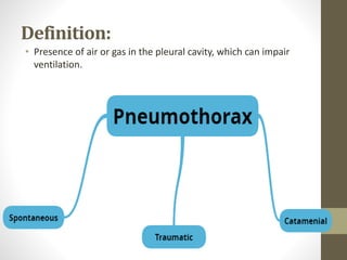
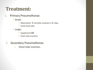
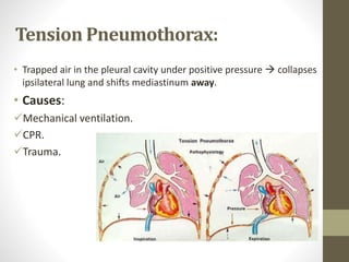
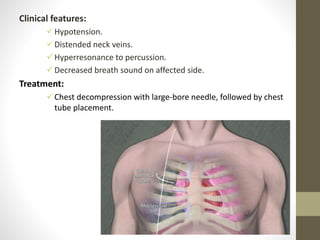
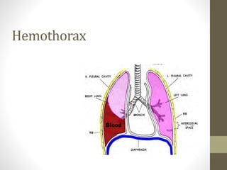
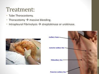
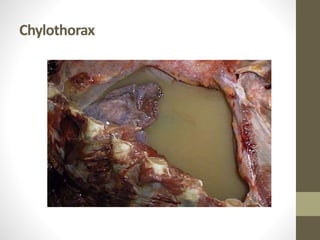
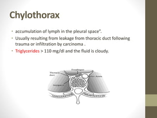
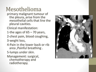
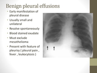
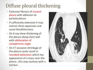
Anatomy & Physiology
of Pleura
Eyad Attoun
Pleura
A thin serous membrane that envelops each lung and folds back to
make a lining for the chest cavity.
It consists of two connected layers visceral and
parietal layer.
Parietal Layer
It lines the thoracic wall (costal pleura), covers the thoracic surface
of the diaphragm (diaphragmatic pleura) and the lateral aspect of
the mediastinum (mediastinal pleura), and extends into the neck
(cervical pleura).
The Parietal pleura is sensitive to pressure, pain, and temperature.
It produces a well localized pain, and is innervated by
the phrenic and intercostal nerves.
The blood supply is derived from the intercostal arteries.
Visceral Layer
It completely covers the outer surfaces of the lungs and extends
into the interlobar fissures.
The visceral pleura is not sensitive to pain, temperature or touch. Its
sensory fibers only detect stretch. It also receives autonomic
innervation from the pulmonary plexus.
Arterial supply is via the bronchial circulation (internal thoracic
arteries).
The visceral layer of each pleura becomes continuous with the
parietal layer by the means of the pleural cuff.
The pleural cuff surrounds the structures entering and leaving the
lung at the hilum of each lung.
The pleural cuff hangs down as a loose fold called the pulmonary
ligament.
Pleural Fluid
The pleural space is a potential space normally contains 5 to 10
ml of clear fluid.
It lubricates the opposing surfaces of the visceral and parietal
pleurae during respiration.
The formation of the fluid results from hydrostatic and osmotic
pressures between the capillaries of the Parietal pleura.
The pleural fluid is normally absorbed into the capillaries of the
visceral pleura.
Any condition that increases the production of the fluid or
impairs the drainage of the fluid results in the abnormal
accumulation of fluid.
Pulmonary Pressures
There are four primary pressures associated with ventilation:
1- Atmospheric Pressure.
2- Intrapleural pressure.
3- Intra-Alveolar Pressure.
4- Transpulmonary pressure.
Atmospheric pressure: At sea level is 760 mm Hg. The lung
pressures are expressed relative to this pressure.
Intrapleural Pressure:
At rest its -4 mmHg, it varies during ventilation but it is always less
than intra-alveolar pressure.
Intrapleural pressure is always negative during normal breathing.
The negative intrapleural pressure is due to three main factors:
a. The elasticity of the lungs.
The abundant elastic tissue in the lungs tends to recoil and pulls the
lung inward.
b. The elasticity of the thoracic wall.
The elastic thoracic wall tends to pull away from the lung, further
enlarging the pleural cavity and creating this negative pressure.
c. The surface tension of the alveolar fluid.
The surface tension of the alveolar fluid tends to pull each of the
alveoli inward and therefore pulls the entire lung inward.
Intra-Alveolar Pressure
Varies with ventilation, during inspiration it is less than
atmospheric pressure, during expiration it is greater than
atmospheric pressure.
At rest it is equal to atmospheric pressure.
When it is equal to atmospheric pressure it is considered to be at
0 mmHg.
Transpulmonary pressure is the difference between the alveolar
pressure and the intrapleural pressure in the lungs.
Pleural effusion and
Empyema
YAZAN ABUHIJLEH
Pleural effusion
-Is the abnormal accumulation of fluid in the pleural space.
-results from increased drainage into the cavity, increased production,
or decreased absorption.
-there are many types of effusion depending on the nature of fluid.
-causes limited expansion of the lungs
Types
1-serous fluid (hydrothorax)
2- pus (empyema)
3-blood (hemothorax)
4- chyle (chylothorax)
5- urine (urinothorax)
Causes
1-transudate :
-heart failure
-liver cirrhosis
-Nephrotic syndrome
-myxedema
Causes
2- Exudate:
-pneumonia
-malignancy
-tuberculosis
-pulmonary embolism
-fungal infection
-myxedema
3-others:
-blood: trauma, aortic dissection, aneurysms
-chyle: fistula
Symptoms
1-shortness of breath
2-cough
3-chest pain
4- others: specific to the underlying cause
-orthopnea and PND: with HF
-night sweats, fever, hemoptysis, weight loss: TB
Signs
1- dullnes on percussion
2- decreased tactile vocal fremitus
3-decreased breath sounds
4- asymmetrical expansion of chest
5- tracheal deviation away from effusion (in severe cases)
Diagnosis
1- chest X-ray:
-loss of costophrenic angle
- atleast 250 ml must accumulate to be seen
2- CT scan
3-thoracocentesis
Empyema (pyothorax)
- Is the accumulation of pus in the pleural cavity.
- Mostly the cause is pneumonia.
-Most common types of bacteria are:
-streptococcus pneumonia
-Staphylococcus aureus
Symptoms
1- cough
2-chest pain
3-shortness of breath
4- fever
5-chills
6-fatigue, loss of apetite, and headache
Diagnosis
1- chest X ray
2- CT scan
3-thoracocentisis
4-culture
5-PCR
6-wbc count
Treatment
1- Antibiotics:
-empiric: penicillin, clindamycin
-for S.aureus and streptococcus pneumonia
2-Drainage of the fluid
THANK YOU
Pneumothorax
Mamoon Saleh
Definition:
• Presence of air or gas in the pleural cavity, which can impair
ventilation.
A. Spontaneous Pneumothorax
1. Primary Pneumothorax:
 Occurs without any lung disease  healthy persons.
 Caused by rupture of blebs.
 Common in tall, lean, young men.
2. SecondaryPneumothorax:
 Occurs as a complication of underlying lung disease.
 Most commonly COPD.
 May occur with Asthma, ILD, TB etc..
 Life threatening.
Primary
Pneumothorax
Secondary
Pneumothorax
Healthy person In diseased person
Caused by rupture of
blebs
Complication of underlying
lung disease
Less
Life threatening Life threatening
B. Traumatic Pneumothorax:
Blunt chest wound.
Rib fracture.
Iatrogenic.
Clinical features:
• Symptoms:
 Ipsilateral chest pain (sudden in onset).
 Dyspnea, Cough.
• Signs:
 Hyperresonance over the chest.
 Decreased breath sound over affected side.
 Decreased or absent tactile fremitus.
 Mediastinal shift toward side of Pneumothorax.
Treatment:
1. Primary Pneumothorax:
Small :
 Observation  normally resolved in 10 days.
 Small chest tube.
Large:
 Supplement O2.
 Chest tube insertion.
2. Secondary Pneumothorax:
 Chest tube insertion.
Tension Pneumothorax:
• Trapped air in the pleural cavity under positive pressure  collapses
ipsilateral lung and shifts mediastinum away.
• Causes:
Mechanical ventilation.
CPR.
Trauma.
Clinical features:
Hypotension.
Distended neck veins.
Hyperresonance to percussion.
Decreased breath sound on affected side.
Treatment:
Chest decompression with large-bore needle, followed by chest
tube placement.
Hemothorax
Definition:
• Presence of blood in the pleural space.
• The source of blood may be the chest wall, lung parenchyma,
heart, or great vessels.
• Causes:
Traumatic hemothorax.
Spontaneous hemothorax.
Iatrogenic hemothorax.
Pathophysiology:
• The physiologic response to the development
of a hemothorax is manifested in two major
areas:
1. Hemodynamic response
2. Respiratory response
Clinical features:
Signs & symptoms:
1- Blunt or penetrating chest trauma.
2- Shock: (Dyspnea, Tachycardia, Tachypnea, Hypotension).
3- Dull to percussion over injured side.
Diagnosis:
• Chest X-ray  upright.
• CT scan.
• Pleural fluid hematocrit.
Treatment:
• Tube Thoracostomy.
• Thoracotomy  massive bleeding.
• Intrapleural Fibrinolysis  streptokinase or urokinase.
Resources:
• STEP-UP to MEDICINE 4th EDITION.
• OXFORD HANDBOOK OF CLINICAL MEDICINE.
• www.medscape.com
Chylothorax & Mesothelioma
Hani Bashir
Chylothorax
Chylothorax
• accumulation of lymph in the pleural space”.
• Usually resulting from leakage from thoracic duct following
trauma or infiltration by carcinoma .
• Triglycerides > 110 mg/dl and the fluid is cloudy.
Etiology Of Chylothorax
 Trauma:
 Surgical
 Nonsurgical
 Tumours :
 Lymphoma
 Metastatic Malignancies
 esophageal cancer
 infections :
 Tuberculosis lymphadenitis , mediastinitis .
 Idiopathic
Clinical Presentation
Insidious onset
 Dyspnea
 Fatigue
 Heaviness in chest
 No fever or chest pain
Diagnosis
 Gross: Milky/Bloody/Yellow
 Centifuge : Fluid does not clear
 Triglycerides : >110mg/dl
 Lipoproteins : Chylomicrons
Treatment
General measures :
 Maintain nutrition
 Treatment of primary disease
Specific measures :
 Repeated thorecentesis
 Low fat diet
 Total parenteral nutrition
 Radiotherapy
 chemotherapy
 chemical pleurodesis
 Pleuroperitoneal shunt
 Surgical ligation
Pleural tumors
Pleural tumors
Primary
Mesothelioma
Pleural Fibroma
Pleural Fibrosarcoma
Pleural Liposarcoma
Primary Pleural Lymphoma
Pleural Synovial Sarcoma
Secondary
Metastasis
Thymoma with pleural invasion
Invasive chest wall tumors
Ewing Sarcoma of chest wall with pleural
invasion
Pericardial Tumors with pleural invasion
• ** Mesothelioma :: is an aggressive primary malignant tumor of the
mesothelium. The overwhelming majority arise from the Pleura ..
• -Given the presence of the mesothelium in different parts of the body, mesothelioma
can arise in various locations :
• 1) peritoneal mesothelioma
• 2) pericardial mesothelioma (heart linning)
• 3) cystic/multicystic mesothelioma
• 4) tunica vaginalis testis mesothelioma
Mesothelioma
Epidemiology
• -Mesothelioma is an uncommon entity and accounts for 5-
28% of all malignancies that involve the pleura..
• - There is a strong association with exposure to Asbestos , but
unlike other asbestos-related lung diseases, it doesn't appear
to be dose dependent..
• -There has been no convincing evidence for an association
with smoking..
• -The annual death rate from mesothelioma has decreased in
the last thirty years. Part of this decrease in the reported rate
may be due to better diagnostic techniques..
Possible risk factors
1) Personal history of asbestos exposure
2) Living with someone who works with asbestos
3) Radiation
4) Viruses - simian virus 40 (SV40)
5) Chronic inflammation
6) Heavy metals - nickel and beryllium
7) Chemical agents - Diethylstilbestrol.
Clinical presentation
• -Typically patients present with dyspnoea and low back non-
pleuritic chest pain.
Other Signs &
Symptoms
• persistent cough
• wight loss in peritoneal mesothelioma
• abdominal edema due to ascites
• obstructed bowel
• anemia
• pyrexia
• difficulty in swallowing
• swelling in neck and face
• difficulty in breathing
• malaise
• hoarseness of voice
• hemoptysis(blood in sputum)
• pneumothorax
• **In severe cases :
• Blood clots in the vein
• Jaundice
• Low blood Sugar level
• Severe ascites
• Pulmonary Embolism
• ** Mostly affects people between the ages of 40 – 70
Diagnosis
• -Diagnosis is difficult to make as it shows similar symptoms of
other medical conditions
• -A chest x-ray or CT scan is the necessary first step in
identifying mesothelioma, which is followed up with a
bronchoscopy.
• -The diagnosis itself requires a biopsy (Histological diagnosis is
difficult and generous (often surgical) pleural biopsies are
needed)  although tumour may invade along the surgical
track following biopsy 
• -Mesotheliomas have a predilection for a direct invasion of
adjacent structures (chest wall, diaphragm and Mediastinal
content) but also frequently metastasize to the contralateral
lung and local nodes
Prognosis
• -The prognosis is poor for all tumor types with a median
overall survival without treatment of 4-12 months, although
small survival advantages have been reported following the
use of chemotherapy.
• -In favourable patient subgroups up to 45% 5-year survival
may be achievable , however even with aggressive multi-
modality therapy overall 5-year survival remains poor (3-
18%) with a median survival time of approximately 18
months .
Treatment
**Treatment includes:
1. Extrapleural pneumonectomy (Surgery)
2. Adjuvant chemotherapy
3. Radiotherapy  may be helpful in preventing tumour growth through
previous chest drain or biopsy sites.
Pleurisy & Asbestosis
Mahmoud Diab
Definition
Pleurisy :
also known as pleuritis is an inflammation of
the pleura. There are many possible causes of pleurisy but
viral infections spreading from the lungs to pleural cavity are
the most common. The inflamed pleural layers rub against
each other every time the lungs expand to breathe in air. This
can cause sharp pain when breathing, also called
pleuritic chest pain.
symptoms:
• Chest pain that worsens when you breathe, cough or sneeze
• Shortness of breath — because you are trying to minimize
breathing in and out
• A cough — only in some cases
• A fever — only in some cases
• pain also might affect your shoulder or back
Causes :
#infection
#autoimmunedisease
#Certain medications such
as(hydralizine,isoniazid)
#Lung tumor near the pleural surface
Diagnosis
diagnosis of pleurisy or another pleural condition is based on
a medical history, physical examinations, and diagnostic
tests. The goals are to rule out other sources of the symptoms
and to find the cause of the pleurisy so that the underlying
disorder can be treated.
On examination ;
1- reduce movement of chest on effected side
2- breath sound maybe reduce in effected side
3- pleural rub which is a crackling sound at the end of
inspiration and beginning of expiration localized in small area
of the chest
Diagnostic tests
1- chest X-ray.
2- Blood Test.
3- ECG .
4- Ultrasound.
5- CT scan.
6- Biopsy.
Traetment and mangement:
• Antibiotics for an infection
• Codeine will be prescribed to control a cough that can exacerbate
pain
• Sometimes, if the pleural fluid is infected or the amount is excessive,
the doctor may drain it through a tube inserted in your chest, a
procedure that requires hospitalization.
• paracitamol
• is a group of minerals with thin microscopic fibers. Because these
fibers are resistant to heat, fire, and chemicals and do not conduct
electricity, asbestos has been and used widely in the construction,
automotive, and other industries.
• Asbestosis, an inflammatory condition of lungs that can cause
shortness of breath, and eventually scarring of the lungs that makes
it hard to breathe.
Aspestosis
Benign pleural plaques
• Common
• Usually symmetrical
• Pain during breathig
• Suspect mesothelioma in the
future.
• With time it become calcify
making them more obvious
on X-ray
Mesothelioma
primary malignant tumour of
the pleura,.arise from the
mesothelial cells that line the
pleural cavities.
Clinical manifestation:
1-the ages of 65 – 70 years,
2-chest pain, blood coughing,
3-weght loss,
4-Pain in the lower back or rib
area ,Painful breathing .
5-lumps under skin.
Management: surgical,
chemotherapy and
radiotherapy.
Benign pleural effusions
• Early manifestation of
pleural disease
• Usually small and
unilateral
• Resolve spontaneously
• Blood stained exudate
• Must exclude
mesothelioma
• Present with feature of
pleurisy ( pleural pain ,
fever , leukocytosis )
Diffuse pleural thickening
• Extensive fibrosis of visceral
pleura with adhesion to
parietal pleura
• if sufficiently extensive it may
restrict chest expansion and
cause breathlessness
• On X-ray show thickening of
the pleura along chest wall
with obliteration of
costophrenic angle
• On CT occasion shrinkage of
the pleura cavity result in
rounded atelectasis which has
appearance of a mass near the
pleura , this may confuse with a
tumour .

Pleural disease

  • 1.
    Anatomy & Physiology ofPleura Eyad Attoun
  • 2.
    Pleura A thin serousmembrane that envelops each lung and folds back to make a lining for the chest cavity. It consists of two connected layers visceral and parietal layer.
  • 3.
    Parietal Layer It linesthe thoracic wall (costal pleura), covers the thoracic surface of the diaphragm (diaphragmatic pleura) and the lateral aspect of the mediastinum (mediastinal pleura), and extends into the neck (cervical pleura). The Parietal pleura is sensitive to pressure, pain, and temperature. It produces a well localized pain, and is innervated by the phrenic and intercostal nerves. The blood supply is derived from the intercostal arteries.
  • 4.
    Visceral Layer It completelycovers the outer surfaces of the lungs and extends into the interlobar fissures. The visceral pleura is not sensitive to pain, temperature or touch. Its sensory fibers only detect stretch. It also receives autonomic innervation from the pulmonary plexus. Arterial supply is via the bronchial circulation (internal thoracic arteries). The visceral layer of each pleura becomes continuous with the parietal layer by the means of the pleural cuff. The pleural cuff surrounds the structures entering and leaving the lung at the hilum of each lung. The pleural cuff hangs down as a loose fold called the pulmonary ligament.
  • 6.
    Pleural Fluid The pleuralspace is a potential space normally contains 5 to 10 ml of clear fluid. It lubricates the opposing surfaces of the visceral and parietal pleurae during respiration. The formation of the fluid results from hydrostatic and osmotic pressures between the capillaries of the Parietal pleura. The pleural fluid is normally absorbed into the capillaries of the visceral pleura. Any condition that increases the production of the fluid or impairs the drainage of the fluid results in the abnormal accumulation of fluid.
  • 7.
    Pulmonary Pressures There arefour primary pressures associated with ventilation: 1- Atmospheric Pressure. 2- Intrapleural pressure. 3- Intra-Alveolar Pressure. 4- Transpulmonary pressure. Atmospheric pressure: At sea level is 760 mm Hg. The lung pressures are expressed relative to this pressure.
  • 8.
    Intrapleural Pressure: At restits -4 mmHg, it varies during ventilation but it is always less than intra-alveolar pressure. Intrapleural pressure is always negative during normal breathing. The negative intrapleural pressure is due to three main factors: a. The elasticity of the lungs. The abundant elastic tissue in the lungs tends to recoil and pulls the lung inward.
  • 9.
    b. The elasticityof the thoracic wall. The elastic thoracic wall tends to pull away from the lung, further enlarging the pleural cavity and creating this negative pressure. c. The surface tension of the alveolar fluid. The surface tension of the alveolar fluid tends to pull each of the alveoli inward and therefore pulls the entire lung inward.
  • 10.
    Intra-Alveolar Pressure Varies withventilation, during inspiration it is less than atmospheric pressure, during expiration it is greater than atmospheric pressure. At rest it is equal to atmospheric pressure. When it is equal to atmospheric pressure it is considered to be at 0 mmHg. Transpulmonary pressure is the difference between the alveolar pressure and the intrapleural pressure in the lungs.
  • 11.
  • 12.
    Pleural effusion -Is theabnormal accumulation of fluid in the pleural space. -results from increased drainage into the cavity, increased production, or decreased absorption. -there are many types of effusion depending on the nature of fluid. -causes limited expansion of the lungs
  • 13.
    Types 1-serous fluid (hydrothorax) 2-pus (empyema) 3-blood (hemothorax) 4- chyle (chylothorax) 5- urine (urinothorax)
  • 14.
    Causes 1-transudate : -heart failure -livercirrhosis -Nephrotic syndrome -myxedema
  • 15.
    Causes 2- Exudate: -pneumonia -malignancy -tuberculosis -pulmonary embolism -fungalinfection -myxedema 3-others: -blood: trauma, aortic dissection, aneurysms -chyle: fistula
  • 17.
    Symptoms 1-shortness of breath 2-cough 3-chestpain 4- others: specific to the underlying cause -orthopnea and PND: with HF -night sweats, fever, hemoptysis, weight loss: TB
  • 18.
    Signs 1- dullnes onpercussion 2- decreased tactile vocal fremitus 3-decreased breath sounds 4- asymmetrical expansion of chest 5- tracheal deviation away from effusion (in severe cases)
  • 19.
    Diagnosis 1- chest X-ray: -lossof costophrenic angle - atleast 250 ml must accumulate to be seen 2- CT scan 3-thoracocentesis
  • 20.
    Empyema (pyothorax) - Isthe accumulation of pus in the pleural cavity. - Mostly the cause is pneumonia. -Most common types of bacteria are: -streptococcus pneumonia -Staphylococcus aureus
  • 22.
    Symptoms 1- cough 2-chest pain 3-shortnessof breath 4- fever 5-chills 6-fatigue, loss of apetite, and headache
  • 23.
    Diagnosis 1- chest Xray 2- CT scan 3-thoracocentisis 4-culture 5-PCR 6-wbc count
  • 24.
    Treatment 1- Antibiotics: -empiric: penicillin,clindamycin -for S.aureus and streptococcus pneumonia 2-Drainage of the fluid
  • 25.
  • 26.
  • 27.
    Definition: • Presence ofair or gas in the pleural cavity, which can impair ventilation.
  • 28.
    A. Spontaneous Pneumothorax 1.Primary Pneumothorax:  Occurs without any lung disease  healthy persons.  Caused by rupture of blebs.  Common in tall, lean, young men.
  • 29.
    2. SecondaryPneumothorax:  Occursas a complication of underlying lung disease.  Most commonly COPD.  May occur with Asthma, ILD, TB etc..  Life threatening.
  • 30.
    Primary Pneumothorax Secondary Pneumothorax Healthy person Indiseased person Caused by rupture of blebs Complication of underlying lung disease Less Life threatening Life threatening
  • 31.
    B. Traumatic Pneumothorax: Bluntchest wound. Rib fracture. Iatrogenic.
  • 32.
    Clinical features: • Symptoms: Ipsilateral chest pain (sudden in onset).  Dyspnea, Cough. • Signs:  Hyperresonance over the chest.  Decreased breath sound over affected side.  Decreased or absent tactile fremitus.  Mediastinal shift toward side of Pneumothorax.
  • 34.
    Treatment: 1. Primary Pneumothorax: Small:  Observation  normally resolved in 10 days.  Small chest tube. Large:  Supplement O2.  Chest tube insertion. 2. Secondary Pneumothorax:  Chest tube insertion.
  • 35.
    Tension Pneumothorax: • Trappedair in the pleural cavity under positive pressure  collapses ipsilateral lung and shifts mediastinum away. • Causes: Mechanical ventilation. CPR. Trauma.
  • 36.
    Clinical features: Hypotension. Distended neckveins. Hyperresonance to percussion. Decreased breath sound on affected side. Treatment: Chest decompression with large-bore needle, followed by chest tube placement.
  • 37.
  • 38.
    Definition: • Presence ofblood in the pleural space. • The source of blood may be the chest wall, lung parenchyma, heart, or great vessels. • Causes: Traumatic hemothorax. Spontaneous hemothorax. Iatrogenic hemothorax.
  • 39.
    Pathophysiology: • The physiologicresponse to the development of a hemothorax is manifested in two major areas: 1. Hemodynamic response 2. Respiratory response
  • 40.
    Clinical features: Signs &symptoms: 1- Blunt or penetrating chest trauma. 2- Shock: (Dyspnea, Tachycardia, Tachypnea, Hypotension). 3- Dull to percussion over injured side.
  • 41.
    Diagnosis: • Chest X-ray upright. • CT scan. • Pleural fluid hematocrit.
  • 42.
    Treatment: • Tube Thoracostomy. •Thoracotomy  massive bleeding. • Intrapleural Fibrinolysis  streptokinase or urokinase.
  • 43.
    Resources: • STEP-UP toMEDICINE 4th EDITION. • OXFORD HANDBOOK OF CLINICAL MEDICINE. • www.medscape.com
  • 44.
  • 45.
  • 46.
    Chylothorax • accumulation oflymph in the pleural space”. • Usually resulting from leakage from thoracic duct following trauma or infiltration by carcinoma . • Triglycerides > 110 mg/dl and the fluid is cloudy.
  • 47.
    Etiology Of Chylothorax Trauma:  Surgical  Nonsurgical  Tumours :  Lymphoma  Metastatic Malignancies  esophageal cancer  infections :  Tuberculosis lymphadenitis , mediastinitis .  Idiopathic
  • 48.
    Clinical Presentation Insidious onset Dyspnea  Fatigue  Heaviness in chest  No fever or chest pain
  • 49.
    Diagnosis  Gross: Milky/Bloody/Yellow Centifuge : Fluid does not clear  Triglycerides : >110mg/dl  Lipoproteins : Chylomicrons
  • 50.
    Treatment General measures : Maintain nutrition  Treatment of primary disease Specific measures :  Repeated thorecentesis  Low fat diet  Total parenteral nutrition  Radiotherapy  chemotherapy  chemical pleurodesis  Pleuroperitoneal shunt  Surgical ligation
  • 51.
  • 52.
    Pleural tumors Primary Mesothelioma Pleural Fibroma PleuralFibrosarcoma Pleural Liposarcoma Primary Pleural Lymphoma Pleural Synovial Sarcoma Secondary Metastasis Thymoma with pleural invasion Invasive chest wall tumors Ewing Sarcoma of chest wall with pleural invasion Pericardial Tumors with pleural invasion
  • 54.
    • ** Mesothelioma:: is an aggressive primary malignant tumor of the mesothelium. The overwhelming majority arise from the Pleura .. • -Given the presence of the mesothelium in different parts of the body, mesothelioma can arise in various locations : • 1) peritoneal mesothelioma • 2) pericardial mesothelioma (heart linning) • 3) cystic/multicystic mesothelioma • 4) tunica vaginalis testis mesothelioma Mesothelioma
  • 55.
    Epidemiology • -Mesothelioma isan uncommon entity and accounts for 5- 28% of all malignancies that involve the pleura.. • - There is a strong association with exposure to Asbestos , but unlike other asbestos-related lung diseases, it doesn't appear to be dose dependent.. • -There has been no convincing evidence for an association with smoking.. • -The annual death rate from mesothelioma has decreased in the last thirty years. Part of this decrease in the reported rate may be due to better diagnostic techniques..
  • 56.
    Possible risk factors 1)Personal history of asbestos exposure 2) Living with someone who works with asbestos 3) Radiation 4) Viruses - simian virus 40 (SV40) 5) Chronic inflammation 6) Heavy metals - nickel and beryllium 7) Chemical agents - Diethylstilbestrol.
  • 57.
    Clinical presentation • -Typicallypatients present with dyspnoea and low back non- pleuritic chest pain. Other Signs & Symptoms • persistent cough • wight loss in peritoneal mesothelioma • abdominal edema due to ascites • obstructed bowel • anemia • pyrexia • difficulty in swallowing • swelling in neck and face • difficulty in breathing • malaise • hoarseness of voice • hemoptysis(blood in sputum) • pneumothorax
  • 58.
    • **In severecases : • Blood clots in the vein • Jaundice • Low blood Sugar level • Severe ascites • Pulmonary Embolism • ** Mostly affects people between the ages of 40 – 70
  • 59.
    Diagnosis • -Diagnosis isdifficult to make as it shows similar symptoms of other medical conditions • -A chest x-ray or CT scan is the necessary first step in identifying mesothelioma, which is followed up with a bronchoscopy. • -The diagnosis itself requires a biopsy (Histological diagnosis is difficult and generous (often surgical) pleural biopsies are needed)  although tumour may invade along the surgical track following biopsy  • -Mesotheliomas have a predilection for a direct invasion of adjacent structures (chest wall, diaphragm and Mediastinal content) but also frequently metastasize to the contralateral lung and local nodes
  • 60.
    Prognosis • -The prognosisis poor for all tumor types with a median overall survival without treatment of 4-12 months, although small survival advantages have been reported following the use of chemotherapy. • -In favourable patient subgroups up to 45% 5-year survival may be achievable , however even with aggressive multi- modality therapy overall 5-year survival remains poor (3- 18%) with a median survival time of approximately 18 months .
  • 61.
    Treatment **Treatment includes: 1. Extrapleuralpneumonectomy (Surgery) 2. Adjuvant chemotherapy 3. Radiotherapy  may be helpful in preventing tumour growth through previous chest drain or biopsy sites.
  • 62.
  • 63.
    Definition Pleurisy : also knownas pleuritis is an inflammation of the pleura. There are many possible causes of pleurisy but viral infections spreading from the lungs to pleural cavity are the most common. The inflamed pleural layers rub against each other every time the lungs expand to breathe in air. This can cause sharp pain when breathing, also called pleuritic chest pain.
  • 64.
    symptoms: • Chest painthat worsens when you breathe, cough or sneeze • Shortness of breath — because you are trying to minimize breathing in and out • A cough — only in some cases • A fever — only in some cases • pain also might affect your shoulder or back Causes : #infection #autoimmunedisease #Certain medications such as(hydralizine,isoniazid) #Lung tumor near the pleural surface
  • 65.
    Diagnosis diagnosis of pleurisyor another pleural condition is based on a medical history, physical examinations, and diagnostic tests. The goals are to rule out other sources of the symptoms and to find the cause of the pleurisy so that the underlying disorder can be treated. On examination ; 1- reduce movement of chest on effected side 2- breath sound maybe reduce in effected side 3- pleural rub which is a crackling sound at the end of inspiration and beginning of expiration localized in small area of the chest
  • 66.
    Diagnostic tests 1- chestX-ray. 2- Blood Test. 3- ECG . 4- Ultrasound. 5- CT scan. 6- Biopsy.
  • 67.
    Traetment and mangement: •Antibiotics for an infection • Codeine will be prescribed to control a cough that can exacerbate pain • Sometimes, if the pleural fluid is infected or the amount is excessive, the doctor may drain it through a tube inserted in your chest, a procedure that requires hospitalization. • paracitamol
  • 68.
    • is agroup of minerals with thin microscopic fibers. Because these fibers are resistant to heat, fire, and chemicals and do not conduct electricity, asbestos has been and used widely in the construction, automotive, and other industries. • Asbestosis, an inflammatory condition of lungs that can cause shortness of breath, and eventually scarring of the lungs that makes it hard to breathe. Aspestosis
  • 69.
    Benign pleural plaques •Common • Usually symmetrical • Pain during breathig • Suspect mesothelioma in the future. • With time it become calcify making them more obvious on X-ray
  • 70.
    Mesothelioma primary malignant tumourof the pleura,.arise from the mesothelial cells that line the pleural cavities. Clinical manifestation: 1-the ages of 65 – 70 years, 2-chest pain, blood coughing, 3-weght loss, 4-Pain in the lower back or rib area ,Painful breathing . 5-lumps under skin. Management: surgical, chemotherapy and radiotherapy.
  • 71.
    Benign pleural effusions •Early manifestation of pleural disease • Usually small and unilateral • Resolve spontaneously • Blood stained exudate • Must exclude mesothelioma • Present with feature of pleurisy ( pleural pain , fever , leukocytosis )
  • 72.
    Diffuse pleural thickening •Extensive fibrosis of visceral pleura with adhesion to parietal pleura • if sufficiently extensive it may restrict chest expansion and cause breathlessness • On X-ray show thickening of the pleura along chest wall with obliteration of costophrenic angle • On CT occasion shrinkage of the pleura cavity result in rounded atelectasis which has appearance of a mass near the pleura , this may confuse with a tumour .