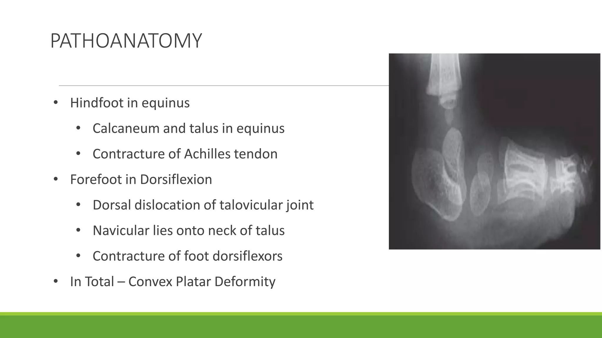
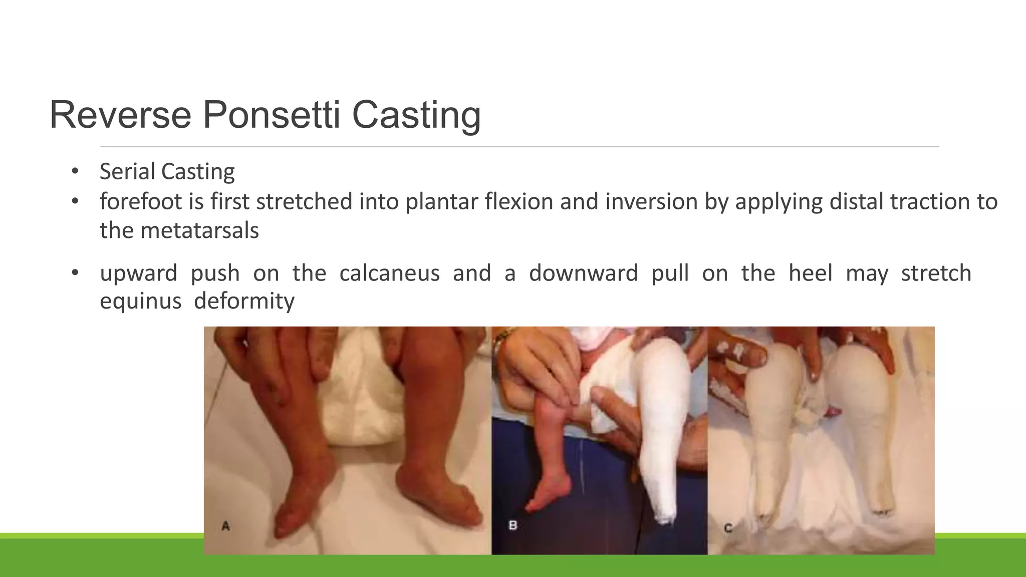
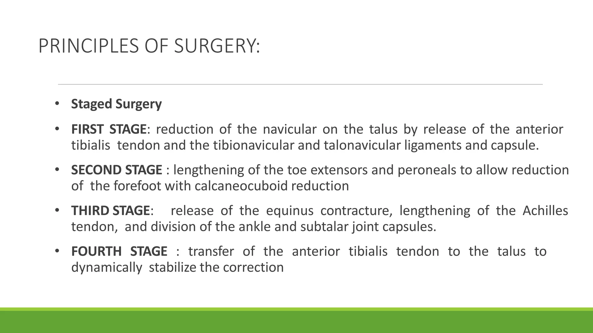
This document discusses several foot conditions including pes planus (flat foot), pes valgus, flexible flat foot, congenital vertical talus, tarsal coalition, and posterior tibial tendon disorder. It provides details on the anatomy of the foot arches, clinical features, causes, treatments and imaging findings for each condition. Common causes of flat foot discussed include flexible flat foot in children, congenital vertical talus, tarsal coalition, and posterior tibial tendon insufficiency in adults. Imaging can help evaluate the degree of deformity and guide treatment, which may include orthotics, casting, or surgery depending on the condition.
Collapse of medial longitudinal arch, with the entire sole of the foot coming into complete or near-complete contact with the ground.
Books Refered :
Text Book Of ANATOMY - Vishram Singh
Joint Structure And Function – Cynthia Norkin
Therapeutic Exrercise – Carolyn Kisner
Orthopaedic Physical Assessment – Magee
Orthopaedic Medicine – L. Ombregt
Campbell’s Operative Orthopaedics
Avascular necrosis (AVN) or Aseptic Necrosis of the hip is caused by a disruption to the hip’s blood supply which results in the deterioration and often collapse of the ball of the thigh bone (femoral head). Early identification and treatment of the condition increases the likelihood that a patient’s hip will recover. Surgery may be required in severe cases to repair or revascularize (restore circulation) the hip or to replace the hip in neglected/end stage cases.
http://www.davidsfeldmanmd.com/specialties/avascular-necrosis-hip
Dr. Anisuddin Bhatti Paediatric Orthopaedic Surgeon DR. Ziauddin University Karachi presented talk on Congenital Vertical Talus at AKU karachi on August 2023 in Orthopaedic Review course. Acknowledged for some text material & photo taken from Published literature.
Basics of patellofemoral instability for postgraduates. Gives brief introduction about patellofemoral joint anatomy, causes, examintaion and treatment for patellofemoral instability
Surgical Approaches to Acetabulum and PelvisBijay Mehta
Important surgical approaches to acetabulum and pelvis are described.
Ilioinguinal approach, Modified Stoppa Approach, Kocher lagenbeck Approach, Ilifemoral approach and extensile approaches are well illustrated and described.
Flu Vaccine Alert in Bangalore Karnatakaaddon Scans
As flu season approaches, health officials in Bangalore, Karnataka, are urging residents to get their flu vaccinations. The seasonal flu, while common, can lead to severe health complications, particularly for vulnerable populations such as young children, the elderly, and those with underlying health conditions.
Dr. Vidisha Kumari, a leading epidemiologist in Bangalore, emphasizes the importance of getting vaccinated. "The flu vaccine is our best defense against the influenza virus. It not only protects individuals but also helps prevent the spread of the virus in our communities," he says.
This year, the flu season is expected to coincide with a potential increase in other respiratory illnesses. The Karnataka Health Department has launched an awareness campaign highlighting the significance of flu vaccinations. They have set up multiple vaccination centers across Bangalore, making it convenient for residents to receive their shots.
To encourage widespread vaccination, the government is also collaborating with local schools, workplaces, and community centers to facilitate vaccination drives. Special attention is being given to ensuring that the vaccine is accessible to all, including marginalized communities who may have limited access to healthcare.
Residents are reminded that the flu vaccine is safe and effective. Common side effects are mild and may include soreness at the injection site, mild fever, or muscle aches. These side effects are generally short-lived and far less severe than the flu itself.
Healthcare providers are also stressing the importance of continuing COVID-19 precautions. Wearing masks, practicing good hand hygiene, and maintaining social distancing are still crucial, especially in crowded places.
Protect yourself and your loved ones by getting vaccinated. Together, we can help keep Bangalore healthy and safe this flu season. For more information on vaccination centers and schedules, residents can visit the Karnataka Health Department’s official website or follow their social media pages.
Stay informed, stay safe, and get your flu shot today!
Couples presenting to the infertility clinic- Do they really have infertility...Sujoy Dasgupta
Dr Sujoy Dasgupta presented the study on "Couples presenting to the infertility clinic- Do they really have infertility? – The unexplored stories of non-consummation" in the 13th Congress of the Asia Pacific Initiative on Reproduction (ASPIRE 2024) at Manila on 24 May, 2024.
Ethanol (CH3CH2OH), or beverage alcohol, is a two-carbon alcohol
that is rapidly distributed in the body and brain. Ethanol alters many
neurochemical systems and has rewarding and addictive properties. It
is the oldest recreational drug and likely contributes to more morbidity,
mortality, and public health costs than all illicit drugs combined. The
5th edition of the Diagnostic and Statistical Manual of Mental Disorders
(DSM-5) integrates alcohol abuse and alcohol dependence into a single
disorder called alcohol use disorder (AUD), with mild, moderate,
and severe subclassifications (American Psychiatric Association, 2013).
In the DSM-5, all types of substance abuse and dependence have been
combined into a single substance use disorder (SUD) on a continuum
from mild to severe. A diagnosis of AUD requires that at least two of
the 11 DSM-5 behaviors be present within a 12-month period (mild
AUD: 2–3 criteria; moderate AUD: 4–5 criteria; severe AUD: 6–11 criteria).
The four main behavioral effects of AUD are impaired control over
drinking, negative social consequences, risky use, and altered physiological
effects (tolerance, withdrawal). This chapter presents an overview
of the prevalence and harmful consequences of AUD in the U.S.,
the systemic nature of the disease, neurocircuitry and stages of AUD,
comorbidities, fetal alcohol spectrum disorders, genetic risk factors, and
pharmacotherapies for AUD.
Lung Cancer: Artificial Intelligence, Synergetics, Complex System Analysis, S...Oleg Kshivets
RESULTS: Overall life span (LS) was 2252.1±1742.5 days and cumulative 5-year survival (5YS) reached 73.2%, 10 years – 64.8%, 20 years – 42.5%. 513 LCP lived more than 5 years (LS=3124.6±1525.6 days), 148 LCP – more than 10 years (LS=5054.4±1504.1 days).199 LCP died because of LC (LS=562.7±374.5 days). 5YS of LCP after bi/lobectomies was significantly superior in comparison with LCP after pneumonectomies (78.1% vs.63.7%, P=0.00001 by log-rank test). AT significantly improved 5YS (66.3% vs. 34.8%) (P=0.00000 by log-rank test) only for LCP with N1-2. Cox modeling displayed that 5YS of LCP significantly depended on: phase transition (PT) early-invasive LC in terms of synergetics, PT N0—N12, cell ratio factors (ratio between cancer cells- CC and blood cells subpopulations), G1-3, histology, glucose, AT, blood cell circuit, prothrombin index, heparin tolerance, recalcification time (P=0.000-0.038). Neural networks, genetic algorithm selection and bootstrap simulation revealed relationships between 5YS and PT early-invasive LC (rank=1), PT N0—N12 (rank=2), thrombocytes/CC (3), erythrocytes/CC (4), eosinophils/CC (5), healthy cells/CC (6), lymphocytes/CC (7), segmented neutrophils/CC (8), stick neutrophils/CC (9), monocytes/CC (10); leucocytes/CC (11). Correct prediction of 5YS was 100% by neural networks computing (area under ROC curve=1.0; error=0.0).
CONCLUSIONS: 5YS of LCP after radical procedures significantly depended on: 1) PT early-invasive cancer; 2) PT N0--N12; 3) cell ratio factors; 4) blood cell circuit; 5) biochemical factors; 6) hemostasis system; 7) AT; 8) LC characteristics; 9) LC cell dynamics; 10) surgery type: lobectomy/pneumonectomy; 11) anthropometric data. Optimal diagnosis and treatment strategies for LC are: 1) screening and early detection of LC; 2) availability of experienced thoracic surgeons because of complexity of radical procedures; 3) aggressive en block surgery and adequate lymph node dissection for completeness; 4) precise prediction; 5) adjuvant chemoimmunoradiotherapy for LCP with unfavorable prognosis.
Tom Selleck Health: A Comprehensive Look at the Iconic Actor’s Wellness Journeygreendigital
Tom Selleck, an enduring figure in Hollywood. has captivated audiences for decades with his rugged charm, iconic moustache. and memorable roles in television and film. From his breakout role as Thomas Magnum in Magnum P.I. to his current portrayal of Frank Reagan in Blue Bloods. Selleck's career has spanned over 50 years. But beyond his professional achievements. fans have often been curious about Tom Selleck Health. especially as he has aged in the public eye.
Follow us on: Pinterest
Introduction
Many have been interested in Tom Selleck health. not only because of his enduring presence on screen but also because of the challenges. and lifestyle choices he has faced and made over the years. This article delves into the various aspects of Tom Selleck health. exploring his fitness regimen, diet, mental health. and the challenges he has encountered as he ages. We'll look at how he maintains his well-being. the health issues he has faced, and his approach to ageing .
Early Life and Career
Childhood and Athletic Beginnings
Tom Selleck was born on January 29, 1945, in Detroit, Michigan, and grew up in Sherman Oaks, California. From an early age, he was involved in sports, particularly basketball. which played a significant role in his physical development. His athletic pursuits continued into college. where he attended the University of Southern California (USC) on a basketball scholarship. This early involvement in sports laid a strong foundation for his physical health and disciplined lifestyle.
Transition to Acting
Selleck's transition from an athlete to an actor came with its physical demands. His first significant role in "Magnum P.I." required him to perform various stunts and maintain a fit appearance. This role, which he played from 1980 to 1988. necessitated a rigorous fitness routine to meet the show's demands. setting the stage for his long-term commitment to health and wellness.
Fitness Regimen
Workout Routine
Tom Selleck health and fitness regimen has evolved. adapting to his changing roles and age. During his "Magnum, P.I." days. Selleck's workouts were intense and focused on building and maintaining muscle mass. His routine included weightlifting, cardiovascular exercises. and specific training for the stunts he performed on the show.
Selleck adjusted his fitness routine as he aged to suit his body's needs. Today, his workouts focus on maintaining flexibility, strength, and cardiovascular health. He incorporates low-impact exercises such as swimming, walking, and light weightlifting. This balanced approach helps him stay fit without putting undue strain on his joints and muscles.
Importance of Flexibility and Mobility
In recent years, Selleck has emphasized the importance of flexibility and mobility in his fitness regimen. Understanding the natural decline in muscle mass and joint flexibility with age. he includes stretching and yoga in his routine. These practices help prevent injuries, improve posture, and maintain mobilit
Ozempic: Preoperative Management of Patients on GLP-1 Receptor Agonists Saeid Safari
Preoperative Management of Patients on GLP-1 Receptor Agonists like Ozempic and Semiglutide
ASA GUIDELINE
NYSORA Guideline
2 Case Reports of Gastric Ultrasound
Factory Supply Best Quality Pmk Oil CAS 28578–16–7 PMK Powder in Stockrebeccabio
Factory Supply Best Quality Pmk Oil CAS 28578–16–7 PMK Powder in Stock
Telegram: bmksupplier
signal: +85264872720
threema: TUD4A6YC
You can contact me on Telegram or Threema
Communicate promptly and reply
Free of customs clearance, Double Clearance 100% pass delivery to USA, Canada, Spain, Germany, Netherland, Poland, Italy, Sweden, UK, Czech Republic, Australia, Mexico, Russia, Ukraine, Kazakhstan.Door to door service
Hot Selling Organic intermediates
- Video recording of this lecture in English language: https://youtu.be/lK81BzxMqdo
- Video recording of this lecture in Arabic language: https://youtu.be/Ve4P0COk9OI
- Link to download the book free: https://nephrotube.blogspot.com/p/nephrotube-nephrology-books.html
- Link to NephroTube website: www.NephroTube.com
- Link to NephroTube social media accounts: https://nephrotube.blogspot.com/p/join-nephrotube-on-social-media.html
Prix Galien International 2024 Forum ProgramLevi Shapiro
June 20, 2024, Prix Galien International and Jerusalem Ethics Forum in ROME. Detailed agenda including panels:
- ADVANCES IN CARDIOLOGY: A NEW PARADIGM IS COMING
- WOMEN’S HEALTH: FERTILITY PRESERVATION
- WHAT’S NEW IN THE TREATMENT OF INFECTIOUS,
ONCOLOGICAL AND INFLAMMATORY SKIN DISEASES?
- ARTIFICIAL INTELLIGENCE AND ETHICS
- GENE THERAPY
- BEYOND BORDERS: GLOBAL INITIATIVES FOR DEMOCRATIZING LIFE SCIENCE TECHNOLOGIES AND PROMOTING ACCESS TO HEALTHCARE
- ETHICAL CHALLENGES IN LIFE SCIENCES
- Prix Galien International Awards Ceremony
The prostate is an exocrine gland of the male mammalian reproductive system
It is a walnut-sized gland that forms part of the male reproductive system and is located in front of the rectum and just below the urinary bladder
Function is to store and secrete a clear, slightly alkaline fluid that constitutes 10-30% of the volume of the seminal fluid that along with the spermatozoa, constitutes semen
A healthy human prostate measures (4cm-vertical, by 3cm-horizontal, 2cm ant-post ).
It surrounds the urethra just below the urinary bladder. It has anterior, median, posterior and two lateral lobes
It’s work is regulated by androgens which are responsible for male sex characteristics
Generalised disease of the prostate due to hormonal derangement which leads to non malignant enlargement of the gland (increase in the number of epithelial cells and stromal tissue)to cause compression of the urethra leading to symptoms (LUTS
3. FLAT FOOT : INTRODUCTION
•Condition in which the medial arch of the foot is diminished or
absent, allowing the entire sole to touch the ground.
Can be
• Asymptomatic/Symptomatic
• Flexible/Rigid/Compensatory
4. INCIDENCE
• 23 % of adult population.
• Of this ,approximately two thirds have a flexible flatfoot.
• Approximately one fourth of flatfeet exhibit a contracture of the
triceps surae associated with an otherwise typical hypermobile
flatfoot
• The remainder of flatfeet are characterized by more rigidity of the
subtalar joint, typically seen with tarsal coalitions.
5. CLINICAL FEATURES
• Medial arch of the foot is depressed
(REPRODUCIBLE/NON
REPRODUCIBLE)
• Heel bone, when viewed from the rear is
everted or in valgus
• Forefoot is abducted relative to the
hindfoot
• “Too many toes sign”
6. ARCHES OF FOOT
• The springboards and shock absorbers of
foot.
• There are three main arches of foot :
• Medial longitudinal arch
• Lateral longitudinal arch
• Transverse arch
7. MEDIAL LONGITUDINAL ARCH
• Arch –Why??
• Segmented structure supports weight best if built in
form of arch
• Highest and most flexible arch
• Acts as shock absorber
• Helps in propulsion of the foot while walking
8. SUPPORTS OF MLA
• The key stone is the talus
• The staples are plantar ligaments,
tendon of tibialis posterior
• The tie beam is made by plantar
aponeurosis, flexor dig. Brevis,
abductor hallucis, flexor hallucis
longus, flexor dig. Longus, flexor
hallucis brevis
• Ant pillar : 3 metatarsal heads
•Post pillar : medial calcaneal tubercle
10. • Common childhood complain
• Arch is usually obscured in an infant’s foot because of subcutaneous
fat.
• Usually disappears between 4 to 10 years when longitudinal arch
develops.
• “usual in infants, common in children, and within the normal range in
adults”- Staheli and Colleagues
FLEXIBLE FLATFOOT
11. • May be associated with ligamentous laxity- look for Beighton score
• Needs to be differentiated from CALCANEOVAVALGUS
• Calcaneovalgus
• Rigid flatfoot
• Incidence-30%
• Packaging disorder
FLEXIBLE FLATFOOT
12. • Painless most of the times.
• Usually noticed by parents, grandparents or assistants in
the shoe shop
• On Inspection:
• excessive eversion during weight bearing,
• the forefoot is abducted, with a midfoot sag with
lowering of the longitudinal arch
• medial column appears longer than the lateral
column
• On Palpation:
• talar head and navicular tuberosity appear to be in
contact with the floor
CLINICAL FEATURES
13. • Movement :
• may have increased mobility of ankle or subtalar joint
• Tests :
• Tip toe test : Inversion of the heels and arch reconstitution
during toe standing
• Jack’s Test/Hubscher’s Test : Dorsiflexing the great toe
restores the arch
CLINICAL FEATURES
14. IMAGING
• Usually not required
• Done to rule out causes of the deformity
other than idiopathy
• BUT ONE CAN VISUALISE FOLLOWING
PARAMETERS WITH ITS AID:
• lateral talus–first metatarsal angle, or
Meary angle
• location of the sag—talonavicular or
naviculocuneiform joint
• degree of plantar flexion of the talus
15. XRAY : MEASUREMENTS
•Calcaneal pitch angle (α): formed by the horizontal line and a
line from the base of heel and inferior cortex of calcaneus, and
less than 20° is considered to represent pes planus.
•Meary’s angle (β) : angle between the lines from the centers of
longitudinal axes of the talus and the first metatarsal. More than
4° is considered as pes planus
• Lateral talocalcaneal angle(γ) : angle formed by the intersection
of the line bisecting the talus with the line along the lower
border of the calcaneus. An angle over 45° indicates hindfoot
valgus, a component of pes planus
•Talonavicular coverage angle (δ) : Angle between a line
connecting the edges of the articular surface of the talus a line
connecting the edges of the articular surface of the navicular ,
greater than 7° indicates lateral talar subluxation
•Talo-first metatarsal angle (dashed line) : formed by drawing a
line through the midaxis of the talus; if this line is angled medial
to the first metatarsal, it indicates pes planus.
16. FLAT FOOT : TREATMENT
• SURGERY VS CONSERVATIVE
• Indications for surgery
• Intractable symptoms unresponsive to shoe or orthotic modifications
• In individuals who are unable to modify the activities that produce pain
17. FLEXIBLE FLATFOOT : TREATMENT
• Conservative Treatment
• No treatment required in an asymptomatic pediatric
patient.
• Education and reassurance are the mainstays.
• If an Achilles tendon contracture is
present- stretching exercises-both active
and passive
18. Role of orthoses
• Traditionally used in all patients
• But there is no scientific
evidence that orthoses and
medial arch supports are
efficacious.
• BUT…in cases of medial arch
pain and fatigue, as well as
cramping at night the orthoses
may be helpful.
19. SURGICAL TREATMENT : OPTIONS
• Arthroereisis- limits the amount of valgus motion
in the subtalar joint by using an interposition peg
• Lateral column lengthening
• Heel cord lengthening
• Imbrication of talonaviculocuneiform complex
• Subtalar fusion - only as salvage procedure.
• Triple arthrodesis
21. CONGENITAL VERTICAL TALUS
• A cause of rigid pes planus
• Characterized by a fixed dorsal dislocation of
the talonavicular joint in conjunction with rigid
hindfoot equinus
• Rocker bottom deformity
• Aka congenital convex pes valgus,
teratologic dorsolateral dislocation of the
talocalcaneonavicular joint
• 1 in 10000 live births
22. ETIOLOGY
• Exact etiology unknown
• Likely causes
• Abnormal variation in muscle fibre size
• Congenital vascular abnormalities
• Arrest in fetal development of foot at 7-12 weeks POG
• Autosomal dominant pattern of inheritance
• Gene mutations (HOXD10 )
23. • 60% associated with other
congenital anamolies
ASSOCIATIONS Associations
Neurological
Discorders
Myelomeningocele-10%
Arthrogryposis-11%
spinal muscular atrophy,
neurofibromatosis,
congenital dislocation of
the hip
Genetic
Trisomy 13,15 ,18
24. PATHOANATOMY
• Hindfoot in equinus
• Calcaneum and talus in equinus
• Contracture of Achilles tendon
• Forefoot in Dorsiflexion
• Dorsal dislocation of talovicular joint
• Navicular lies onto neck of talus
• Contracture of foot dorsiflexors
• In Total – Convex Platar Deformity
25. LIGAMENTOUS CHANGES:
• CONTRACTED ONES: tibionavicular portion of the superficial deltoid, bifurcated
ligament, calcaneofibular ligament, and the interosseous talocalcaneal ligaments
• ATTENUATED ONES: spring ligament
TENDONS AND MUSCLE CHANGES:
• CONTRACTURES OF : tibialis anterior, long toe extensors, peroneus brevis, and triceps
surae
• Posterior tibial and peroneal tendons may be displaced anteriorly so that they
act as dorsiflexors rather than plantar flexors.
PATHOANATOMY
26. CLINICAL FEATURES
ON INSPECTION:
• a rocker bottom foot, the apex of which is at the talar head
• callosities may be seen
• hindfoot foot is everted into a valgus
• forefoot is abducted and dorsiflexed
ON PALPATION:
• a contracted achilles tendon
• peroneal and anterior tibialis tendons are taut
• navicular is palpable as it lies on the talar neck
ON MOVEMENT: passive correction of deformity is impossible
27. IMAGING: XRAY
• LATERAL PROJECTION:
• Neutral
• Maximum Dorsiflexion
• Maximum Plantarflexion
• Differentiate from Oblique Talus- Talus aligns
with 1st metatarsal in maximum plantarflexion
29. TREATMENT
EARLIER BELIEF :
• Major reconstructive surgery was necessary to correct the deformities
• But resulted in substantial complications- STIFFNESS
RECENT BELIEF : Serial casting (described by Dobb)
• to stretch the contracted dorsal and lateral soft tissues
• gradually reduce the talonavicular joint followed by
• Minimally invasive procedures for final correction.
30. Reverse Ponsetti Casting
• Serial Casting
• forefoot is first stretched into plantar flexion and inversion by applying distal traction to
the metatarsals
• upward push on the calcaneus and a downward pull on the heel may stretch
equinus deformity
31. PRINCIPLES OF SURGERY:
• Staged Surgery
• FIRST STAGE: reduction of the navicular on the talus by release of the anterior
tibialis tendon and the tibionavicular and talonavicular ligaments and capsule.
• SECOND STAGE : lengthening of the toe extensors and peroneals to allow reduction
of the forefoot with calcaneocuboid reduction
• THIRD STAGE: release of the equinus contracture, lengthening of the Achilles
tendon, and division of the ankle and subtalar joint capsules.
• FOURTH STAGE : transfer of the anterior tibialis tendon to the talus to
dynamically stabilize the correction
32. TARSAL COALITION
• An abnormal connection between two or more bones of the foot
• Produce pain and limitation of foot motion.
• Incidence varies from 0.03% to 1.0%.
• 50 to 60% of tarsal coalitions are bilateral.
• Tarsal coalition, rigid pes planus, and peroneal muscle spasm - components of
peroneal spastic pes planus.
33. TYPESOFTARSALCOALITIONS
• Calcaneonavicular: most common form but less symptomatic
• Talocalcaneal: more symptomatic form
• Other rare forms : calcaneocuboid, naviculocuboid, naviculocuneiform, or
massive tarsal coalition
• Etiology: Failure of normal segmentation of fetal tarsal
• Autosomal dominant inheritance
36. CLINICALFEATURES
Symptoms :
• Usually become symptomatic around12-16 yrs of age
• Pain-
• often over the tarsal sinus, beneath the medial malleolus, along the arch
of the foot, or occasionally on the dorsum of the foot
• exacerbated by vigorous sports activities
• Stiffness of the hindfoot
• Frequent ankle sprains
• Progressive deformity of foot: flat foot
37. Signs
• Flat foot appearance , with external rotation of foot , and abduction
of forefoot
• Restricted ROM of hindfoot ( subtalar inversion and eversion)
• Joint motion is more preserved in calcaneonavicular
coalition
• Increased foot progression angle,
• Loss of hindfoot inversion occurs during a toe rise
38. IMAGING..
• X-ray : views usually performed are :
• 45 degree lateral to medial oblique view: to
visualise calcaneonavicular coalition
• Harris axial view : to visualise talocalcaneal
coalition across medial subtalar joint
• Lateral view of foot : to see for elongated
anterior projection of the calcaneus, the so-
called anteater’s nose, an anterior beak on the
talus
39. IMAGING
CT SCAN:
• Best imaging modality for the diagnosis of coalition
• Denotes extent and type of coalition
• Based on CT, KUMAR et al .classified coaitions into : type I- osseous, type II-
cartilaginous, type III- fibrous
• *non osseous are more symptomatic
MRI: useful in fibrous coalitions and when CT is nondiagnostic
40. TREATMENT
Options include :
• Conservative treatment:
• use of a firm orthosis,
• 4- to 6-week period of immobilization in a short-leg walking cast
• Surgery :
• Indication : failure to relieve symptoms from a trial of conservative
treatment
• Resection of coalition and interposition of soft tissue in gap
• Limited hindfoot fusion
• Triple arthrodesis- useful in cases of degenerative changes
42. ACCESSORY NAVICULAR
• First described by Bauhin in 1605
• Aka accessory scaphoid, accessory navicular,
prehallux, and os tibiale externum
• a congenital anomaly in which the
tuberosity of the navicular develops from a
secondary center of ossification and located
on the medial aspect of the arch in
association with the navicular.
43. ACCESSORY NAVICULAR AND FLAT FOOT
Kidner’s hypothesis : Flat foot in presence of an accessory navicular had one of
three causes:
• Alteration of the line of pull of the posterior tibial tendon
• Forcing of the posterior tibial tendon by the accessory navicular to
become more of an adductor than a supinator of the forefoot, thereby
decreasing support for the longitudinal arch;
• Impingement of the accessory navicular against the medial malleolus as the
foot adducts, which tends to keep the foot in an abducted position and thus
partially flattens the longitudinal arch.
44. TYPES
• Three types described by COUGHLIN
• Type I : small, not attached to navicular, probably sesamoid in tibialis posterior
• Type II: definite part of the body of the navicular, separated by cartilaginous plate
45. Type III : united by a bony ridge, producing a cornuate navicular.
46. CLINICAL FEATURES
• Asymptomatic –most of the time
• Can become symptomatic in childhood or early adulthood
• In children, the symptoms are usually caused by pressure of the accessory bone
against the shoe.
• Progressive flattening of the longitudinal arch.
• In adults, symptoms usually develop after trauma to the foot, often
resulting from a twisting injury.
48. TREATMENT
NON SURGICAL OPTIONS:
◦ In cases of asymptomatic incidental findings- reassurance
◦ Shoe changes to reduce pressure over the area
◦ In acutely symptomatic cases after an injury - immobilization in a below-knee
walking cast, followed by the use of a longitudinal arch support
◦ Occasionally use of steroid may provide a relief
49. SURGICAL OPTION : THE KIDNER PROCEDURE
• Excision of the accessory navicular with or
without the plication of posterior tibial
tendon.
• Posterior tibial tendon is detached from the
insertion on navicular and rerouted in
plantar to dorsal direction and sutured on
itself or surrounding periosteum.
• Rerouting is necessary only when there is
pes planus.
50. POSTERIOR TIBIAL TENDON INSUFFICIENCY(PTTI)
• Most Common cause of adult flat
foot
• The main functions of posterior
tibial tendon are:
• plantar flexion of ankle ,
• inversion of foot
• stabilization of the medial
longitudinal arch
51. PATHOGENESIS
STAGE 1: TIB POST INSUFFICIENCY-CHRONIC OVERLOAD,
MICROTRAUMA, INFLAMMATION
STAGE 2 : PTT TEAR- WATERSHED AREA
FAILURE OF STATIC STABILIZERS
STAGE 3:ARCH COLLAPSE
ARTHRITIS
52. CLASSIFICATION
• Originally developed by Johnson and Strom in 1989
• Modified bty Myerson et al.
STAGES FEATURES
I TENOSYNOVITIS; NO DEFORMITY ,TOE RAISE TESTS POSSIBLE
II LOSS OF PTT FUNCTION;HIND FOOT VALGUS, BUT FLEXIBLE
III FIXED HINDFOOT DEFORMITY (VALGUS);DEGEN. CHANGES MAY BE
SEEN
IV VALGUS POSITIONING AND INCONGRUENCY OF ANKLE JOINT
INCLUDING STAGE III FEATURES
53. PTTI : RISK FACTORS
•Obesity
•Pre-Existing Flat foot
•Diabetes
•Increasing age
•Corticosteroid Use
•Seronegative Inflammatory disorders
54. CLINICAL FEATURES
STAGE I : Inflammation
• Pain-initially medially but later on localised to lateral side, Swelling
• Tenderness over Tib post
• Loss of medial longitudinal arch
• Can do single heel test
STAGE II : Tib post rupture
• Pain, swelling
• Heel Valgus Deformity-Flexible
• Can’t do Single heel raise test , but can do double heel raise
55. STAGE III : Fixed Deformity
• Pain-both medial and lateral side
• Fixed flat foot
• Stiff Subtalar joint
56. IMAGING
• X RAY:
• Provides inferences to MLA loss, forefoot abduction,
• Helps in ruling out the other causes of MLA loss
• But, may be normal even with complete rupture of tendon
• USG
• To look for PTT Rupture
• MRI
• To see for the peritendinous fluid collection, cystic degeneration and distorted
anatomy
57. TREATMENT
STAGES TREATMENT OPTIONS
STAGE I • Rest, NSAIDs, Physiotherapy
• Corticosteroid injection
• Orthosis
• Rarely tenosynovectomy
STAGE II • Orthotic devices, Physiotherapy
• Surgical reconstruction-FDL/FHL transfer to augment PTT
• Spring ligament repair/reconstruction,
• Lateral column lengthening
58. Contd..
STAGES TREATMENT OPTIONS
STAGE
III
• Orthotic devices
• Arthrodeses-isolated talonavicular, talonavicular and subtalar
arthrodesis, triple arthrodesis
STAGE
IV
• Orthotic treatment
• Arthrodeses- ankle/tibiotalocalcaneal/triple
• Ankle arthroplasty - if hindfoot deformity can be corrected
59. SUMMARY
• Pes planus - presentation of various pathologies - leading to alteration medial
longitudinal arch support.
• Most important step for management - find out whether it is flexible or rigid.
• Understanding the pathoanatomy of condition requires the knowledge of
biomechanics of feet and anatomical variations in foot.
• Patient may present with pain or deformity of foot .
• Treatment options vary from mere counselling to very difficult procedures like
extensive soft tissue release and bony alignment.
Asymptomatic-incidental finding on examination
Symptomatic- ranges from mild pain to decreases ROM to severe pain
Bones of MLA : Calcaneum, talus, navicular, three cuneiforms and first three metatarsals
Congenital Muscular torticollis ,DDH
-Meary’s Angle – usually 0 degree, flat foot – if angle greater than 4° convex downward is considered a flat foot, 15° - 30° moderate flat foot, and greater than 30° severe flat foot
-Calcaneal pitch – Normal 20-30, , 18 –flatfoot
Lateral (A) and anteroposterior (B) weight-bearing radiographs of a 13-year-old girl after arthroereisis procedure. Lateral (A) and anteroposterior (B) views show the optimal localization of the arthroereisis implant in the sinus tarsi in the subtalar joint between the talus and calcaneus.
UCBL Orthoses – University of California Biomechanics Laboratory orthoses – for flat foot
Skeletal anatomy is characteristic
NAVICULAR:
articulates with the dorsal aspect of the neck of the talus and is locked there
proximal articular surface is tilted plantarward
Head of TALUS -
hourglass shape ,equinus position
longitudinal axis is almost the same as that of the tibia, and
only the posterior one third of its superior articular surface articulates with the tibia.
CALCANEUM:
displaced posterolaterally in relation to the talus
in contact with the distal end of the fibula
tilted into equinus
Tibialis Posterior : Origin: Posterior fibula,tibia and interosseous membrane
Insertion: Tendon is divided into 3 limbs :
Anterior limb: inserts onto navicular tuberosity and 1st cuneiform
Middle limb: inserts onto 2nd and 3rd cuneiforms , cuboids and metatarsals 2-4
Posterior Limb : inserts onto sustentaculum tali anteriorly