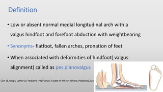
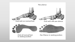
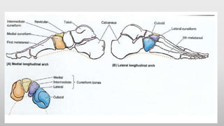
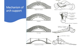
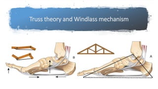
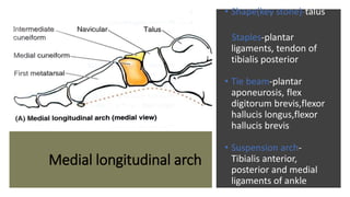
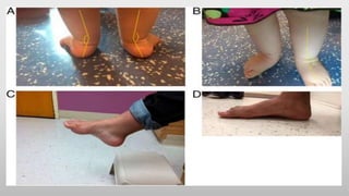
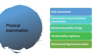
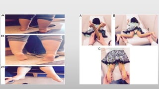
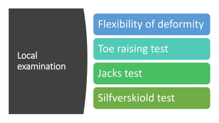
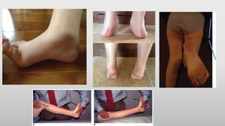
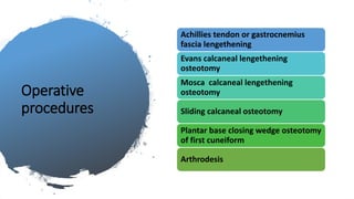
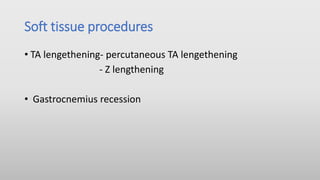
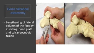
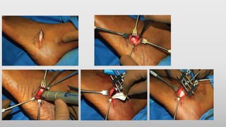
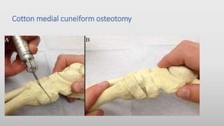
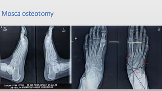
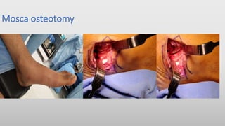
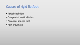
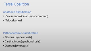
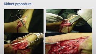
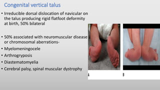
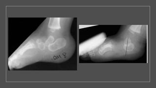
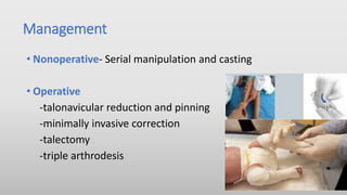
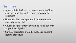
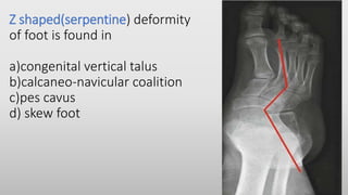
Planovalgus foot
Presenter-Dr Roshan yadav
JR Orthopaedics
AIIMS , New Delhi
Definition
• Low or absent normal medial longitudinal arch with a
valgus hindfoot and forefoot abduction with weightbearing
• Synonyms- flatfoot, fallen arches, pronation of feet
• When associated with deformities of hindfoot( valgus
alignment) called as pes planovalgus
Carr JB, Yang S, Lather LA.Pediatric Pes Planus: AState-of-the-Art Review. Pediatrics.2016
Anatomy Of
The Arches Of
Foot
A) Two longitudinal arches
• Medial longitudinal arch
• Lateral longitudinal arch
B) Transverse arch
• Integrity of bony arches
maintained by passive factors
and dynamic supports
Mechanism of
arch support
Truss theory and Windlass mechanism
Medial longitudinal arch
• Shape(key stone)-talus
Staples-plantar
ligaments, tendon of
tibialis posterior
• Tie beam-plantar
aponeurosis, flex
digitorum brevis,flexor
hallucis longus,flexor
hallucis brevis
• Suspension arch-
Tibialis anterior,
posterior and medial
ligaments of ankle
Lateral longitudinal Arch
• Shape- key stone is cuboid
• Staples-long and short plantar ligaments
• Tie beam- plantar aponeurosis, abductor digitiminimi, flexor
digitorum brevis, flexor digitorum longus(lateral part)
• Suspension arch-peroneus longus and brevis
Transverse arch
• Shape-wedge shaped
cuneiform and bases of
metatarsals
• Staples-deep transverse
ligaments and plantar
ligaments,short muscles of
foot
• Tie beam- tendon of peroneus
longus
• Suspension arch-peroneus
longus and brevis
SUSPENDING THE ARCH FROM ABOVE
• Medial longtitudinal arch: Tibialis anterior, Tibialis posterior,
medial ligament of ankle joint
• Lateral longtitudinal arch: Peroneus longus, Peroneus brevis
• Transverse arch: Peroneus longus
Functions of arched foot
• Supports body weight in
upright posture
• Acts as a lever to propel
the body forwards in
walking, running and
jumping
• Acts as a shock absorber
• Concavity of the arches
protects the soft tissues
of the sole against
pressure
Development
• In infants and toddlers, the longitudinal arch is not
developed and flat feet are normal.
• Flatfoot in infants is actually a fatfoot as the
excessive amount of fat obscures the arches
• The arch develops in childhood by the age of
10 years
Gould N, Moreland M,AlvarezR,etal. Developmentof the arch201child’s 4
•Flatfeet were present in 97% of 18 months old, 54% in 3 year
olds and 26% in 6 year olds and 4% at 10 years old
•Boys were more likely to be flatfooted than girls(52% versus
36%)
•Flatfeet were present in 62% obese , 52% overweight
Pfeiffer M, Kotz R, Ledl T, et al. Prevalence of flat foot in preschool-aged children. Pediatrics 2006
A significant difference in the prevalence of flatfoot occurred between; under-
weight (13.9%), normal-weight (16.1%), overweight (26.9%), and obese
(30.8%)children. Conclusion: The increasing prevalence of childhood obesity is
one of the most serious health challenges across the globe, and a positive
correlation between increased BMI and flatfoot was present.
Causes of Flat foot
Paedatric
flatfoot
Adult
flatfoot
Clinical features
Presenting Symptoms
• Parental concern
• Abnormal appearance of foot
• Pain
• Difficulties with shoewear,
walking, running
History
Usually bilateral and asymptomatic, painless
Ask for developmental and family history, previous medical history
Age of child and any history of trauma
Pain at rest,numbness,weakness,polyarticular pain,constitutional
symptoms
Limping,inability to bear weight
Physical
examination
Gait assessment
Generalised musculoskeletal
examination
Rotational profiles of legs
Tendoachillies tightness
Generalised ligamentous laxity
Local
examination
Flexibility of deformity
Toe raising test
Jacks test
Silfverskiold test
Radiology
Xrays –
Anteroposterior
and lateral views,
Harris-Beath
view
Computed
tomography
MRI
Anteroposterior
view foot
• Fig A-
talocalcaneal(kite
angle)
• Fig B- talo-first
metatarsal relation
• Fig-C- talonavicular
coverage angle
Lateral view
• A- Meary’s angle
• B- Calcaneal
pitch(inclination)
• C- Calaneus-first
metatarsal angle
Cyma line
• Fig A- “S” shape formed by outline of calcaneocuboid
and talonavicular joints- this continuous line is Cyma
line
• Fig-B- in flatfoot ”S” shape is displaced –anterior break
in Cyma line
Lateral
view
• Fig A- In flatfoot, sinus tarsi of subtalar joint is not well
visualized and termed as “obliterated”
• Fig B- In Cavus foot, sinus tarsi is very well visualized and may
be termed as “bullethole”
CT and MRI
• Computed tomography is the gold standard for assessment
of tarsal coalitions
• Coalition type ,extent and secondary degenerative changes
can be visualised
• MRI- abnormalities to posterior tibial tendon, other
ligaments within foot
Carr JB, Yang S,Lather LA.Pediatric Pes Planus: AState-of-the-Art Review. Pediatrics. 2016
Treatment
0-7 years old:
• No treatment unless very strong family
history of persistent flatfeet
• Counselling and reassurance of parents
Carr JB, Yang S, Lather LA.Pediatric Pes Planus: AState-of-the-Art Review.
Pediatrics. 2016
Conservative
management
• Counselling, weight control
and psychological support
Physiotherapy-
-gastrocnemius stretching ,
tendoachillies stretching
-tibialis posterior and
intrinsic muscle
strengethening
Orthotics
• Custom made inserts-
UCBL(University of California
Laboratory)
• Soft inserts - Plastazote
They concluded there remains uncertainty about the effectiveness of foot orthosis in
paediatric flexible pes planus
Surgical treatment
Indications
• Persistent pain and ulceration or callus under the head of the plantar
flexed talus and interferes with normal activity
• Rigid and painful flatfoot
• To prevent progression in neuropathic(charcot joint)
• Tibialis posterior dysfunction
Rodriguez N, Choung DJ, Dobbs MB; Rigid pediatric pes planovalgus: surgical treatment options. Clin Podiatr Med Surg. 2010 Jan
Operative
procedures
Achillies tendon or gastrocnemius
fascia lengethening
Evans calcaneal lengethening
osteotomy
Mosca calcaneal lengethening
osteotomy
Sliding calcaneal osteotomy
Plantar base closing wedge osteotomy
of first cuneiform
Arthrodesis
Soft tissue procedures
• TA lengethening- percutaneous TA lengethening
- Z lengthening
• Gastrocnemius recession
Tendoachillies lengethening is a safe procedure when used in conjunction of
hindfoot fusion procedures for flatfoot deformities
Evans calcaneal
osteotomy
• Lengthening of lateral
column of the foot by
inserting bone graft
and calcaneocuboid
fusion
Cotton medial cuneiform osteotomy
Mosca osteotomy
Mosca osteotomy
• Usually done after the age of 12
• Triple arthrodesis tend to have a high (50%) failure rate
in children under 10 years of age
• Contraindicated in young children (less than 10-12 yrs)
because the procedure limits foot growth
Triple Arthrodesis
Joints fused are:
• Subtalar joint
• Calcaneo cuboid
joint
• Talo navicular joint
Triple arthrodesis
Complications of Surgery
• Nonunion
• Degenerative joint disease
• Avascular necrosis
• Lateral instability
• Stiff foot
Posterior tibial tendon dysfunction
• The most common cause of acquired adult flatfoot is posterior tibial
tendon dysfunction
• Anatomy
Etiology
• Spontaneous rupture
• Trauma
• Inflammatory
disorders, collagen
disorders
• Presence of
accessory navicular
bone
Examination
• Inspection-
- flatfoot , swelling along PTT, fullness around medial aspect of
ankle,lateral skin wrinkling
-hindfoot valgus, too many toes sign
• Rom- ankle, subtalar, midfoot
• Specific tests-heel rise test
Treatment
• Stage 1- Conservative management and tenosynovectomy
• Stage 2- Flexor digitorum longus tendon transfer combined with
medial displacement calcaneal osteotomy
• Advanced stages- subtalar arthrodesis , triple arthrodesis
Types
Flexible –on weight bearing
arch disappears and on non
weight bearing it reappears
Rigid – acceptable medial
longitudinal arch is not
seen even on non weight
bearing with reduced or
absent motion in
midfoot,hindfoot
Causes of rigid flatfoot
• Tarsal coalition
• Congenital vertical talus
• Peroneal spastic foot
• Post traumatic
Tarsal Coalition
Anatomic classification
• Calcaneonavicular (most common)
• Talocalcaneal
Pathoanatomic classification
• Fibrous (syndesmosis)
• Cartilaginous(synchondrosis)
• Osseous(synostosis)
Examination
• Inspection- hindfoot valgus,
forefoot abduction , pes planus
• Range of motion- limited
subtalar motion, heelcord
contractures, arch doesnot
reconstitute on toe standing
•Radiography- Ap,lateral
45 degrees internal
oblique view,
harris beath view
•CT scan , MRI
Calcaneal-
navicular
coalition
• Calcanealnavicular coalition – anteater
nose sign
Talocalcaneal coalition- C sign , talar beaking
Accessory navicular bone
• It is a normal variant located in
posteromedial aspect of foot, proximal
to navicular bone
• Often incidental, most patients are
asymptomatic/ medial arch pain
• Examination-swelling, tenderness of
medial aspect of foot
Radiography
• Special view - 45 degree
external oblique
• Antero-Posterior view and
lateral weight bearing views
of the foot should be taken
to evaluate other
deformities
Radiographic
classification
• Type 1- Sesamoid bone in
the substance of tibialis
posterior tendon
• Type 2- Separate
accessory bone attached
to native navicular via
synchrondosis
• Type 3- Complete bony
enlargement
Treatment
Nonoperative
• Activity restriction , shoe modification, analgesics
• Cast immobilization
Operative
• Excision of accessory navicular (kidner procedure)
Kidner procedure
Congenital vertical talus
• Irreducible dorsal dislocation of navicular on
the talus producing rigid flatfoot deformity
at birth, 50% bilateral
• 50% associated with neuromuscular disease
or chromosomal aberrations-
• Myelomeningocele
• Arthrogryposis
• Diastematomyelia
• Cerebral palsy, spinal muscular dystrophy
Management
• Nonoperative- Serial manipulation and casting
• Operative
-talonavicular reduction and pinning
-minimally invasive correction
-talectomy
-triple arthrodesis
Summary
• Hypermobile flatfoot is a normal variant of foot
structure and doesnot require prophylactic
treatment
• Nonoperative management in adolescents is
generally successful
• Causes of rigid flatfoot should be ruled out with
proper investigation
• Surgical correction should emphasize on joint
sparing procedure
Thank you
Z shaped(serpentine) deformity
of foot is found in
a)congenital vertical talus
b)calcaneo-navicular coalition
c)pes cavus
d) skew foot
What is the normal value of Meary’s angle?
a) 10-15 degrees
b) zero degree
c) 30-45 degrees
d) 15-30 degrees

Pes planus seminar

  • 1.
    Planovalgus foot Presenter-Dr Roshanyadav JR Orthopaedics AIIMS , New Delhi
  • 2.
    Definition • Low orabsent normal medial longitudinal arch with a valgus hindfoot and forefoot abduction with weightbearing • Synonyms- flatfoot, fallen arches, pronation of feet • When associated with deformities of hindfoot( valgus alignment) called as pes planovalgus Carr JB, Yang S, Lather LA.Pediatric Pes Planus: AState-of-the-Art Review. Pediatrics.2016
  • 4.
    Anatomy Of The ArchesOf Foot A) Two longitudinal arches • Medial longitudinal arch • Lateral longitudinal arch B) Transverse arch • Integrity of bony arches maintained by passive factors and dynamic supports
  • 6.
  • 7.
    Truss theory andWindlass mechanism
  • 8.
    Medial longitudinal arch •Shape(key stone)-talus Staples-plantar ligaments, tendon of tibialis posterior • Tie beam-plantar aponeurosis, flex digitorum brevis,flexor hallucis longus,flexor hallucis brevis • Suspension arch- Tibialis anterior, posterior and medial ligaments of ankle
  • 9.
    Lateral longitudinal Arch •Shape- key stone is cuboid • Staples-long and short plantar ligaments • Tie beam- plantar aponeurosis, abductor digitiminimi, flexor digitorum brevis, flexor digitorum longus(lateral part) • Suspension arch-peroneus longus and brevis
  • 10.
    Transverse arch • Shape-wedgeshaped cuneiform and bases of metatarsals • Staples-deep transverse ligaments and plantar ligaments,short muscles of foot • Tie beam- tendon of peroneus longus • Suspension arch-peroneus longus and brevis
  • 11.
    SUSPENDING THE ARCHFROM ABOVE • Medial longtitudinal arch: Tibialis anterior, Tibialis posterior, medial ligament of ankle joint • Lateral longtitudinal arch: Peroneus longus, Peroneus brevis • Transverse arch: Peroneus longus
  • 12.
    Functions of archedfoot • Supports body weight in upright posture • Acts as a lever to propel the body forwards in walking, running and jumping • Acts as a shock absorber • Concavity of the arches protects the soft tissues of the sole against pressure
  • 13.
    Development • In infantsand toddlers, the longitudinal arch is not developed and flat feet are normal. • Flatfoot in infants is actually a fatfoot as the excessive amount of fat obscures the arches • The arch develops in childhood by the age of 10 years Gould N, Moreland M,AlvarezR,etal. Developmentof the arch201child’s 4
  • 14.
    •Flatfeet were presentin 97% of 18 months old, 54% in 3 year olds and 26% in 6 year olds and 4% at 10 years old •Boys were more likely to be flatfooted than girls(52% versus 36%) •Flatfeet were present in 62% obese , 52% overweight Pfeiffer M, Kotz R, Ledl T, et al. Prevalence of flat foot in preschool-aged children. Pediatrics 2006
  • 15.
    A significant differencein the prevalence of flatfoot occurred between; under- weight (13.9%), normal-weight (16.1%), overweight (26.9%), and obese (30.8%)children. Conclusion: The increasing prevalence of childhood obesity is one of the most serious health challenges across the globe, and a positive correlation between increased BMI and flatfoot was present.
  • 16.
    Causes of Flatfoot Paedatric flatfoot Adult flatfoot
  • 17.
    Clinical features Presenting Symptoms •Parental concern • Abnormal appearance of foot • Pain • Difficulties with shoewear, walking, running
  • 18.
    History Usually bilateral andasymptomatic, painless Ask for developmental and family history, previous medical history Age of child and any history of trauma Pain at rest,numbness,weakness,polyarticular pain,constitutional symptoms Limping,inability to bear weight
  • 20.
    Physical examination Gait assessment Generalised musculoskeletal examination Rotationalprofiles of legs Tendoachillies tightness Generalised ligamentous laxity
  • 22.
    Local examination Flexibility of deformity Toeraising test Jacks test Silfverskiold test
  • 24.
    Radiology Xrays – Anteroposterior and lateralviews, Harris-Beath view Computed tomography MRI
  • 25.
    Anteroposterior view foot • FigA- talocalcaneal(kite angle) • Fig B- talo-first metatarsal relation • Fig-C- talonavicular coverage angle
  • 26.
    Lateral view • A-Meary’s angle • B- Calcaneal pitch(inclination) • C- Calaneus-first metatarsal angle
  • 28.
    Cyma line • FigA- “S” shape formed by outline of calcaneocuboid and talonavicular joints- this continuous line is Cyma line • Fig-B- in flatfoot ”S” shape is displaced –anterior break in Cyma line
  • 29.
    Lateral view • Fig A-In flatfoot, sinus tarsi of subtalar joint is not well visualized and termed as “obliterated” • Fig B- In Cavus foot, sinus tarsi is very well visualized and may be termed as “bullethole”
  • 30.
    CT and MRI •Computed tomography is the gold standard for assessment of tarsal coalitions • Coalition type ,extent and secondary degenerative changes can be visualised • MRI- abnormalities to posterior tibial tendon, other ligaments within foot Carr JB, Yang S,Lather LA.Pediatric Pes Planus: AState-of-the-Art Review. Pediatrics. 2016
  • 31.
    Treatment 0-7 years old: •No treatment unless very strong family history of persistent flatfeet • Counselling and reassurance of parents Carr JB, Yang S, Lather LA.Pediatric Pes Planus: AState-of-the-Art Review. Pediatrics. 2016
  • 32.
    Conservative management • Counselling, weightcontrol and psychological support Physiotherapy- -gastrocnemius stretching , tendoachillies stretching -tibialis posterior and intrinsic muscle strengethening
  • 33.
    Orthotics • Custom madeinserts- UCBL(University of California Laboratory) • Soft inserts - Plastazote
  • 34.
    They concluded thereremains uncertainty about the effectiveness of foot orthosis in paediatric flexible pes planus
  • 35.
    Surgical treatment Indications • Persistentpain and ulceration or callus under the head of the plantar flexed talus and interferes with normal activity • Rigid and painful flatfoot • To prevent progression in neuropathic(charcot joint) • Tibialis posterior dysfunction Rodriguez N, Choung DJ, Dobbs MB; Rigid pediatric pes planovalgus: surgical treatment options. Clin Podiatr Med Surg. 2010 Jan
  • 36.
    Operative procedures Achillies tendon orgastrocnemius fascia lengethening Evans calcaneal lengethening osteotomy Mosca calcaneal lengethening osteotomy Sliding calcaneal osteotomy Plantar base closing wedge osteotomy of first cuneiform Arthrodesis
  • 37.
    Soft tissue procedures •TA lengethening- percutaneous TA lengethening - Z lengthening • Gastrocnemius recession
  • 38.
    Tendoachillies lengethening isa safe procedure when used in conjunction of hindfoot fusion procedures for flatfoot deformities
  • 39.
    Evans calcaneal osteotomy • Lengtheningof lateral column of the foot by inserting bone graft and calcaneocuboid fusion
  • 41.
  • 42.
  • 43.
  • 44.
    • Usually doneafter the age of 12 • Triple arthrodesis tend to have a high (50%) failure rate in children under 10 years of age • Contraindicated in young children (less than 10-12 yrs) because the procedure limits foot growth
  • 45.
    Triple Arthrodesis Joints fusedare: • Subtalar joint • Calcaneo cuboid joint • Talo navicular joint
  • 46.
  • 47.
    Complications of Surgery •Nonunion • Degenerative joint disease • Avascular necrosis • Lateral instability • Stiff foot
  • 48.
    Posterior tibial tendondysfunction • The most common cause of acquired adult flatfoot is posterior tibial tendon dysfunction • Anatomy
  • 49.
    Etiology • Spontaneous rupture •Trauma • Inflammatory disorders, collagen disorders • Presence of accessory navicular bone
  • 50.
    Examination • Inspection- - flatfoot, swelling along PTT, fullness around medial aspect of ankle,lateral skin wrinkling -hindfoot valgus, too many toes sign • Rom- ankle, subtalar, midfoot • Specific tests-heel rise test
  • 52.
    Treatment • Stage 1-Conservative management and tenosynovectomy • Stage 2- Flexor digitorum longus tendon transfer combined with medial displacement calcaneal osteotomy • Advanced stages- subtalar arthrodesis , triple arthrodesis
  • 53.
    Types Flexible –on weightbearing arch disappears and on non weight bearing it reappears Rigid – acceptable medial longitudinal arch is not seen even on non weight bearing with reduced or absent motion in midfoot,hindfoot
  • 54.
    Causes of rigidflatfoot • Tarsal coalition • Congenital vertical talus • Peroneal spastic foot • Post traumatic
  • 55.
    Tarsal Coalition Anatomic classification •Calcaneonavicular (most common) • Talocalcaneal Pathoanatomic classification • Fibrous (syndesmosis) • Cartilaginous(synchondrosis) • Osseous(synostosis)
  • 56.
    Examination • Inspection- hindfootvalgus, forefoot abduction , pes planus • Range of motion- limited subtalar motion, heelcord contractures, arch doesnot reconstitute on toe standing
  • 57.
    •Radiography- Ap,lateral 45 degreesinternal oblique view, harris beath view •CT scan , MRI
  • 58.
  • 59.
    Talocalcaneal coalition- Csign , talar beaking
  • 60.
    Accessory navicular bone •It is a normal variant located in posteromedial aspect of foot, proximal to navicular bone • Often incidental, most patients are asymptomatic/ medial arch pain • Examination-swelling, tenderness of medial aspect of foot
  • 61.
    Radiography • Special view- 45 degree external oblique • Antero-Posterior view and lateral weight bearing views of the foot should be taken to evaluate other deformities
  • 62.
    Radiographic classification • Type 1-Sesamoid bone in the substance of tibialis posterior tendon • Type 2- Separate accessory bone attached to native navicular via synchrondosis • Type 3- Complete bony enlargement
  • 63.
    Treatment Nonoperative • Activity restriction, shoe modification, analgesics • Cast immobilization Operative • Excision of accessory navicular (kidner procedure)
  • 64.
  • 65.
    Congenital vertical talus •Irreducible dorsal dislocation of navicular on the talus producing rigid flatfoot deformity at birth, 50% bilateral • 50% associated with neuromuscular disease or chromosomal aberrations- • Myelomeningocele • Arthrogryposis • Diastematomyelia • Cerebral palsy, spinal muscular dystrophy
  • 67.
    Management • Nonoperative- Serialmanipulation and casting • Operative -talonavicular reduction and pinning -minimally invasive correction -talectomy -triple arthrodesis
  • 69.
    Summary • Hypermobile flatfootis a normal variant of foot structure and doesnot require prophylactic treatment • Nonoperative management in adolescents is generally successful • Causes of rigid flatfoot should be ruled out with proper investigation • Surgical correction should emphasize on joint sparing procedure
  • 70.
  • 71.
    Z shaped(serpentine) deformity offoot is found in a)congenital vertical talus b)calcaneo-navicular coalition c)pes cavus d) skew foot
  • 72.
    What is thenormal value of Meary’s angle? a) 10-15 degrees b) zero degree c) 30-45 degrees d) 15-30 degrees