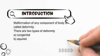
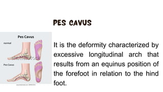
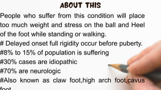
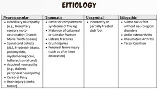
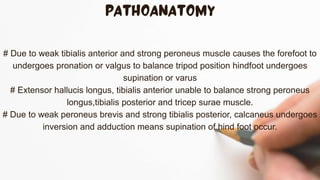
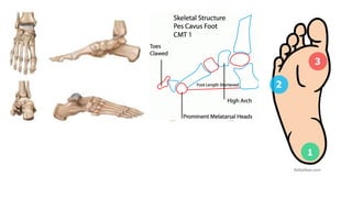
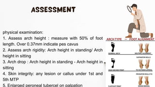
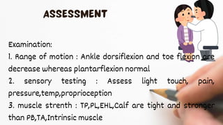
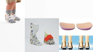
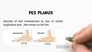
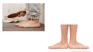
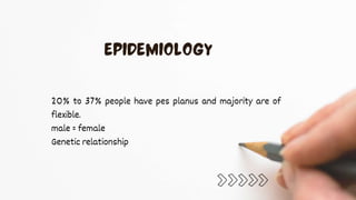
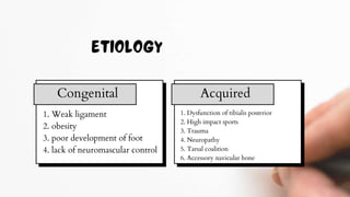
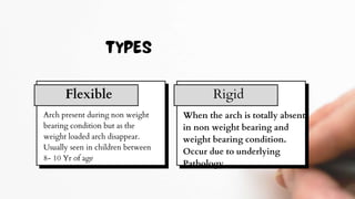
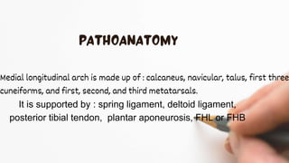
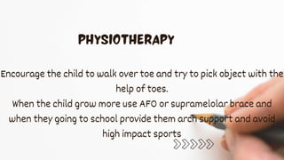
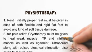
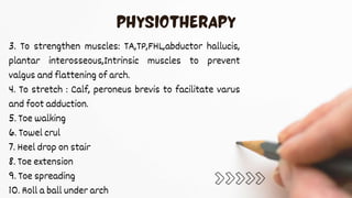
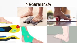
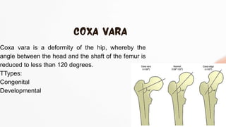
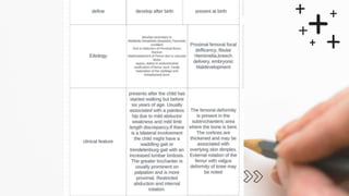
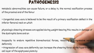
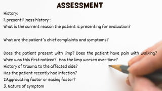
1. pes cavus
2. pes planus
3.Coxa vara
Malformation of any component of body is
called deformity.
There are two types of deformity
a) congenital
b) aquired
It is the deformity characterized by
excessive longitudinal arch that
results from an equinus position of
the forefoot in relation to the hind
foot.
People who suffer from this condition will place
too much weight and stress on the ball and Heel
of the foot while standing or walking.
# Delayed onset full rigidity occur before puberty.
#8% to 15% of population is suffering
#30% cases are idiopathic
#70% are neurologic
#Also known as claw foot,high arch foot,cavus
# Due to weak tibialis anterior and strong peroneus muscle causes the forefoot to
undergoes pronation or valgus to balance tripod position hindfoot undergoes
supination or varus
# Extensor hallucis longus, tibialis anterior unable to balance strong peroneus
longus,tibialis posterior and tricep surae muscle.
# Due to weak peroneus brevis and strong tibialis posterior, calcaneus undergoes
inversion and adduction means supination of hind foot occur.
•During gait the first ray strike first before heel strike and
interphalangeal joints are flexed while MTP are extended.
Note IP joints are flexed due to FDL and MTP joint are
extended due to EDL.
• Initially the deformity is soft but rigid as the disease progress.
Heavy callus and plantar ulcers are common on 1st and 5th
metatarsal head due to high plantar pressure and reduced
sensation in these area. This can leads to infection and
amputation.
During gait normally the forefoot undergoes pronation
and midfoot joint unlock to enable the foot to absorb
shock and toward the end of stance phase the foot and
the lower leg muscle induces elevation of arch and cause
the midfoot to lock this create a rigid lever which propel
th foot forward. In person with pes cavus the mid foot
remain locked and elevated arch throughout the stance
phase this result in reduced shock absorption with
increase risk of fracture.
Types
Pes cavovarus
: most common
: seen with neurological &
idiopathic cases
: characterized by
forefoot varus,plantar
flexed 1st ray, claw toe
Pes calcaneovarus
: usually due to
poliomyelitis
: characterised by
calocalcaneal dorsiflexion,
forefoot plantarflexion
Pes cavus
: characterized by
normal calcaneus
with plantarflexed
forefoot
1. Elevated medial longitudinal arch
2. Plantarflexed first ray
3. Claw toe deformity
4. Metatarsalgia
5. Pain under first metatarsal
6. Plantarfascitis
7. Painfull callosites
8. Ankle arthritis
9. Achilles tendinitis
10. Ankle arthritis
11. Keratosis
12. Lateral Ankle instability
13. Hind foot varus
14. Forefoot plantar flexed
15. Lower limb stress fracture
16. It band friction syndrome
17. 5th metatarsal fracture
1. Demographics
2. history of present illness
a) when did the Symptoms start and how they progressed
since onset?
b) was development of pes cavus idiopathic? Was it linked
to any neurological,congenital,traumatic injury?
c) any treatment taken and course of treatment
3. past medicalhistory
a) which medicine and what therapy you take in past?
4. Aggravating & reliving factor
5. nature of symptom :
constant,intermittent,sharp,dull
6. pattern of symptom : symptom present throughout
day or night and change in symptom due to weather
7. occupational history
8. environmentalhistory
physical examination:
1. Assess arch height : measure with 50% of foot
length. Over 0.37mm indicate pes cavus
2. Assess arch rigidity: Arch height in standing/ Arch
height in sitting
3. Arch drop : Arch height in standing - Arch height in
sitting
4. Skin integrity: any lesion or callus under 1st and
5th MTP
5. Enlarged peroneal tubercel on palpation
Examination:
1. Range of motion : Ankle dorsiflexion and toe flexion are
decrease whereas plantarflexion normal
2. sensory testing : Assess light touch, pain,
pressure,temp,proprioception
3. muscle strenth : TP,PL,EHL,Calf are tight and stronger
than PB,TA,Intrinsic muscle
4. Gait analysis: usually pes cavus patient walk with
excessive supination
5. Balance assessment: static and dynamic balance
6. fuctional mobility : Time up and go test
7. Neuro motor development : peabody developmentalscale
8. Nerve testing & reflex testing : plantar reflex
9. peek -a-boo sign
Normal calcaneal pitch 17°-
32°
Meary angle Normal range : 0° more than 4° is consider cavus
Normal Talocalcaneal angle:
25° - 45°, less than 20° is
consider cavus
Goal
Decrease pain
Goal
Improve strength of weak muscle
Goal
Improve ROM
Goal
Improve gait/balance
Goal
Return to recreational activity
Rehabilitation include :
1. stretching of tight structure : Gastrocnemius, plantar fascia,tibialisposterior,
peroneus longus stretching should be provided
2. strengthening of weak muscle : Tibialis anterior, peroneus brevis
strengthening must be done
3. Orthotic support: To reduce ulcer formation and callosities lateral border
insoles are used to decrease pressure on lateral aspect. It has been
suggested that it reduce pain and pressure only in adult population but not
effective in growing population.
4. serial casting and night splints : casting along with night splints are more
beneficia. It continouslystretch the tight structure and provide foot in neutral
position.
5. pain relief : TENS along with hydrocolator pack is
used in rigid case's while cryo is used in infants and
in neurological cases to reduce spasticity. But not
more than 5min .
6. Gait and balance : walking with splints and insoles
must be encouraged. In case of neurological defict
assistive devices can also be used.
7. patient education: position changing and stretching
must be done regularly and must avoid high impact
https://www.ncbi.nlm.nih.gov/books/NBK556016/
https://www.physio-pedia.com/Pes_Cavus
https://en.m.wikipedia.org/wiki/Pes_cavus
https://now.aapmr.org/pes-cavus/
research paper & orthopedic books
Disorder of foot characterized by loss of medial
longitudinal arch . Also known as flat foot
20% to 37% people have pes planus and majority are of
flexible.
male = female
Genetic relationship
1. Weak ligament
2. obesity
3. poor development of foot
4. lack of neuromascular control
Congenital Acquired
1. Dysfunction of tibialis posterior
2. High impact sports
3. Trauma
4. Neuropathy
5. Tarsal coalition
6. Accessory navicular bone
Arch present during non weight
bearing condition but as the
weight loaded arch disappear.
Usually seen in children between
8- 10 Yr of age
Flexible Rigid
When the arch is totally absent
in non weight bearing and
weight bearing condition.
Occur due to underlying
Pathology
Medial longitudinal arch is made up of : calcaneus, navicular, talus, first three
cuneiforms, and first, second, and third metatarsals.
It is supported by : spring ligament, deltoid ligament,
posterior tibial tendon, plantar aponeurosis, FHL or FHB
Usually TP and calonavicular ligaments weakness
contribute in pes planus.
TP invert the hind foot to lock the transverse tarsal joint
during standing to provide rigidity to foot but in case of
weak TP inversion not occur properly which leads to strong
pull of peroneal muscle cause the eversion of hind foot and
pronation occur Therefore medial longitudinal arch become
flatten.
History :
1. Demographics
2. cheif complaint : pain, swelling, pressure ulcers
•ask about timing of Symptoms
• severity of Symptoms
• is arch normal in non weight bearing position
• was it present during birth or after an high impact
activity
3. Past medical history: Hypertension, Diabetes, arthritis,
sensory neuropathy
4. Familyhistory of pes planus
5. environmental history
Physical examination:
1. examine the arch with or without weight bearing
2. check for posterior view of foot to show ' too many
toe' sign
3. palpate TP, PF for tenderness and swelling
4. collapse medial longitudinal arch
5. valgus deformityin heel and forefoot abduction
Reduce
pain
Increase foot flexibility
Build normal
arch
Avoid soft tissue damage
Improve foot muscle strength
Patient reassurance
Management strategy:
Infants : Initially casting is done which to maintain the
hind foot in varus direction and fore foot adducted
and supinated at the same time medial longitudinal
arch is shaped by the cast till 2-3months
after 2-3months when cast is removed stretching of
calf must be done with ankle rom .
Encourage walking on sand when the child begin to
Encourage the child to walk over toe and try to pick object with the
help of toes.
When the child grow more use AFO or supramelolar brace and
when they going to school provide them arch support and avoid
high impact sports
1. Rest : Initially proper rest must be given in
case of both flexible and rigid flat feet to
avoid any kind of soft tissue damage.
2. for pain relief: Cryotherapy must be given
to heal weak muscle TP and Intrinsic
muscle as well as ligament. Ultrasound
along with pulsed electrical stimulation also
3. To strengthen muscles: TA,TP,FHL,abductor hallucis,
plantar interosseous,Intrinsic muscles to prevent
valgus and flattening of arch.
4. To stretch : Calf, peroneus brevis to facilitate varus
and foot adduction.
5. Toe walking
6. Towel crul
7. Heel drop on stair
8. Toe extension
9. Toe spreading
10. Roll a ball under arch
https://www.theseus.fi/handle/10024/807633
https://www.physio-
pedia.com/Pes_Planushttps://www.ncbi.nlm.nih.gov/
books/NBK430802/
https://radiopaedia.org/articles/pes-planus
https://en.m.wikipedia.org/wiki/Flat_feet
Coxa vara is a deformity of the hip, whereby the
angle between the head and the shaft of the femur is
reduced to less than 120 degrees.
TTypes:
Congenital
Developmental
Metabolic abnormalities can cause failure, or a delay in, the normal ossification process
of the proximal end of the femur
• Congenital coxa vara is believed to be the result of a primary ossification defect in the
inferior femoral neck on which
physiologic shearing stresses are applied during weight-bearing.This results in damage of
the dystrophic bone and an
incapacity to endure repetitive biomechanical forces, resulting in progressive varus
deformity
• Progression of coxa vara deformity can increase the shearing force at the hypertrophic
cell layer of the epiphyseal plate by
History:
1. present illness history :
What is the current reason the patient is presenting for evaluation?
What are the patient’s chief complaints and symptoms?
Does the patient present with limp? Does the patient have pain with walking?
When was this first noticed? Has the limp worsen over time?
History of trauma to the affected side?
Has the patient recently had infection?
2Aggravating factor or easing factor?
3. Nature of symptom
5. Past medical history:
Family history of coxa vara? Traumatic birth history?
Askaboutany other comorbidity like Ricket,osteogenesis
imperfecta,osteoporosis
Any medication or surgical procedure had you taken?
6. Social/Occupational history :
Does the patient participate in competitive sports?
what physical task are frequently performed?
7. Environmental history : stairs,no of floor,who live with patient any
caregiver? Any modification necessary?
physical examination:
Grater trochanter is prominent and easily Palpable
In post operatively examine any incision site for
infection,erythema,edema,increase temp?
Examination:
1. Range of motion : Assess active and passive ROM of lower extremity
joints bilaterally using goniometer. Note any asymmetry
- Hip abduction is limited due to a decreased articulo-trochanteric
distance
- Internal rotation is limited due to decrease femoral anteversion
2. Muscle strength : Most often, there is significant muscle weakness
in the involved hip, particularly the hip abductor
3.Gait Analysis : –Assess gait for symmetry and note any abnormalities or
complaints of pain
–“Waddling” gait or a pain-free limp is most notable
–Positive Trendelenburg sign (can be bilateral)
–Assess the need for assistive devices pre- and postoperatively, especially if there
are any weight-bearingrestrictions
–Use 6-minute walk test (6MWT) for distance
4. Balance:
5. Posture Analysis: –Does the patient sit or stand asymmetrically
due to leg-length discrepancy or pain?
–Increased lumbar lordosis and genu valgum is often present with
developmental coxa vara affecting both extremities
Special Test :
1. criag Test : patient is in prone with knee flexed 90°, examine rotate
the hip so that when grater trochenter is more prominent than
measure with goniometer, The degree anteversion , 15° is normal .
More than 15° indicate anteversion and less than 15°
2. Nelaton line : draw between Asis and
ischeal tuberosity. If the head of Femur is
lower than this line it indicate coxa vara.
3. Galeazzi sign : patient lie supine with hip
45°flexed and knee 90° flexed and both foot
heel at the same level then check the
shortening of femur.
Goal
1. Decrease postoperative
pain,edema and swelling
2.improve ROM
3. improve muscle strength
4.improve balance
Goal
5. Improve walking pattern
6. Increase functional mobility
This condition require combination of both physiotherapy or
surgical approach to treat.
1. To decrease pain,edema: Use cryotherapy, STM
2. increase hip internal rot and abduction : carefully stretch
the hip abductor, internal rotators,adductors,hamstrings
and AROM
3. Improve strength: Flexors, extensors, abductors and
internal rotators
4. Improve balance and proprioception: single leg
stance,parallel bars, obstacles walking
5. Gait Training: walking drill, step up, stair climbing,
Treadmill walking
6. Functional movement: lunges,step up, kicking
ball
7. pelvic stability : pelvic tilt on swiss ball .
https://scholar.google.com/scholar?hl=en&as_sdt=0
%2C5&q=physiotherapy+treatment+of+coxa+vara&b
tnG=#d=gs_qabs&t=1712637525998&u=%23p%3D
Wnx6r3IXdK0J
https://www.orthobullets.com/pediatrics/4041/develop
mental-coxa-vara
https://radiopaedia.org/articles/coxa-vara
https://en.m.wikipedia.org/wiki/Coxa_vara
Bagga I B, Raghuveer R, Singh S (March 06, 2024)
Physical Therapy Interventions: A Case Report of
Building Strength, Confidence, and Mobility
Ortho deformity  physiotherapy treatment

Ortho deformity physiotherapy treatment

  • 2.
    1. pes cavus 2.pes planus 3.Coxa vara
  • 3.
    Malformation of anycomponent of body is called deformity. There are two types of deformity a) congenital b) aquired
  • 4.
    It is thedeformity characterized by excessive longitudinal arch that results from an equinus position of the forefoot in relation to the hind foot.
  • 5.
    People who sufferfrom this condition will place too much weight and stress on the ball and Heel of the foot while standing or walking. # Delayed onset full rigidity occur before puberty. #8% to 15% of population is suffering #30% cases are idiopathic #70% are neurologic #Also known as claw foot,high arch foot,cavus
  • 7.
    # Due toweak tibialis anterior and strong peroneus muscle causes the forefoot to undergoes pronation or valgus to balance tripod position hindfoot undergoes supination or varus # Extensor hallucis longus, tibialis anterior unable to balance strong peroneus longus,tibialis posterior and tricep surae muscle. # Due to weak peroneus brevis and strong tibialis posterior, calcaneus undergoes inversion and adduction means supination of hind foot occur.
  • 9.
    •During gait thefirst ray strike first before heel strike and interphalangeal joints are flexed while MTP are extended. Note IP joints are flexed due to FDL and MTP joint are extended due to EDL. • Initially the deformity is soft but rigid as the disease progress. Heavy callus and plantar ulcers are common on 1st and 5th metatarsal head due to high plantar pressure and reduced sensation in these area. This can leads to infection and amputation.
  • 10.
    During gait normallythe forefoot undergoes pronation and midfoot joint unlock to enable the foot to absorb shock and toward the end of stance phase the foot and the lower leg muscle induces elevation of arch and cause the midfoot to lock this create a rigid lever which propel th foot forward. In person with pes cavus the mid foot remain locked and elevated arch throughout the stance phase this result in reduced shock absorption with increase risk of fracture.
  • 11.
    Types Pes cavovarus : mostcommon : seen with neurological & idiopathic cases : characterized by forefoot varus,plantar flexed 1st ray, claw toe Pes calcaneovarus : usually due to poliomyelitis : characterised by calocalcaneal dorsiflexion, forefoot plantarflexion Pes cavus : characterized by normal calcaneus with plantarflexed forefoot
  • 12.
    1. Elevated mediallongitudinal arch 2. Plantarflexed first ray 3. Claw toe deformity 4. Metatarsalgia 5. Pain under first metatarsal 6. Plantarfascitis 7. Painfull callosites 8. Ankle arthritis 9. Achilles tendinitis 10. Ankle arthritis 11. Keratosis 12. Lateral Ankle instability 13. Hind foot varus 14. Forefoot plantar flexed 15. Lower limb stress fracture 16. It band friction syndrome 17. 5th metatarsal fracture
  • 13.
    1. Demographics 2. historyof present illness a) when did the Symptoms start and how they progressed since onset? b) was development of pes cavus idiopathic? Was it linked to any neurological,congenital,traumatic injury? c) any treatment taken and course of treatment 3. past medicalhistory a) which medicine and what therapy you take in past?
  • 14.
    4. Aggravating &reliving factor 5. nature of symptom : constant,intermittent,sharp,dull 6. pattern of symptom : symptom present throughout day or night and change in symptom due to weather 7. occupational history 8. environmentalhistory
  • 15.
    physical examination: 1. Assessarch height : measure with 50% of foot length. Over 0.37mm indicate pes cavus 2. Assess arch rigidity: Arch height in standing/ Arch height in sitting 3. Arch drop : Arch height in standing - Arch height in sitting 4. Skin integrity: any lesion or callus under 1st and 5th MTP 5. Enlarged peroneal tubercel on palpation
  • 16.
    Examination: 1. Range ofmotion : Ankle dorsiflexion and toe flexion are decrease whereas plantarflexion normal 2. sensory testing : Assess light touch, pain, pressure,temp,proprioception 3. muscle strenth : TP,PL,EHL,Calf are tight and stronger than PB,TA,Intrinsic muscle
  • 17.
    4. Gait analysis:usually pes cavus patient walk with excessive supination 5. Balance assessment: static and dynamic balance 6. fuctional mobility : Time up and go test 7. Neuro motor development : peabody developmentalscale 8. Nerve testing & reflex testing : plantar reflex 9. peek -a-boo sign
  • 18.
    Normal calcaneal pitch17°- 32° Meary angle Normal range : 0° more than 4° is consider cavus
  • 19.
    Normal Talocalcaneal angle: 25°- 45°, less than 20° is consider cavus
  • 20.
    Goal Decrease pain Goal Improve strengthof weak muscle Goal Improve ROM Goal Improve gait/balance Goal Return to recreational activity
  • 21.
    Rehabilitation include : 1.stretching of tight structure : Gastrocnemius, plantar fascia,tibialisposterior, peroneus longus stretching should be provided 2. strengthening of weak muscle : Tibialis anterior, peroneus brevis strengthening must be done 3. Orthotic support: To reduce ulcer formation and callosities lateral border insoles are used to decrease pressure on lateral aspect. It has been suggested that it reduce pain and pressure only in adult population but not effective in growing population. 4. serial casting and night splints : casting along with night splints are more beneficia. It continouslystretch the tight structure and provide foot in neutral position.
  • 23.
    5. pain relief: TENS along with hydrocolator pack is used in rigid case's while cryo is used in infants and in neurological cases to reduce spasticity. But not more than 5min . 6. Gait and balance : walking with splints and insoles must be encouraged. In case of neurological defict assistive devices can also be used. 7. patient education: position changing and stretching must be done regularly and must avoid high impact
  • 24.
  • 25.
    Disorder of footcharacterized by loss of medial longitudinal arch . Also known as flat foot
  • 27.
    20% to 37%people have pes planus and majority are of flexible. male = female Genetic relationship
  • 28.
    1. Weak ligament 2.obesity 3. poor development of foot 4. lack of neuromascular control Congenital Acquired 1. Dysfunction of tibialis posterior 2. High impact sports 3. Trauma 4. Neuropathy 5. Tarsal coalition 6. Accessory navicular bone
  • 29.
    Arch present duringnon weight bearing condition but as the weight loaded arch disappear. Usually seen in children between 8- 10 Yr of age Flexible Rigid When the arch is totally absent in non weight bearing and weight bearing condition. Occur due to underlying Pathology
  • 30.
    Medial longitudinal archis made up of : calcaneus, navicular, talus, first three cuneiforms, and first, second, and third metatarsals. It is supported by : spring ligament, deltoid ligament, posterior tibial tendon, plantar aponeurosis, FHL or FHB
  • 31.
    Usually TP andcalonavicular ligaments weakness contribute in pes planus. TP invert the hind foot to lock the transverse tarsal joint during standing to provide rigidity to foot but in case of weak TP inversion not occur properly which leads to strong pull of peroneal muscle cause the eversion of hind foot and pronation occur Therefore medial longitudinal arch become flatten.
  • 32.
    History : 1. Demographics 2.cheif complaint : pain, swelling, pressure ulcers •ask about timing of Symptoms • severity of Symptoms • is arch normal in non weight bearing position • was it present during birth or after an high impact activity
  • 33.
    3. Past medicalhistory: Hypertension, Diabetes, arthritis, sensory neuropathy 4. Familyhistory of pes planus 5. environmental history
  • 34.
    Physical examination: 1. examinethe arch with or without weight bearing 2. check for posterior view of foot to show ' too many toe' sign 3. palpate TP, PF for tenderness and swelling 4. collapse medial longitudinal arch 5. valgus deformityin heel and forefoot abduction
  • 36.
    Reduce pain Increase foot flexibility Buildnormal arch Avoid soft tissue damage Improve foot muscle strength Patient reassurance
  • 38.
    Management strategy: Infants :Initially casting is done which to maintain the hind foot in varus direction and fore foot adducted and supinated at the same time medial longitudinal arch is shaped by the cast till 2-3months after 2-3months when cast is removed stretching of calf must be done with ankle rom . Encourage walking on sand when the child begin to
  • 39.
    Encourage the childto walk over toe and try to pick object with the help of toes. When the child grow more use AFO or supramelolar brace and when they going to school provide them arch support and avoid high impact sports
  • 40.
    1. Rest :Initially proper rest must be given in case of both flexible and rigid flat feet to avoid any kind of soft tissue damage. 2. for pain relief: Cryotherapy must be given to heal weak muscle TP and Intrinsic muscle as well as ligament. Ultrasound along with pulsed electrical stimulation also
  • 41.
    3. To strengthenmuscles: TA,TP,FHL,abductor hallucis, plantar interosseous,Intrinsic muscles to prevent valgus and flattening of arch. 4. To stretch : Calf, peroneus brevis to facilitate varus and foot adduction. 5. Toe walking 6. Towel crul 7. Heel drop on stair 8. Toe extension 9. Toe spreading 10. Roll a ball under arch
  • 43.
  • 44.
    Coxa vara isa deformity of the hip, whereby the angle between the head and the shaft of the femur is reduced to less than 120 degrees. TTypes: Congenital Developmental
  • 46.
    Metabolic abnormalities cancause failure, or a delay in, the normal ossification process of the proximal end of the femur • Congenital coxa vara is believed to be the result of a primary ossification defect in the inferior femoral neck on which physiologic shearing stresses are applied during weight-bearing.This results in damage of the dystrophic bone and an incapacity to endure repetitive biomechanical forces, resulting in progressive varus deformity • Progression of coxa vara deformity can increase the shearing force at the hypertrophic cell layer of the epiphyseal plate by
  • 47.
    History: 1. present illnesshistory : What is the current reason the patient is presenting for evaluation? What are the patient’s chief complaints and symptoms? Does the patient present with limp? Does the patient have pain with walking? When was this first noticed? Has the limp worsen over time? History of trauma to the affected side? Has the patient recently had infection? 2Aggravating factor or easing factor? 3. Nature of symptom
  • 48.
    5. Past medicalhistory: Family history of coxa vara? Traumatic birth history? Askaboutany other comorbidity like Ricket,osteogenesis imperfecta,osteoporosis Any medication or surgical procedure had you taken? 6. Social/Occupational history : Does the patient participate in competitive sports? what physical task are frequently performed? 7. Environmental history : stairs,no of floor,who live with patient any caregiver? Any modification necessary? physical examination: Grater trochanter is prominent and easily Palpable In post operatively examine any incision site for infection,erythema,edema,increase temp?
  • 49.
    Examination: 1. Range ofmotion : Assess active and passive ROM of lower extremity joints bilaterally using goniometer. Note any asymmetry - Hip abduction is limited due to a decreased articulo-trochanteric distance - Internal rotation is limited due to decrease femoral anteversion 2. Muscle strength : Most often, there is significant muscle weakness in the involved hip, particularly the hip abductor
  • 50.
    3.Gait Analysis :–Assess gait for symmetry and note any abnormalities or complaints of pain –“Waddling” gait or a pain-free limp is most notable –Positive Trendelenburg sign (can be bilateral) –Assess the need for assistive devices pre- and postoperatively, especially if there are any weight-bearingrestrictions –Use 6-minute walk test (6MWT) for distance 4. Balance:
  • 51.
    5. Posture Analysis:–Does the patient sit or stand asymmetrically due to leg-length discrepancy or pain? –Increased lumbar lordosis and genu valgum is often present with developmental coxa vara affecting both extremities Special Test : 1. criag Test : patient is in prone with knee flexed 90°, examine rotate the hip so that when grater trochenter is more prominent than measure with goniometer, The degree anteversion , 15° is normal . More than 15° indicate anteversion and less than 15°
  • 52.
    2. Nelaton line: draw between Asis and ischeal tuberosity. If the head of Femur is lower than this line it indicate coxa vara. 3. Galeazzi sign : patient lie supine with hip 45°flexed and knee 90° flexed and both foot heel at the same level then check the shortening of femur.
  • 54.
    Goal 1. Decrease postoperative pain,edemaand swelling 2.improve ROM 3. improve muscle strength 4.improve balance Goal 5. Improve walking pattern 6. Increase functional mobility
  • 55.
    This condition requirecombination of both physiotherapy or surgical approach to treat. 1. To decrease pain,edema: Use cryotherapy, STM 2. increase hip internal rot and abduction : carefully stretch the hip abductor, internal rotators,adductors,hamstrings and AROM 3. Improve strength: Flexors, extensors, abductors and internal rotators
  • 56.
    4. Improve balanceand proprioception: single leg stance,parallel bars, obstacles walking 5. Gait Training: walking drill, step up, stair climbing, Treadmill walking 6. Functional movement: lunges,step up, kicking ball 7. pelvic stability : pelvic tilt on swiss ball .
  • 57.