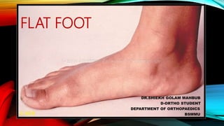
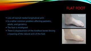
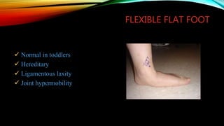
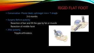
FLAT FOOT
DR.SHIEKH GOLAM MAHBUB
D-ORTHO STUDENT
DEPARTMENT OF ORTHOPAEDICS
BSMMU
WHAT IS FLAT FOOT ?
Also known as Pes planus or Fallen arches.
Medial border of the foot is abnormally in contact
with the floor during weight bearing.
Low or absent medial longitudinal arch.
When associated with deformities of the hind, mid
and fore foot – pes plano valgus
DIVISION OF FOOT
Forefoot
Midfoot
Rearfoot/Hindfoot
Forefoot
5 MT’s
– Proximally 1-3 articulate with cuneiforms
– Proximally 4-5 articulate with cuboid
– Bases articulate with:
Phalanges
Midfoot
– Navicular
– 3 Cuneiforms
– Cuboid
Hindfoot (Rearfoot)
• Subtalor Joint
– Talus and calcaneus articulation
– Individual Bone Formation
• Calcaneus
– Calcaneal Tuberosity
– Sustentaculum Tali
• Inferior Talus
– Three facets
– Five functional articulation
ARCHES OF THE FOOT
Medial Longitudinal Arch
Lateral Longitudinal Arch
Transverse Arch
Medial Longitudinal Arch
Calcaneus
Talus
Navicular
1-3 cuneiforms
1-3 MT’s
• Ligament Support
Plantar Calcaneonavicular
Long Plantar Ligament
Deltoid
Plantar fascia
• Muscular Support
Intrinsic
Abductor Hallucis
Flexor Digitorum Brevis
Extrinsic
Tibialis Posterior
Flexor Hallucis Longus
Flexor Digitorum Longus
Tibialis Anterior
Flexor Digitorm Longus
Lateral longitudinal Arch
• Composed of
Calcaneus
Cuboid
4-5th MT’s
• Ligament Support
Long & Short Plantar
Plantar Fascia
Transverse Arch
• Formed By:
Ligament Support
Intermetatarsal Ligaments
Plantar Fascia
Muscle Support
All intrinsic muscles
Extrinisic
Tibialis Posterior
Tibialis Anterior
Peroneus Longus
FLAT FOOT
 Loss of normal medial longitudinal arch.
 Is a rather common problem affecting pediatric,
adults, and geriatrics.
 The foot is misaligned
 There is displacement of the hindfoot bones forcing
a lowering of the natural arch of the foot.
Normal
ASSOCIATED ABNORMALITY
 Heel valgus
 Mild subluxation of subtalar joint
 Eversion of calcaneus
 Lateral angulation at the midtarsal joint
 Supination of forefoot
 Shortened Achilles tendon
TYPES
Congenital
Flexible flat foot
Rigid flat foot
Acquired
Osseus (# talus or calcaneous)
Ligamentous
Postural or static
Arthritic
FLEXIBLE FLAT FOOT
 There is an arch with no pressure on the foot.
 Upon standing there is a loss of the height of the arc.
 The foot can be put back into its “normal” position
during not weight bearing.
 Jack’s test: Restored arch by dorsiflexing great toe.
No Weight
Weight
FLEXIBLE FLAT FOOT
 Normal in toddlers
 Hereditary
 Ligamentous laxity
 Joint hypermobility
RIGID/STIFF FLAT FOOT
 The foot has no arch on or off the ground.
 Cannot be manually forced back into it’s normal
position.
 Causes-
Congenital vertical talus
Tarsal coalition
Inflammatory joint disorder
Neurological disorder
CLINICAL ASSESMENT
• History-Neonatal problems, family history
• Heel position
• Tiptoe test
• Gait
• Jack’s test
• Sign of arthritis
• Spine,hip & knee
RADIOLOGICAL FEATURES
X-ray: AP, Lateral & oblique-
Medial displacement of talus
Beaking of head of talus
Narrowing of TC joint
CN bars-C sign
Flattening of arc
Meary’s angle - between long axis of talus and long axis
of first metatarsal on a standing lateral Xray
0 degrees – normal
0 – 15 degrees – mild
15 – 30 degrees – moderate
> 30 degrees – severe
Calcaneal pitch - angle between the plantar surface of the calcaneum and horizontal on a
lateral x-ray
Normal 15 degrees , in flat foot is decreased
May be 0 or negative in case of tightened TA
The talocalcaneal angle on an AP view is a
marker of hind foot valgus
Talus much more vertical than normal
WHAT IS HAPPENING?
The talus (ankle bone) is displaced from its
normal position on the hindfoot or tarsal bones.
It falls off its normal alignment with the hind
foot bones.
The talus turns inward and
the foot turns outward.
Normal Alignment
• Talus is sitting on
top of the hind
foot bones.
• Sinus tarsi (natural
spaced between
the ankle & heel
bone ) is in an
“open” position.
Abnormal Alignment
• Talus is not sitting
where it is supposed to
be on the heel bone.
• This partially collapses
the sinus tarsi.
• Partial talotarsal joint
dislocation is present.
• Excessively abnormal
forces are acting on the
foot.
Calcaneus
Calcaneus
TREATMENT
It depends on the type of “flat” foot
Flexible
Rigid
TREATMENT PLAN
Upto 2 years- no treatment
2 to 3 years-
Orthopaedic shoes with Thomas heels, medial heel wedges (1/8 to 3/16 inch), and
navicular pads
3 to 9 years-
Asymtomatic –Parent education
Symtomtic- Orthopaedics shoes, Custom prosthesis
10 to 14 years-
Asymtomatic –No treatment
Symtomtic- molded orthroses
FLEXIBLE FLAT FOOT
Exercise- strengthen muscles
“Special” Shoes
Arch Supports/Orthotics-
Extra-Osseous TaloTarsal
Stabilization (EOTTS)
Reconstructive Hindfoot
Surgery
SURGICAL OPTIONS
• Miller’s procedure
• Modified Hoke Miller procedure
• Durham pes planus plasty
• Tripple arthrodesis
• Post. displacement osteotomy of calcaneum
• Anterior calcaneal lengthening-distraction wedge osteotomy
• Kidner’s operation(Accessory navicular)
DURHAM
PLASTY FOR
PES PLANUS
A, Incision.
B, Elevation of
posterior tibial tendon.
C, Elevation of
osteoperiosteal flap
from proximal to distal.
D, Arthrodesis of
navicular–first
cuneiform joint.
E, Extent of
arthrodesis resection
through midfoot.
F, Internal fixation of
navicular–first
cuneiform joint.
•
pull the posterior tibial
tendon taut into its
prepared bed on the
plantar surface of the
waist of the navicular,
and tie the suture
dorsally
RIGID FLAT FOOT
• Conservative- Plaster 6wks; splintage( iron+ T strap)-
3-6 months
• Surgery-Before puberty
Resection of bar and fill the gap by fat or muscle
Resection of middle facet
• After puberty
Tripple arthrodesis
TRIPLE ARTHRODESIS
• A, Lateral skin incision over
sinus tarsi.
• B, Suggested plane of
arthrodesis of calcaneocuboid
and talonavicular joints.
• C, Suggested plane of removal
of talocalcaneal joint–posterior
facet.
• D, Medial skin incision.
• E, Medial aspect of
talonavicular joint and
suggested planes of osteotomy.
• F, Final position of
talonavicular, calcaneocuboid,
and talocalcaneal joints and
internal fixation with
Steinmann pins.
TAKE HOME MESSAGE
 Correction of flexible pes planus for disabling pain and after failure of conservative
management, not for cosmetic reasons only.
 Loss of inversion and eversion of the foot.
 Arthrodeses for relieving painful pes planus have been most successful.
 Sinus tarsi implants are at medical crossroads.
Surgeons are studying their results and modifying operative techniques and
implant design.
“Our feet are no more alike than our
faces” British Medical Journal
THANK YOU

Flat foot By Dr.Mahbub

  • 1.
    FLAT FOOT DR.SHIEKH GOLAMMAHBUB D-ORTHO STUDENT DEPARTMENT OF ORTHOPAEDICS BSMMU
  • 2.
    WHAT IS FLATFOOT ? Also known as Pes planus or Fallen arches. Medial border of the foot is abnormally in contact with the floor during weight bearing. Low or absent medial longitudinal arch. When associated with deformities of the hind, mid and fore foot – pes plano valgus
  • 3.
  • 4.
    Forefoot 5 MT’s – Proximally1-3 articulate with cuneiforms – Proximally 4-5 articulate with cuboid – Bases articulate with: Phalanges Midfoot – Navicular – 3 Cuneiforms – Cuboid
  • 5.
    Hindfoot (Rearfoot) • SubtalorJoint – Talus and calcaneus articulation – Individual Bone Formation • Calcaneus – Calcaneal Tuberosity – Sustentaculum Tali • Inferior Talus – Three facets – Five functional articulation
  • 6.
    ARCHES OF THEFOOT Medial Longitudinal Arch Lateral Longitudinal Arch Transverse Arch
  • 7.
    Medial Longitudinal Arch Calcaneus Talus Navicular 1-3cuneiforms 1-3 MT’s • Ligament Support Plantar Calcaneonavicular Long Plantar Ligament Deltoid Plantar fascia
  • 8.
    • Muscular Support Intrinsic AbductorHallucis Flexor Digitorum Brevis Extrinsic Tibialis Posterior Flexor Hallucis Longus Flexor Digitorum Longus Tibialis Anterior Flexor Digitorm Longus
  • 9.
    Lateral longitudinal Arch •Composed of Calcaneus Cuboid 4-5th MT’s • Ligament Support Long & Short Plantar Plantar Fascia
  • 10.
    Transverse Arch • FormedBy: Ligament Support Intermetatarsal Ligaments Plantar Fascia Muscle Support All intrinsic muscles Extrinisic Tibialis Posterior Tibialis Anterior Peroneus Longus
  • 11.
    FLAT FOOT  Lossof normal medial longitudinal arch.  Is a rather common problem affecting pediatric, adults, and geriatrics.  The foot is misaligned  There is displacement of the hindfoot bones forcing a lowering of the natural arch of the foot. Normal
  • 12.
    ASSOCIATED ABNORMALITY  Heelvalgus  Mild subluxation of subtalar joint  Eversion of calcaneus  Lateral angulation at the midtarsal joint  Supination of forefoot  Shortened Achilles tendon
  • 13.
    TYPES Congenital Flexible flat foot Rigidflat foot Acquired Osseus (# talus or calcaneous) Ligamentous Postural or static Arthritic
  • 14.
    FLEXIBLE FLAT FOOT There is an arch with no pressure on the foot.  Upon standing there is a loss of the height of the arc.  The foot can be put back into its “normal” position during not weight bearing.  Jack’s test: Restored arch by dorsiflexing great toe. No Weight Weight
  • 15.
    FLEXIBLE FLAT FOOT Normal in toddlers  Hereditary  Ligamentous laxity  Joint hypermobility
  • 16.
    RIGID/STIFF FLAT FOOT The foot has no arch on or off the ground.  Cannot be manually forced back into it’s normal position.  Causes- Congenital vertical talus Tarsal coalition Inflammatory joint disorder Neurological disorder
  • 17.
    CLINICAL ASSESMENT • History-Neonatalproblems, family history • Heel position • Tiptoe test • Gait • Jack’s test • Sign of arthritis • Spine,hip & knee
  • 18.
    RADIOLOGICAL FEATURES X-ray: AP,Lateral & oblique- Medial displacement of talus Beaking of head of talus Narrowing of TC joint CN bars-C sign Flattening of arc Meary’s angle - between long axis of talus and long axis of first metatarsal on a standing lateral Xray 0 degrees – normal 0 – 15 degrees – mild 15 – 30 degrees – moderate > 30 degrees – severe
  • 19.
    Calcaneal pitch -angle between the plantar surface of the calcaneum and horizontal on a lateral x-ray Normal 15 degrees , in flat foot is decreased May be 0 or negative in case of tightened TA The talocalcaneal angle on an AP view is a marker of hind foot valgus Talus much more vertical than normal
  • 20.
    WHAT IS HAPPENING? Thetalus (ankle bone) is displaced from its normal position on the hindfoot or tarsal bones. It falls off its normal alignment with the hind foot bones. The talus turns inward and the foot turns outward.
  • 21.
    Normal Alignment • Talusis sitting on top of the hind foot bones. • Sinus tarsi (natural spaced between the ankle & heel bone ) is in an “open” position. Abnormal Alignment • Talus is not sitting where it is supposed to be on the heel bone. • This partially collapses the sinus tarsi. • Partial talotarsal joint dislocation is present. • Excessively abnormal forces are acting on the foot. Calcaneus Calcaneus
  • 22.
    TREATMENT It depends onthe type of “flat” foot Flexible Rigid
  • 23.
    TREATMENT PLAN Upto 2years- no treatment 2 to 3 years- Orthopaedic shoes with Thomas heels, medial heel wedges (1/8 to 3/16 inch), and navicular pads 3 to 9 years- Asymtomatic –Parent education Symtomtic- Orthopaedics shoes, Custom prosthesis 10 to 14 years- Asymtomatic –No treatment Symtomtic- molded orthroses
  • 24.
    FLEXIBLE FLAT FOOT Exercise-strengthen muscles “Special” Shoes Arch Supports/Orthotics- Extra-Osseous TaloTarsal Stabilization (EOTTS) Reconstructive Hindfoot Surgery
  • 25.
    SURGICAL OPTIONS • Miller’sprocedure • Modified Hoke Miller procedure • Durham pes planus plasty • Tripple arthrodesis • Post. displacement osteotomy of calcaneum • Anterior calcaneal lengthening-distraction wedge osteotomy • Kidner’s operation(Accessory navicular)
  • 26.
    DURHAM PLASTY FOR PES PLANUS A,Incision. B, Elevation of posterior tibial tendon. C, Elevation of osteoperiosteal flap from proximal to distal. D, Arthrodesis of navicular–first cuneiform joint. E, Extent of arthrodesis resection through midfoot. F, Internal fixation of navicular–first cuneiform joint.
  • 27.
    • pull the posteriortibial tendon taut into its prepared bed on the plantar surface of the waist of the navicular, and tie the suture dorsally
  • 28.
    RIGID FLAT FOOT •Conservative- Plaster 6wks; splintage( iron+ T strap)- 3-6 months • Surgery-Before puberty Resection of bar and fill the gap by fat or muscle Resection of middle facet • After puberty Tripple arthrodesis
  • 29.
    TRIPLE ARTHRODESIS • A,Lateral skin incision over sinus tarsi. • B, Suggested plane of arthrodesis of calcaneocuboid and talonavicular joints. • C, Suggested plane of removal of talocalcaneal joint–posterior facet. • D, Medial skin incision. • E, Medial aspect of talonavicular joint and suggested planes of osteotomy. • F, Final position of talonavicular, calcaneocuboid, and talocalcaneal joints and internal fixation with Steinmann pins.
  • 30.
    TAKE HOME MESSAGE Correction of flexible pes planus for disabling pain and after failure of conservative management, not for cosmetic reasons only.  Loss of inversion and eversion of the foot.  Arthrodeses for relieving painful pes planus have been most successful.  Sinus tarsi implants are at medical crossroads. Surgeons are studying their results and modifying operative techniques and implant design.
  • 31.
    “Our feet areno more alike than our faces” British Medical Journal THANK YOU