PERITONITIS
Mrs. D. Melba Sahaya Sweety RN,RM
PhD Nursing , MSc (Pediatric Nursing), B.Sc Nursing
Associate Professor
Enam Nursing College,
Savar, Bangladesh.
1
INTRODUCTION
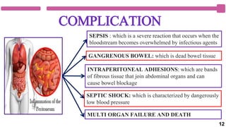
Peritonitis is an inflammation of the
peritoneum, the tissue that lines the
inner wall of the abdomen and covers
and supports most of your abdominal
organs. Peritonitis is usually caused by
infection from bacteria or fungi.
Left untreated, peritonitis can rapidly
spread into the blood (sepsis) and to
other organs, resulting in multiple organ
failure and death.
2
01
DEFINITION
Peritonitis is inflammation of the peritoneum
which is the serous membrane lining the
abdominal cavity and covering the viscera
usually it occur due to Bacterial infection.
- Brunner and Suddarth
3
TYPES OF PERITONITIS
Primary Peritonitis also called spontaneous
bacterial peritonitis (SBP), occurs as a spontaneous
bacterial infection of ascitic fluid. This occurs most
commonly in adult patients with liver failure
Secondary Peritonitis, occurs secondary to perforation
of abdominal organs with spillage that infects the
serous peritoneum.
Tertiary Peritonitis occurs as a result of
suprainfection in a patient who is immuno
compromised. Tuberculosis peritonitis in a patient with
AIDS is an example of tertiary peritonitis. 4
ETIOLOGY
PRIMARY
PERITONITIS
BACTERIAL INFECTION: Escherichia
Coli, Klebsiella, Proteus, Pseudomonas,
Streptococcus species, Myobacterial
infection and FUNGAL INFECTION
SECONDARY
PERITONITIS
Perforated ulcer – a severe, untreated ulcer
can sometimes burn through the wall of the
stomach or duodenum, allowing digestive
juices and food to leak into the abdominal
cavity.
Abdominal surgery – infection is a risk of
any type of major surgery
5
ETIOLOGY
SECONDARY
PERITONITIS
Perforated bowel – the intestines can be damaged
and perforated by a range of conditions, including
diverticulitis and inflammatory diseases such as
Crohn’s disease.
Burst appendix – the appendix is a thin tail
growing out of the large intestine.
Perforated gall bladder –A severe infection
(cholecystitis) can cause the gall bladder to burst.
Pancreatitis – an inflamed pancreas can directly
cause inflammation in the abdomen.
Ectopic pregnancy – the fertilised egg and grows
inside the slim fallopian tube instead of the uterus.
The tube ruptures in around one out of five cases.
6
ETIOLOGY
SECONDARY
PERITONITIS
Necrotising enterocolitis – a condition that
sometimes prompts peritonitis.
Blood infection – can be caused by a range of
conditions, including cirrhosis of the liver, some
forms of kidney disease and appendicitis.
Dialysis – bacteria on peritoneal dialysis
equipment can enter the abdominal cavity.
Stab wound – bacteria from a knife or other sharp
object enters the abdominal cavity.
TERTIARY
PERITONITIS
HIV, Tuberculosis peritonitis
7
PATHOPHYSIOLOGY
Due to Etiological factors
Leakage of contents from abdominal organs in to the abdominal cavity
Bacterial proliferation ,Edema of the tissues
Fluid in the peritoneal cavity becomes turbid with increasing amounts of
protein, WBC, cellular debris and blood.
exudation of fluid develops in
a short time
The immediate response of the intestinal tract is hypermotality. Soon
followed by paralytic illus with an accumulation of air and fluid in the bowel.
8
2
3 4
5
1
CLINICAL MANIFESTATION
Severe and constant
abdominal pain
Fever, chills
Nausea and
Vomiting,
Loss of
appetite Constipation
or Inability to
pass gas or
stool
Low blood
pressure
Thirst, Low
urine output
Fatigue ,
confusion
Diarrhea
Abdominal
Bloating or
distension
Shock
9
DIAGNOSTIC EVALUATION
History Collection :about the nature of pain , past
surgical history, and medical history
Physical Examination : the abdomen is hard and
painful. There are no bowel movements or sounds.
Blood tests. Complete blood count (CBC), can measure
WBC and RBC count. A high WBC count usually signals
inflammation or infection. A low RBC count may
indicate intra-abdominal bleeding. A blood culture can
help to identify the bacteria causing the infection or
inflammation.
10
DIAGNOSTIC EVALUATION
Abdominal X- ray :Show air and fluid levels as
well as distended bowel loop,
Abdominal Ultrasound : May reveal abscesses.
CT and MRI. May used for diagnosis of intra-
abdominal abscesses
Peritoneal Aspiration and culture and sensitivity
studies. May reveal infection and identify the
causative organisms.
11
COMPLICATION
SEPSIS : which is a severe reaction that occurs when the
bloodstream becomes overwhelmed by infectious agents
SEPTIC SHOCK: which is characterized by dangerously
low blood pressure
INTRAPERITONEAL ADHESIONS: which are bands
of fibrous tissue that join abdominal organs and can
cause bowel blockage
GANGRENOUS BOWEL: which is dead bowel tissue
MULTI ORGAN FAILURE AND DEATH
12
MANAGEMENT
MEDICAL MANAGEMENT
•Fluid colloid and electrolyte replacement is the major focus
of medical management
•Administration of several liters of an isotonic solution as
prescribed ( hypovolemia occurs because massive amount of
fluid and electrolyte move from the intestinal lumen in to the
peritoneal cavity )
•Oxygen therapy by nasal cannula or mask generally
promotes adequate oxygen
•Intestinal intubation and suction assist in relieving
abdominal distention and in promoting intestinal function.
DRUG THERAPY
•Analgesic
medication to
relieve pain
•Antiemetic for
nausea and vomiting
•Antibiotic therapy
– broad spectrum
antibiotic are given
IV until the specific
organism causing
the infection
13
MANAGEMENT
SURGICAL MANAGEMENT
Repair the ruptured organ and wash out the abdominal
cavity of blood and pus.
Ultrasound and CT guided peritoneal drainage of
abdominal and extra peritoneal abscesses has allowed for
avoidance or delay of surgical therapy until the acute septic
process has subsided.
NURSING MANAGEMENT
Blood pressure monitoring :- by arterial line if shock is present
Input and output monitoring ;- accurate according to all intake and output could help in the
assessment of fluid replacement.
 I/v fluid ;- administers and closely monitors I/v fluids .
Drainage monitoring ;- monitor and record the character of the drainage post operatively.
GI function should be monitored to assess response to interventions
14
NURSING DIAGNOSIS
Acute pain related to peritoneal irritation as manifested by
report of pain.
Deficient Fluid Volume related to Fluid shifts from
extracellular, intravascular, and interstitial compartments into
intestines or peritoneal space as manifested by decreased urine
output.
Risk for shock related to septicemia or hypovolemia possibly
evidenced by low Blood pressure.
Anxiety related to diagnostic test and therapeutic procedures as
evidenced by facial expression
15
16

Peritonitis.pptx

  • 1.
    PERITONITIS Mrs. D. MelbaSahaya Sweety RN,RM PhD Nursing , MSc (Pediatric Nursing), B.Sc Nursing Associate Professor Enam Nursing College, Savar, Bangladesh. 1
  • 2.
    INTRODUCTION Peritonitis is aninflammation of the peritoneum, the tissue that lines the inner wall of the abdomen and covers and supports most of your abdominal organs. Peritonitis is usually caused by infection from bacteria or fungi. Left untreated, peritonitis can rapidly spread into the blood (sepsis) and to other organs, resulting in multiple organ failure and death. 2
  • 3.
    01 DEFINITION Peritonitis is inflammationof the peritoneum which is the serous membrane lining the abdominal cavity and covering the viscera usually it occur due to Bacterial infection. - Brunner and Suddarth 3
  • 4.
    TYPES OF PERITONITIS PrimaryPeritonitis also called spontaneous bacterial peritonitis (SBP), occurs as a spontaneous bacterial infection of ascitic fluid. This occurs most commonly in adult patients with liver failure Secondary Peritonitis, occurs secondary to perforation of abdominal organs with spillage that infects the serous peritoneum. Tertiary Peritonitis occurs as a result of suprainfection in a patient who is immuno compromised. Tuberculosis peritonitis in a patient with AIDS is an example of tertiary peritonitis. 4
  • 5.
    ETIOLOGY PRIMARY PERITONITIS BACTERIAL INFECTION: Escherichia Coli,Klebsiella, Proteus, Pseudomonas, Streptococcus species, Myobacterial infection and FUNGAL INFECTION SECONDARY PERITONITIS Perforated ulcer – a severe, untreated ulcer can sometimes burn through the wall of the stomach or duodenum, allowing digestive juices and food to leak into the abdominal cavity. Abdominal surgery – infection is a risk of any type of major surgery 5
  • 6.
    ETIOLOGY SECONDARY PERITONITIS Perforated bowel –the intestines can be damaged and perforated by a range of conditions, including diverticulitis and inflammatory diseases such as Crohn’s disease. Burst appendix – the appendix is a thin tail growing out of the large intestine. Perforated gall bladder –A severe infection (cholecystitis) can cause the gall bladder to burst. Pancreatitis – an inflamed pancreas can directly cause inflammation in the abdomen. Ectopic pregnancy – the fertilised egg and grows inside the slim fallopian tube instead of the uterus. The tube ruptures in around one out of five cases. 6
  • 7.
    ETIOLOGY SECONDARY PERITONITIS Necrotising enterocolitis –a condition that sometimes prompts peritonitis. Blood infection – can be caused by a range of conditions, including cirrhosis of the liver, some forms of kidney disease and appendicitis. Dialysis – bacteria on peritoneal dialysis equipment can enter the abdominal cavity. Stab wound – bacteria from a knife or other sharp object enters the abdominal cavity. TERTIARY PERITONITIS HIV, Tuberculosis peritonitis 7
  • 8.
    PATHOPHYSIOLOGY Due to Etiologicalfactors Leakage of contents from abdominal organs in to the abdominal cavity Bacterial proliferation ,Edema of the tissues Fluid in the peritoneal cavity becomes turbid with increasing amounts of protein, WBC, cellular debris and blood. exudation of fluid develops in a short time The immediate response of the intestinal tract is hypermotality. Soon followed by paralytic illus with an accumulation of air and fluid in the bowel. 8
  • 9.
    2 3 4 5 1 CLINICAL MANIFESTATION Severeand constant abdominal pain Fever, chills Nausea and Vomiting, Loss of appetite Constipation or Inability to pass gas or stool Low blood pressure Thirst, Low urine output Fatigue , confusion Diarrhea Abdominal Bloating or distension Shock 9
  • 10.
    DIAGNOSTIC EVALUATION History Collection:about the nature of pain , past surgical history, and medical history Physical Examination : the abdomen is hard and painful. There are no bowel movements or sounds. Blood tests. Complete blood count (CBC), can measure WBC and RBC count. A high WBC count usually signals inflammation or infection. A low RBC count may indicate intra-abdominal bleeding. A blood culture can help to identify the bacteria causing the infection or inflammation. 10
  • 11.
    DIAGNOSTIC EVALUATION Abdominal X-ray :Show air and fluid levels as well as distended bowel loop, Abdominal Ultrasound : May reveal abscesses. CT and MRI. May used for diagnosis of intra- abdominal abscesses Peritoneal Aspiration and culture and sensitivity studies. May reveal infection and identify the causative organisms. 11
  • 12.
    COMPLICATION SEPSIS : whichis a severe reaction that occurs when the bloodstream becomes overwhelmed by infectious agents SEPTIC SHOCK: which is characterized by dangerously low blood pressure INTRAPERITONEAL ADHESIONS: which are bands of fibrous tissue that join abdominal organs and can cause bowel blockage GANGRENOUS BOWEL: which is dead bowel tissue MULTI ORGAN FAILURE AND DEATH 12
  • 13.
    MANAGEMENT MEDICAL MANAGEMENT •Fluid colloidand electrolyte replacement is the major focus of medical management •Administration of several liters of an isotonic solution as prescribed ( hypovolemia occurs because massive amount of fluid and electrolyte move from the intestinal lumen in to the peritoneal cavity ) •Oxygen therapy by nasal cannula or mask generally promotes adequate oxygen •Intestinal intubation and suction assist in relieving abdominal distention and in promoting intestinal function. DRUG THERAPY •Analgesic medication to relieve pain •Antiemetic for nausea and vomiting •Antibiotic therapy – broad spectrum antibiotic are given IV until the specific organism causing the infection 13
  • 14.
    MANAGEMENT SURGICAL MANAGEMENT Repair theruptured organ and wash out the abdominal cavity of blood and pus. Ultrasound and CT guided peritoneal drainage of abdominal and extra peritoneal abscesses has allowed for avoidance or delay of surgical therapy until the acute septic process has subsided. NURSING MANAGEMENT Blood pressure monitoring :- by arterial line if shock is present Input and output monitoring ;- accurate according to all intake and output could help in the assessment of fluid replacement.  I/v fluid ;- administers and closely monitors I/v fluids . Drainage monitoring ;- monitor and record the character of the drainage post operatively. GI function should be monitored to assess response to interventions 14
  • 15.
    NURSING DIAGNOSIS Acute painrelated to peritoneal irritation as manifested by report of pain. Deficient Fluid Volume related to Fluid shifts from extracellular, intravascular, and interstitial compartments into intestines or peritoneal space as manifested by decreased urine output. Risk for shock related to septicemia or hypovolemia possibly evidenced by low Blood pressure. Anxiety related to diagnostic test and therapeutic procedures as evidenced by facial expression 15
  • 16.