PERITONITIS
BY:_
REGIN GEORGE
Dept. of Radiography and
Imaging Technology
Definition
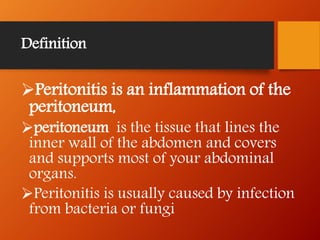
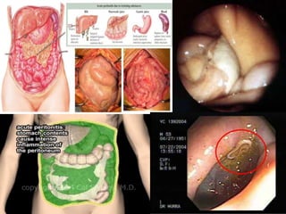
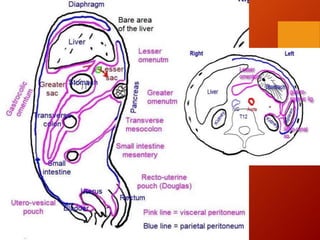
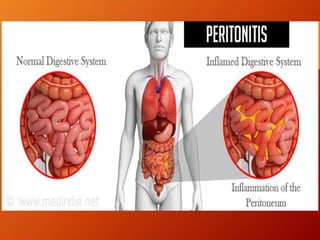
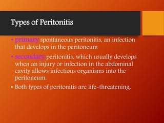
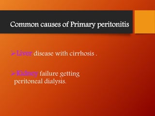
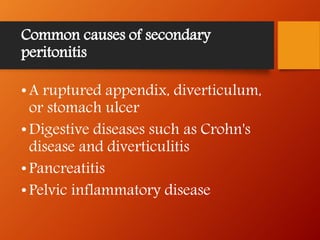
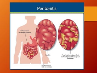
Peritonitis is an inflammation of the
peritoneum,
peritoneum is the tissue that lines the
inner wall of the abdomen and covers
and supports most of your abdominal
organs.
Peritonitis is usually caused by infection
from bacteria or fungi
Peritoneum is the largest serous
membrane of the body
Consist two part
1. parietal layer: part attaches to cavity
wall
2. Visceral layer: part that covers and
attaches to the organ inside this cavity
Each layer consist of areolar connective
tissue covered by mesothelium (simple
squamous epithelium)
Mesothelium secretes serous fluid
(watery lubricating fluid that allowed
organ to glide easily over one and other or
to slide again the wall of cavities)
Symptoms of Peritonitis
The first symptoms of peritonitis are
typically poor appetite and nausea and a
dull abdominal ache that quickly turns
into persistent, severe abdominal pain,
which is worsened by any movement.
Other signs and symptoms related to
peritonitis may includes:
Abdominal tenderness or distention
Chills
Fever
Fluid in the abdomen
Not passing any urine, or passing significantly
less urine than usual.
Difficulty passing gas or
having a bowel movement
Vomiting
Types of Peritonitis
• primary spontaneous peritonitis, an infection
that develops in the peritoneum
• secondary peritonitis, which usually develops
when an injury or infection in the abdominal
cavity allows infectious organisms into the
peritoneum.
• Both types of peritonitis are life-threatening.
Common causes of Primary peritonitis
Liver disease with cirrhosis .
Kidney failure getting
peritoneal dialysis.
Common causes of secondary
peritonitis
•A ruptured appendix, diverticulum,
or stomach ulcer
•Digestive diseases such as Crohn's
disease and diverticulitis
•Pancreatitis
•Pelvic inflammatory disease
•Perforations of the stomach,
intestine, gallbladder, or appendix
•Surgery
•Trauma to the abdomen, such as an
injury from a knife or gunshot wound
General Examination
CBC Count and Other Blood Studies
Urinalysis
Stool Sample
Peritoneal Fluid Analysis
Bedside Reagent Strips
Radiological Investigation
Radiography
Ultrasonography
CT scan
MRI
Ultrasonography
 Ultrasonography is a more sensitive
technique than clinical judgment in
diagnosing peritonitis.
 Ultrasonography may be a useful
diagnosing modality in patients with
peritonitis in whom the clinical cause is
unclear.
Contrast CT- Peritoneography
(traditional method/Old method)
 provides the highest resolution in the
delineation of anatomic details and the
demonstration of extra peritoneal fluid.
contrast CT peritoneography was also used to
evaluate the functional surface area of the
peritoneum with stereological methods
Drawback: CT peritoneography time-
consuming
MRI
MRI (it is comparitively better than CT)
MRI using contrast medium has been reported
to offer multiplanar imaging capabilities for the
evaluation of PD-related complications
When MRI is used, water is observed as
hyperintense with T2-weighted, turbo
spin-echo techniques. Therefore, the high
signal intensity of water or electrolyte
solutions should highlight normal and
pathologic anatomic features of the
peritoneal cavity.
Treatments and drugs
Antibiotic therapy
Surgical treatment
Blood transfusion/ Peritoneal
dialysis
Prevention
• Wash your hands, including underneath your
fingernails and between your fingers, before
touching the catheter.
• Clean the skin around the catheter with an
antiseptic every day.
• Store your supplies in a sanitary area.
• Wear a surgical mask during your dialysis
fluid exchanges.
• If you have pets, don't sleep with them.
• Talk with your dialysis care team about proper
care for your peritoneal dialysis catheter.
ThanK YoU

Peritonitis

  • 1.
    PERITONITIS BY:_ REGIN GEORGE Dept. ofRadiography and Imaging Technology
  • 2.
    Definition Peritonitis is aninflammation of the peritoneum, peritoneum is the tissue that lines the inner wall of the abdomen and covers and supports most of your abdominal organs. Peritonitis is usually caused by infection from bacteria or fungi
  • 4.
    Peritoneum is thelargest serous membrane of the body Consist two part 1. parietal layer: part attaches to cavity wall 2. Visceral layer: part that covers and attaches to the organ inside this cavity
  • 6.
    Each layer consistof areolar connective tissue covered by mesothelium (simple squamous epithelium) Mesothelium secretes serous fluid (watery lubricating fluid that allowed organ to glide easily over one and other or to slide again the wall of cavities)
  • 8.
    Symptoms of Peritonitis Thefirst symptoms of peritonitis are typically poor appetite and nausea and a dull abdominal ache that quickly turns into persistent, severe abdominal pain, which is worsened by any movement. Other signs and symptoms related to peritonitis may includes:
  • 9.
    Abdominal tenderness ordistention Chills Fever Fluid in the abdomen Not passing any urine, or passing significantly less urine than usual. Difficulty passing gas or having a bowel movement Vomiting
  • 10.
    Types of Peritonitis •primary spontaneous peritonitis, an infection that develops in the peritoneum • secondary peritonitis, which usually develops when an injury or infection in the abdominal cavity allows infectious organisms into the peritoneum. • Both types of peritonitis are life-threatening.
  • 11.
    Common causes ofPrimary peritonitis Liver disease with cirrhosis . Kidney failure getting peritoneal dialysis.
  • 12.
    Common causes ofsecondary peritonitis •A ruptured appendix, diverticulum, or stomach ulcer •Digestive diseases such as Crohn's disease and diverticulitis •Pancreatitis •Pelvic inflammatory disease
  • 13.
    •Perforations of thestomach, intestine, gallbladder, or appendix •Surgery •Trauma to the abdomen, such as an injury from a knife or gunshot wound
  • 14.
    General Examination CBC Countand Other Blood Studies Urinalysis Stool Sample Peritoneal Fluid Analysis Bedside Reagent Strips
  • 15.
  • 16.
    Ultrasonography  Ultrasonography isa more sensitive technique than clinical judgment in diagnosing peritonitis.  Ultrasonography may be a useful diagnosing modality in patients with peritonitis in whom the clinical cause is unclear.
  • 17.
    Contrast CT- Peritoneography (traditionalmethod/Old method)  provides the highest resolution in the delineation of anatomic details and the demonstration of extra peritoneal fluid. contrast CT peritoneography was also used to evaluate the functional surface area of the peritoneum with stereological methods Drawback: CT peritoneography time- consuming
  • 18.
    MRI MRI (it iscomparitively better than CT) MRI using contrast medium has been reported to offer multiplanar imaging capabilities for the evaluation of PD-related complications
  • 19.
    When MRI isused, water is observed as hyperintense with T2-weighted, turbo spin-echo techniques. Therefore, the high signal intensity of water or electrolyte solutions should highlight normal and pathologic anatomic features of the peritoneal cavity.
  • 20.
    Treatments and drugs Antibiotictherapy Surgical treatment Blood transfusion/ Peritoneal dialysis
  • 21.
    Prevention • Wash yourhands, including underneath your fingernails and between your fingers, before touching the catheter. • Clean the skin around the catheter with an antiseptic every day. • Store your supplies in a sanitary area.
  • 22.
    • Wear asurgical mask during your dialysis fluid exchanges. • If you have pets, don't sleep with them. • Talk with your dialysis care team about proper care for your peritoneal dialysis catheter.
  • 25.