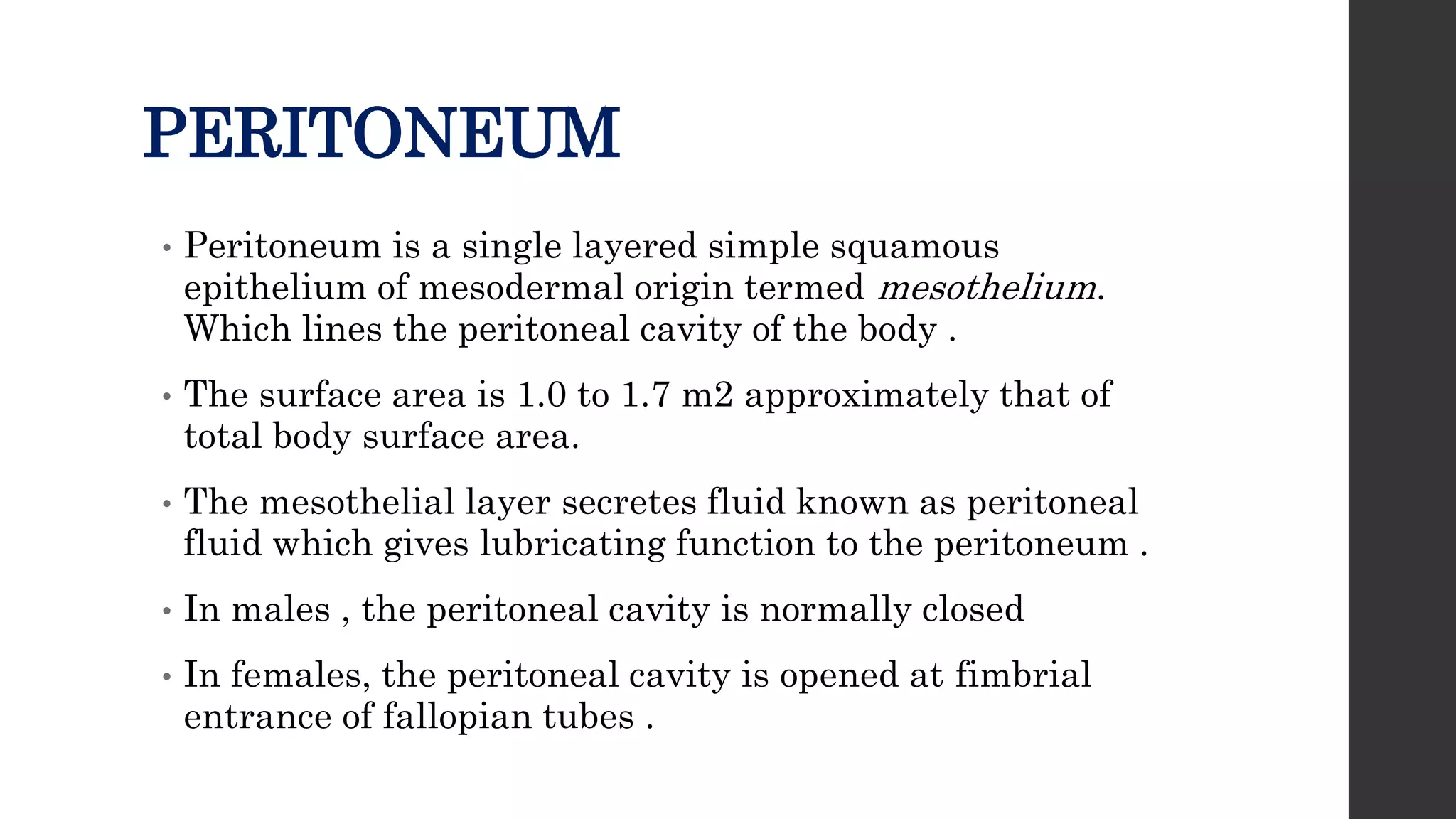
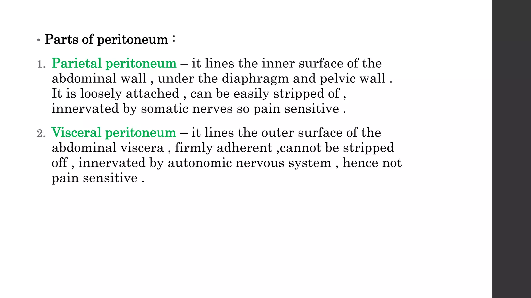
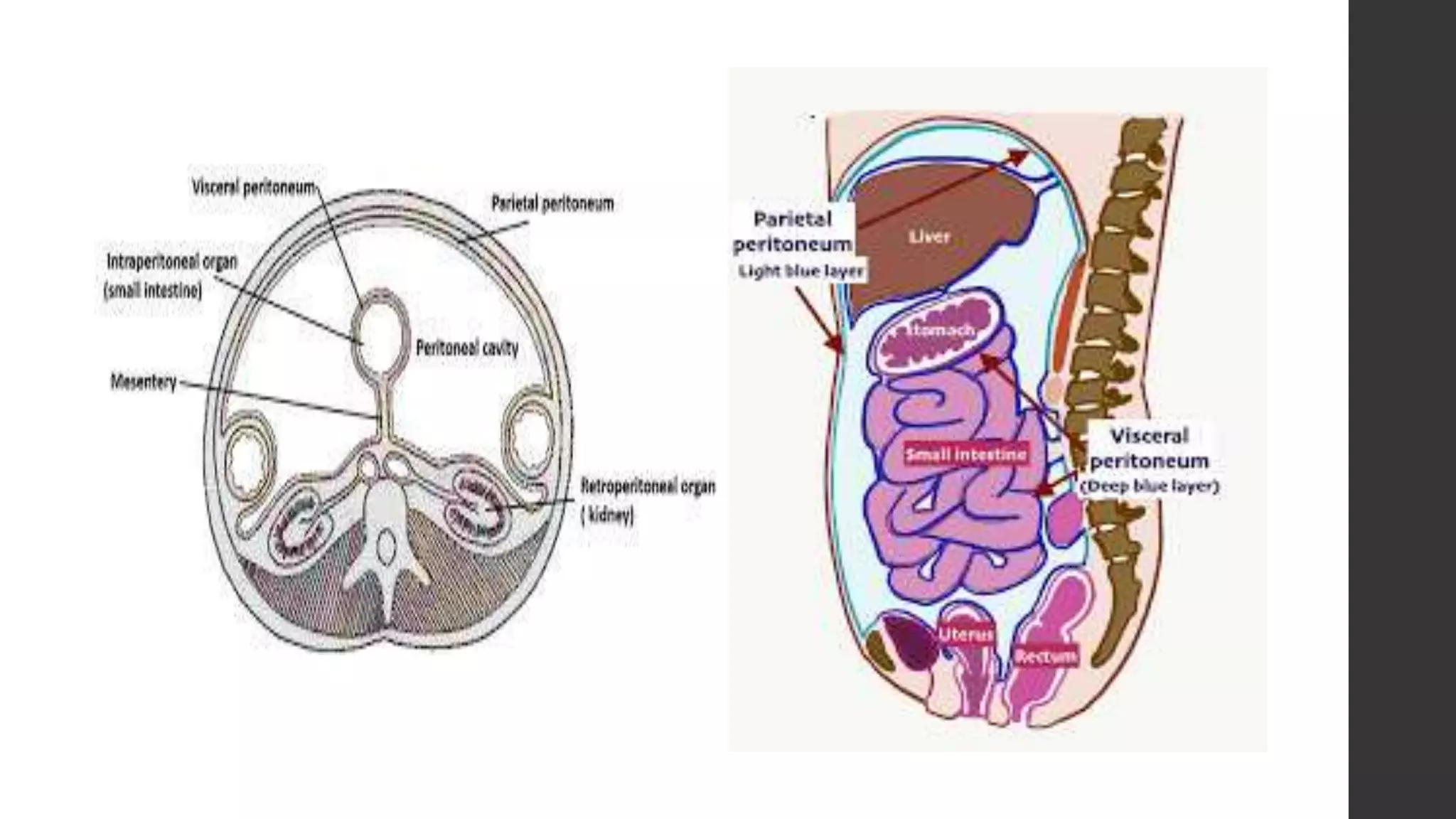
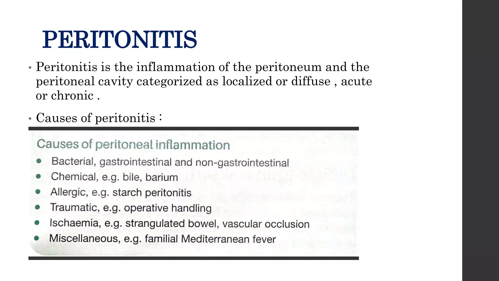
SlideShare a Scribd company logo
PERITONITIS AND
INTRA ABDOMINAL
ABSCESS
PRESENTER : DR B. SNEHA
JUNIOR RESIDENT
MOERATOR : DR SALINI RAO
ASSISTANT PROFESSOR
PERITONEUM
• Peritoneum is a single layered simple squamous
epithelium of mesodermal origin termed mesothelium.
Which lines the peritoneal cavity of the body .
• The surface area is 1.0 to 1.7 m2 approximately that of
total body surface area.
• The mesothelial layer secretes fluid known as peritoneal
fluid which gives lubricating function to the peritoneum .
• In males , the peritoneal cavity is normally closed
• In females, the peritoneal cavity is opened at fimbrial
entrance of fallopian tubes .
• Parts of peritoneum :
1. Parietal peritoneum – it lines the inner surface of the
abdominal wall , under the diaphragm and pelvic wall .
It is loosely attached , can be easily stripped of ,
innervated by somatic nerves so pain sensitive .
2. Visceral peritoneum – it lines the outer surface of the
abdominal viscera , firmly adherent ,cannot be stripped
off , innervated by autonomic nervous system , hence not
pain sensitive .
Peritoneal cavity
• It is the potential space between the parietal and visceral
peritoneum .
• Normally it contains 100ml or clear straw colored fluid
secreted by mesothelial cells .
• Spaces in the peritoneal cavity :
• peritoneal cavity being the largest cavity in the body it is
divided into different spaces by the ligaments and
mesenteries.
• They are 11 ligaments and mesenteries :
1. Falciform ligament
2. Coronary ligament
3. Gastrohepatic ligament
4. Hepatoduodenal ligament
5. Phrenicolic ligament
6. Splenorenal ligament
7. Gastrosplenic ligament
8. Duodenocolic ligament
9. Gastrocolic ligament
10. Transverse mesocolon
11. Small bowel mesentery
• Intra abdominal spaces are 9 in number they are :
1. Right and left subphrenic
2. Subhepatic
3. Lesser sac
4. Supra mesenteric
5. Infra mesenteric
6. Left and right paracolic gutters
7. Pelvis
• Functions of peritoneum
PERITONITIS
• Peritonitis is the inflammation of the peritoneum and the
peritoneal cavity categorized as localized or diffuse , acute
or chronic .
• Causes of peritonitis :
• Primary peritonitis – this occurs due to bacterial , chlamydial,
fungal or mycobacterial infection in the absence of perforation or
inflammation of GI or GU tract .
• Secondary peritonitis –this occurs in settings of GI or GU tract
perforation or inflammation with common causes including acute
appendicitis , colonic diverticulitis and pelvic inflammatory
disease.
• Tertiary peritonitis – it occurs after any abdominal surgeries,
which are usually severe and the patient may go into SIRS or
MODS early, it is defined as recurrent or persistent intra
abdominal infection after an adequate treatment for primary or
secondary peritonitis usually after 48 hrs.
• Paths to peritoneal infection :
1. Gastrointestinal perforation – perforated ulcer, appendix,
diverticulum.
2. Transmural translocation (no perforation) – pancreatitis ,
ischemic bowel , primary bacterial peritonitis .
3. Exogenous contamination – drains , open surgery , trauma ,
peritoneal dialysis.
4. Female genital tract infection- pelvic inflammatory disease
5. Hematogenous spread( rare) – septicemia
Localised peritonitis
• In this peritonitis localized area of peritoneum has become
inflamed .
• If parietal peritoneum is involved , the patient complains of pain
somatic pain in the area affected . Vital signs may be normal ,
but tachycardia and pyrexia are common .
• The characteristic signs are involuntary guarding ( reflex
abdominal wall contraction to reduce further peritoneal
irritation) and rebound tenderness .
• Collectively these signs and symptoms are called as peritonism
and the patient is described as peritonitic.
• If the inflammation occurs to the peritoneum under diaphragm, there
is shoulder tip pain ( phrenic pain ).
• If the inflammation occurs in pelvic peritoneum due to inflamed
appendix, salpingitis, abdominal signs may ne limited but there is
deep seated tenderness detected by doing digital rectal or vaginal
examination.
• Diagnosis can be done by doing CT , usg , lab biomarkers.
• Aim of the treatment is to remove the underlying cause.
• During surgery in localized peritonitis , there is inflamed peritoneum
which appears reddened, thickened and has velvety texture. Plaques
of yellow/white fibrin may also be found , which causes the looping of
intestines and mesentery to adhere to the intestines.
Diffuse (generalized) peritonitis
• It is a life threatening condition .
• In this not just focal regions , but the whole of the peritoneum of that
region is inflamed.
• It arises due to pressure related perforations of the viscus( obstructed
colon) , when large volumes of blood enter the peritoneal cavity ( ruptured
aortic aneurysm), or due to perforated duodenal ulcer , or anastomotic
leak.
• Patient describes acute or gradual onset pain , it may be localized initially
and then becomes diffuse.
• The patient is gravely ill looking ( Hippocratic facies) , and usually lies
still as possible to minimize the fluid movement within peritoneal cavity.
• The abdominal muscle undergoes reflex contraction and feels , board like
rigidity on palpation.
• Generalised ileus occurs and the abdomen becomes over
distended .
• Vitals are deranged , patient in advance cases is hypotensive,
tachycardia and pyrexia present
• At first patient may seem confused , drowsy but if cause not
treated patient will loose consciousness.
• Investigations and treatment must be done rapidly , as the
salvage time available is limited.
• An erect chest x ray is useful to identify subdiaphragmatic gas.
X-ray chest demonstrating air under diaphragm on
right side in intraperitoneal perforation
clinical features of peritonitis:
• Abdominal pain, worse on movement , coughing and deep
respiration.
• Constitutional upset, anorexia, malaise, fever, lassitude
• Gastrointestinal upset , nausea, vomiting
• Pyrexia may be present or absent
• Raised pulse rate
• Tenderness, guarding , rigidity
• Pain or tenderness in per rectal or per vaginal examination.
• Septic shock ,SIRS, MODS.
• MANAGEMENT OF PERITONITIS:
• General care of patient
• Correction of fluid and electrolyte balance
• Insertion of nasogastric tube and urinary catheter
• Broad spectrum antibiotic therapy
• Analgesia
• Vital support system
• Surgical treatment of cause :
• Source control by removal or exclusion of the cause.
• Peritoneal lavage, with drainage.
Acute bacterial peritonitis
• It most commonly arises due to perforation of viscus of
the alimentary tract.
• Other routes of infection can include the female genital
tract and exogenous contamination
• Some times may include spontaneous peritonitis with
streptococcal , pneumococcal or haemophilus influenza
Non gastrointestinal causes of acute
bacterial peritonitis
• Pelvic infection via the fallopian tubes is most commonly
responsible
• Most commonly affecting organism is chlamydia species
and gonococci.
• These organisms lead to thinning of cervical mucosa and
allow bacteria from vagina to pass into the uterus and
oviducts, causing infection and inflammation.
Biliary peritonitis
• It is most commonly seen after cholecystectomy and arises from
slippage of clip off from the cystic duct , drainage of bile from
accessory cystic duct or perforation of the common bile or hepatic
duct.
• It can also arise after hepatectomy or duodenal surgery, blunt or
penetrating hepatobiliary or duodenal trauma.
• In severe contamination the patient will be extremely unwell and
urgent intervention required.
• Localised collections are treated by percutaneous insertion of drain
followed by ERCP to identify the source of bile leak. ERCP enables
placement of stent across source of leak .
• If there is diffuse or high volume contamination, surgical exploration
with aim being lavage and drainage.
Spontaneous bacterial peritonitis
• It is some times called as primary bacterial peritonitis.
• It is an acute bacterial infection of ascitic fluid , it is rare except in
patient’s with cirrhosis.
• Clinical features as of peritonitis with worsening of liver , renal
function , hepatic encephalopathy and GI bleed.
• Diagnosis is made by paracentesis of ascitic fluid: in which neutrophil
count of ascitic fluid >250/mm. culture of ascitic fluid is negative . If
culture positive most common pathogens include gram negative
bacteria usually E.Coli, and gram positive cocci ( streptococci and
enterococci)
• Treatment of SBP should be initiated immediately after diagnosis, with
empirical treatment. Choice of antibiotic is third generation
cephalosporins, cefotaxime and alternatively quinolones such as
ciprofloxacin can be used.
Primary pneumococcal peritonitis
• The incidence is very low and now its rare
• It may complicate the nephrotic syndrome and cirrhosis in the
children , may also affect the normal healthy children .
• In girls the route of infection is via the vagina and fallopian
tubes , whereas blood borne secondary to respiratory or middle
ear disease is also possible.
• It is usually sudden onset with pain in the lower abdomen ,
raise of temperature with frequent vomitings.
• After 24 -48 hrs profuse diarrhea s the characteristic feature.,
associated with increased frequency of micturition.
• After starting antibiotics , we need to correct the
electrolyte imbalance, dehydration , early surgery is
required.
• Laparotomy or laparoscopy may be useful .
Tuberculous peritonitis
• Intraabdominal tuberculosis is more common in the resource
poor countries , however its incidence is increasing even in the
resource rich countries due to migration and
immunosuppression.
• Mycobacterium avium intracellulare is more prevalent worldwide
with widespread increase in HIV co infection. .
• TB can spread to the peritoneum through gastrointestinal tract
via the mesenteric lymph nodes or through the blood , from
pulmonary TB, and the fallopian tubes
• Clinical features:
• Acute and chronic forms
• Abdominal pain , sweats , malaise, and weight loss are
frequent
• Ascites common, may be loculated, abdominal distension.
• Caseating peritoneal nodules are seen , they distinguish
from metastatic carcinoma and fat necrosis of
pancreatitis.
• Diagnosis:
• Abdominal ultrasonography, CT abdomen
• Ascitic fluid is typically straw coloured exudate with protein >25-
30g/l, with white cells >500/ml, lymphocytes >40%
• Laparoscopy and peritoneal biopsy can be done.
• Gene Xpert MTB/RIF assay and interferon gamma release assay is
diagnostic.
• Management:
• Supportive (nutrition, and hydration)
• With systemic anti TB therapy
• Surgery may be required for specific complication such as intestinal
obstruction
Familial mediterranean fever
• Also known as familial paroxysmal polyserositis, autosomal
recessive inherited inherited autoinflammatory syndrome.
• Associated with mutations in MEFV gene found in Arab,
Armenian and Jewish people
• It is characterized by abdominal pain , tenderness mild
pyrexia.
• Duration of attack is 24 hrs with complete remission but
exacerbation in regular interval
• Peritoneum is inflamed in the splenic and gall bladder vicinity
, and the treatment is colchicine during attack.
INTRAABDOMINAL /INTRA
PERITONEAL ABSCESS
• Intraperitoneal abscess is the collection of pus in the
peritoneal cavity, it normally arises secondary to another
pathology.
• Inflammation of any viscus , if unresolved will lead to
hypersecretion of peritoneal fluid, which later progresses
to frank pus.
• Hence abscess formation commonly accompanies
inflammation of abdominal viscus and based on location
of organs.
• Site of abscess formation :
Appendicitis- pelvic abscess
Infections of female genital organs- pelvic abscess
Diverticulitis- left paracolic and pelvic abscess
Complications of gall bladder disease- sub hepatic and right sub
phrenic abscess
Pancreatitis may result in lesser sac abscess
Inadequately drained peritonitis , anastomotic leak and internal
fistula causes intra loop abscess.
• Clinical features of an abdominal / pelvic abscess:
• Symptoms:
Malaise , lethargy, failure to recover from surgery as expected
Anorexia and weight loss
Sweats, rigors
Abdominal / pelvic pain.
Symptoms from local irritation , shoulder tip / hiccoughs(
subphrenic), diarrhoea and mucus ( pelvic), nausea and vomiting (
any upper abdominal).
• Signs:
Increased temperature and pulse, swinging pyrexia
Localised abdominal tenderness, mass formation.
• Investigations :
Plain x-ray erect abdomen- elevation of hemidiaphragm , air
fluid levels , soft tissue masses, obliteration of psoas
shadow.
Ultrasound of abdomen
CT scan of abdomen and pelvis is the investigation of choice.
• Treatment
• Adequate fluid and electrolyte management ,resuscitation and
support
• Any abscess less than 5 cm in diameter , normally resolves
with intravenous antibiotics .
• As the antibiotics take effect , there is decrease in the fever
spikes , monitoring of CRP levels should be done serially to
see the disease progression .
• Any abscess greater than 5cm in diameter requires
percutaneous aspiration /drainage or surgical intervention
• Prerequisites of percutaneous drainage :
Anatomically safe route
Done in well defined unilocular abscess cavity
Needs surgical and radiological evaluation
Surgical backup if technical failure
• Complications with percutaneous drainage :
Enterocutaneous fistula formation,
Bacteraemia , sepsis
Vascular injury
Enteric puncture
• Surgical drainage :
Done if failure of percutaneous drainage
Diffuse infection
Content of the abscess is too thick
Access is impossible
• Surgical approach , it can done can be done by laparoscopically or
open approach
• Bowel may be matted and difficult to separate to reach the abscess.
all the regions of peritoneal cavity should be accessed for drainage of
any residual collections.
• Entire small intestines along with the mesentery should be exposed
to ensure that there are no residual interloop abscess.
THANK YOU

More Related Content

Similar to peritonitis and intra abdominal abscess.pptx

Small intestine/Intestinal obstruction/crohns disease/ileostomy/viscous organ...
Small intestine/Intestinal obstruction/crohns disease/ileostomy/viscous organ...Small intestine/Intestinal obstruction/crohns disease/ileostomy/viscous organ...
Small intestine/Intestinal obstruction/crohns disease/ileostomy/viscous organ...
RajeevPandit10
 
Diverticular disease of colon.pptx
Diverticular disease of colon.pptxDiverticular disease of colon.pptx
Diverticular disease of colon.pptx
Pradeep Pande
 
Esophageal injury
Esophageal injuryEsophageal injury
Esophageal injury
DENNIS MIRITI
 
Appendicitis
AppendicitisAppendicitis
Appendicitis
piyushparashar13
 
Hirschsprung disease
Hirschsprung diseaseHirschsprung disease
Hirschsprung disease
Kawther Alsadady
 
Intestinal obstruction.pptx
Intestinal obstruction.pptxIntestinal obstruction.pptx
Intestinal obstruction.pptx
MintesinotDesalegn
 
PERITONITIS.pptx
PERITONITIS.pptxPERITONITIS.pptx
PERITONITIS.pptx
Dr. Steven Mlosa
 
11 Peritoneum, Acute Abdomen, Intestinal Obstruction.pptx
11 Peritoneum, Acute Abdomen, Intestinal Obstruction.pptx11 Peritoneum, Acute Abdomen, Intestinal Obstruction.pptx
11 Peritoneum, Acute Abdomen, Intestinal Obstruction.pptx
Ivwananjisikombe1
 
Intra- abdominal infections MMED1 2022_Edited.ppt
Intra- abdominal infections MMED1 2022_Edited.pptIntra- abdominal infections MMED1 2022_Edited.ppt
Intra- abdominal infections MMED1 2022_Edited.ppt
Mkindi Mkindi
 
Appendix
AppendixAppendix
Appendix
KonarkThakkar
 
Rafdhi - Colon, Rectum, & Anus.pptx
Rafdhi - Colon, Rectum, & Anus.pptxRafdhi - Colon, Rectum, & Anus.pptx
Rafdhi - Colon, Rectum, & Anus.pptx
ariel740821
 
Peritonitis.ppt
Peritonitis.pptPeritonitis.ppt
Peritonitis.ppt
YohelioPriawanSibu
 
Abdominal tuberculosis
Abdominal tuberculosisAbdominal tuberculosis
abdominaltuberculosis-181221163344.asddfpdf
abdominaltuberculosis-181221163344.asddfpdfabdominaltuberculosis-181221163344.asddfpdf
abdominaltuberculosis-181221163344.asddfpdf
DrYaqoobBahar
 
intra- abdominal infections MMED1 2022.ppt
intra- abdominal infections  MMED1  2022.pptintra- abdominal infections  MMED1  2022.ppt
intra- abdominal infections MMED1 2022.ppt
Mkindi Mkindi
 
INTESTINAL OBSTRUCTION & PERITONITIS.pptx
INTESTINAL OBSTRUCTION & PERITONITIS.pptxINTESTINAL OBSTRUCTION & PERITONITIS.pptx
INTESTINAL OBSTRUCTION & PERITONITIS.pptx
Fridahchungu
 
Congenital Abnormalities.pptx
Congenital Abnormalities.pptxCongenital Abnormalities.pptx
Congenital Abnormalities.pptx
Johnmvula3
 
APPENDICITIS.pptx
APPENDICITIS.pptxAPPENDICITIS.pptx
APPENDICITIS.pptx
MuhammedBaldeh5
 
Liver abcess
Liver abcessLiver abcess
Liver abcess
syed ubaid
 
appendicitis-191217094731 (1).pdf
appendicitis-191217094731 (1).pdfappendicitis-191217094731 (1).pdf
appendicitis-191217094731 (1).pdf
ssuser07e0b1
 

Similar to peritonitis and intra abdominal abscess.pptx (20)

Small intestine/Intestinal obstruction/crohns disease/ileostomy/viscous organ...
Small intestine/Intestinal obstruction/crohns disease/ileostomy/viscous organ...Small intestine/Intestinal obstruction/crohns disease/ileostomy/viscous organ...
Small intestine/Intestinal obstruction/crohns disease/ileostomy/viscous organ...
 
Diverticular disease of colon.pptx
Diverticular disease of colon.pptxDiverticular disease of colon.pptx
Diverticular disease of colon.pptx
 
Esophageal injury
Esophageal injuryEsophageal injury
Esophageal injury
 
Appendicitis
AppendicitisAppendicitis
Appendicitis
 
Hirschsprung disease
Hirschsprung diseaseHirschsprung disease
Hirschsprung disease
 
Intestinal obstruction.pptx
Intestinal obstruction.pptxIntestinal obstruction.pptx
Intestinal obstruction.pptx
 
PERITONITIS.pptx
PERITONITIS.pptxPERITONITIS.pptx
PERITONITIS.pptx
 
11 Peritoneum, Acute Abdomen, Intestinal Obstruction.pptx
11 Peritoneum, Acute Abdomen, Intestinal Obstruction.pptx11 Peritoneum, Acute Abdomen, Intestinal Obstruction.pptx
11 Peritoneum, Acute Abdomen, Intestinal Obstruction.pptx
 
Intra- abdominal infections MMED1 2022_Edited.ppt
Intra- abdominal infections MMED1 2022_Edited.pptIntra- abdominal infections MMED1 2022_Edited.ppt
Intra- abdominal infections MMED1 2022_Edited.ppt
 
Appendix
AppendixAppendix
Appendix
 
Rafdhi - Colon, Rectum, & Anus.pptx
Rafdhi - Colon, Rectum, & Anus.pptxRafdhi - Colon, Rectum, & Anus.pptx
Rafdhi - Colon, Rectum, & Anus.pptx
 
Peritonitis.ppt
Peritonitis.pptPeritonitis.ppt
Peritonitis.ppt
 
Abdominal tuberculosis
Abdominal tuberculosisAbdominal tuberculosis
Abdominal tuberculosis
 
abdominaltuberculosis-181221163344.asddfpdf
abdominaltuberculosis-181221163344.asddfpdfabdominaltuberculosis-181221163344.asddfpdf
abdominaltuberculosis-181221163344.asddfpdf
 
intra- abdominal infections MMED1 2022.ppt
intra- abdominal infections  MMED1  2022.pptintra- abdominal infections  MMED1  2022.ppt
intra- abdominal infections MMED1 2022.ppt
 
INTESTINAL OBSTRUCTION & PERITONITIS.pptx
INTESTINAL OBSTRUCTION & PERITONITIS.pptxINTESTINAL OBSTRUCTION & PERITONITIS.pptx
INTESTINAL OBSTRUCTION & PERITONITIS.pptx
 
Congenital Abnormalities.pptx
Congenital Abnormalities.pptxCongenital Abnormalities.pptx
Congenital Abnormalities.pptx
 
APPENDICITIS.pptx
APPENDICITIS.pptxAPPENDICITIS.pptx
APPENDICITIS.pptx
 
Liver abcess
Liver abcessLiver abcess
Liver abcess
 
appendicitis-191217094731 (1).pdf
appendicitis-191217094731 (1).pdfappendicitis-191217094731 (1).pdf
appendicitis-191217094731 (1).pdf
 

More from madhurikakarnati

class acute abdomen other causes.pdf PPT.pdf
class acute abdomen other causes.pdf PPT.pdfclass acute abdomen other causes.pdf PPT.pdf
class acute abdomen other causes.pdf PPT.pdf
madhurikakarnati
 
Kavya_Award paper.pptx
Kavya_Award paper.pptxKavya_Award paper.pptx
Kavya_Award paper.pptx
madhurikakarnati
 
esophagus.pdf
esophagus.pdfesophagus.pdf
esophagus.pdf
madhurikakarnati
 
Neoplasms of oesophagus.pptx
Neoplasms of oesophagus.pptxNeoplasms of oesophagus.pptx
Neoplasms of oesophagus.pptx
madhurikakarnati
 
NEOPLASMS OF THYROID slide share.pptx
NEOPLASMS OF THYROID slide share.pptxNEOPLASMS OF THYROID slide share.pptx
NEOPLASMS OF THYROID slide share.pptx
madhurikakarnati
 
GINA GUIDELINES LONGTERM MANAGEMENT OF ASTHMA.pdf
GINA GUIDELINES LONGTERM MANAGEMENT OF ASTHMA.pdfGINA GUIDELINES LONGTERM MANAGEMENT OF ASTHMA.pdf
GINA GUIDELINES LONGTERM MANAGEMENT OF ASTHMA.pdf
madhurikakarnati
 
SURGICAL ANATOMY OF STOMACH AND DUODENUM dr. bruhath^final. .pdf
SURGICAL ANATOMY OF STOMACH AND DUODENUM dr. bruhath^final. .pdfSURGICAL ANATOMY OF STOMACH AND DUODENUM dr. bruhath^final. .pdf
SURGICAL ANATOMY OF STOMACH AND DUODENUM dr. bruhath^final. .pdf
madhurikakarnati
 

More from madhurikakarnati (7)

class acute abdomen other causes.pdf PPT.pdf
class acute abdomen other causes.pdf PPT.pdfclass acute abdomen other causes.pdf PPT.pdf
class acute abdomen other causes.pdf PPT.pdf
 
Kavya_Award paper.pptx
Kavya_Award paper.pptxKavya_Award paper.pptx
Kavya_Award paper.pptx
 
esophagus.pdf
esophagus.pdfesophagus.pdf
esophagus.pdf
 
Neoplasms of oesophagus.pptx
Neoplasms of oesophagus.pptxNeoplasms of oesophagus.pptx
Neoplasms of oesophagus.pptx
 
NEOPLASMS OF THYROID slide share.pptx
NEOPLASMS OF THYROID slide share.pptxNEOPLASMS OF THYROID slide share.pptx
NEOPLASMS OF THYROID slide share.pptx
 
GINA GUIDELINES LONGTERM MANAGEMENT OF ASTHMA.pdf
GINA GUIDELINES LONGTERM MANAGEMENT OF ASTHMA.pdfGINA GUIDELINES LONGTERM MANAGEMENT OF ASTHMA.pdf
GINA GUIDELINES LONGTERM MANAGEMENT OF ASTHMA.pdf
 
SURGICAL ANATOMY OF STOMACH AND DUODENUM dr. bruhath^final. .pdf
SURGICAL ANATOMY OF STOMACH AND DUODENUM dr. bruhath^final. .pdfSURGICAL ANATOMY OF STOMACH AND DUODENUM dr. bruhath^final. .pdf
SURGICAL ANATOMY OF STOMACH AND DUODENUM dr. bruhath^final. .pdf
 

Recently uploaded

Mule 4.6 & Java 17 Upgrade | MuleSoft Mysore Meetup #46
Mule 4.6 & Java 17 Upgrade | MuleSoft Mysore Meetup #46Mule 4.6 & Java 17 Upgrade | MuleSoft Mysore Meetup #46
Mule 4.6 & Java 17 Upgrade | MuleSoft Mysore Meetup #46
MysoreMuleSoftMeetup
 
PART A. Introduction to Costumer Service
PART A. Introduction to Costumer ServicePART A. Introduction to Costumer Service
PART A. Introduction to Costumer Service
PedroFerreira53928
 
Operation Blue Star - Saka Neela Tara
Operation Blue Star   -  Saka Neela TaraOperation Blue Star   -  Saka Neela Tara
Operation Blue Star - Saka Neela Tara
Balvir Singh
 
Unit 2- Research Aptitude (UGC NET Paper I).pdf
Unit 2- Research Aptitude (UGC NET Paper I).pdfUnit 2- Research Aptitude (UGC NET Paper I).pdf
Unit 2- Research Aptitude (UGC NET Paper I).pdf
Thiyagu K
 
The Art Pastor's Guide to Sabbath | Steve Thomason
The Art Pastor's Guide to Sabbath | Steve ThomasonThe Art Pastor's Guide to Sabbath | Steve Thomason
The Art Pastor's Guide to Sabbath | Steve Thomason
Steve Thomason
 
GIÁO ÁN DẠY THÊM (KẾ HOẠCH BÀI BUỔI 2) - TIẾNG ANH 8 GLOBAL SUCCESS (2 CỘT) N...
GIÁO ÁN DẠY THÊM (KẾ HOẠCH BÀI BUỔI 2) - TIẾNG ANH 8 GLOBAL SUCCESS (2 CỘT) N...GIÁO ÁN DẠY THÊM (KẾ HOẠCH BÀI BUỔI 2) - TIẾNG ANH 8 GLOBAL SUCCESS (2 CỘT) N...
GIÁO ÁN DẠY THÊM (KẾ HOẠCH BÀI BUỔI 2) - TIẾNG ANH 8 GLOBAL SUCCESS (2 CỘT) N...
Nguyen Thanh Tu Collection
 
Ethnobotany and Ethnopharmacology ......
Ethnobotany and Ethnopharmacology ......Ethnobotany and Ethnopharmacology ......
Ethnobotany and Ethnopharmacology ......
Ashokrao Mane college of Pharmacy Peth-Vadgaon
 
The Roman Empire A Historical Colossus.pdf
The Roman Empire A Historical Colossus.pdfThe Roman Empire A Historical Colossus.pdf
The Roman Empire A Historical Colossus.pdf
kaushalkr1407
 
The Challenger.pdf DNHS Official Publication
The Challenger.pdf DNHS Official PublicationThe Challenger.pdf DNHS Official Publication
The Challenger.pdf DNHS Official Publication
Delapenabediema
 
2024.06.01 Introducing a competency framework for languag learning materials ...
2024.06.01 Introducing a competency framework for languag learning materials ...2024.06.01 Introducing a competency framework for languag learning materials ...
2024.06.01 Introducing a competency framework for languag learning materials ...
Sandy Millin
 
special B.ed 2nd year old paper_20240531.pdf
special B.ed 2nd year old paper_20240531.pdfspecial B.ed 2nd year old paper_20240531.pdf
special B.ed 2nd year old paper_20240531.pdf
Special education needs
 
How to Break the cycle of negative Thoughts
How to Break the cycle of negative ThoughtsHow to Break the cycle of negative Thoughts
How to Break the cycle of negative Thoughts
Col Mukteshwar Prasad
 
Model Attribute Check Company Auto Property
Model Attribute  Check Company Auto PropertyModel Attribute  Check Company Auto Property
Model Attribute Check Company Auto Property
Celine George
 
Fish and Chips - have they had their chips
Fish and Chips - have they had their chipsFish and Chips - have they had their chips
Fish and Chips - have they had their chips
GeoBlogs
 
Additional Benefits for Employee Website.pdf
Additional Benefits for Employee Website.pdfAdditional Benefits for Employee Website.pdf
Additional Benefits for Employee Website.pdf
joachimlavalley1
 
Palestine last event orientationfvgnh .pptx
Palestine last event orientationfvgnh .pptxPalestine last event orientationfvgnh .pptx
Palestine last event orientationfvgnh .pptx
RaedMohamed3
 
Chapter 3 - Islamic Banking Products and Services.pptx
Chapter 3 - Islamic Banking Products and Services.pptxChapter 3 - Islamic Banking Products and Services.pptx
Chapter 3 - Islamic Banking Products and Services.pptx
Mohd Adib Abd Muin, Senior Lecturer at Universiti Utara Malaysia
 
Unit 8 - Information and Communication Technology (Paper I).pdf
Unit 8 - Information and Communication Technology (Paper I).pdfUnit 8 - Information and Communication Technology (Paper I).pdf
Unit 8 - Information and Communication Technology (Paper I).pdf
Thiyagu K
 
Synthetic Fiber Construction in lab .pptx
Synthetic Fiber Construction in lab .pptxSynthetic Fiber Construction in lab .pptx
Synthetic Fiber Construction in lab .pptx
Pavel ( NSTU)
 
Introduction to Quality Improvement Essentials
Introduction to Quality Improvement EssentialsIntroduction to Quality Improvement Essentials
Introduction to Quality Improvement Essentials
Excellence Foundation for South Sudan
 

Recently uploaded (20)

Mule 4.6 & Java 17 Upgrade | MuleSoft Mysore Meetup #46
Mule 4.6 & Java 17 Upgrade | MuleSoft Mysore Meetup #46Mule 4.6 & Java 17 Upgrade | MuleSoft Mysore Meetup #46
Mule 4.6 & Java 17 Upgrade | MuleSoft Mysore Meetup #46
 
PART A. Introduction to Costumer Service
PART A. Introduction to Costumer ServicePART A. Introduction to Costumer Service
PART A. Introduction to Costumer Service
 
Operation Blue Star - Saka Neela Tara
Operation Blue Star   -  Saka Neela TaraOperation Blue Star   -  Saka Neela Tara
Operation Blue Star - Saka Neela Tara
 
Unit 2- Research Aptitude (UGC NET Paper I).pdf
Unit 2- Research Aptitude (UGC NET Paper I).pdfUnit 2- Research Aptitude (UGC NET Paper I).pdf
Unit 2- Research Aptitude (UGC NET Paper I).pdf
 
The Art Pastor's Guide to Sabbath | Steve Thomason
The Art Pastor's Guide to Sabbath | Steve ThomasonThe Art Pastor's Guide to Sabbath | Steve Thomason
The Art Pastor's Guide to Sabbath | Steve Thomason
 
GIÁO ÁN DẠY THÊM (KẾ HOẠCH BÀI BUỔI 2) - TIẾNG ANH 8 GLOBAL SUCCESS (2 CỘT) N...
GIÁO ÁN DẠY THÊM (KẾ HOẠCH BÀI BUỔI 2) - TIẾNG ANH 8 GLOBAL SUCCESS (2 CỘT) N...GIÁO ÁN DẠY THÊM (KẾ HOẠCH BÀI BUỔI 2) - TIẾNG ANH 8 GLOBAL SUCCESS (2 CỘT) N...
GIÁO ÁN DẠY THÊM (KẾ HOẠCH BÀI BUỔI 2) - TIẾNG ANH 8 GLOBAL SUCCESS (2 CỘT) N...
 
Ethnobotany and Ethnopharmacology ......
Ethnobotany and Ethnopharmacology ......Ethnobotany and Ethnopharmacology ......
Ethnobotany and Ethnopharmacology ......
 
The Roman Empire A Historical Colossus.pdf
The Roman Empire A Historical Colossus.pdfThe Roman Empire A Historical Colossus.pdf
The Roman Empire A Historical Colossus.pdf
 
The Challenger.pdf DNHS Official Publication
The Challenger.pdf DNHS Official PublicationThe Challenger.pdf DNHS Official Publication
The Challenger.pdf DNHS Official Publication
 
2024.06.01 Introducing a competency framework for languag learning materials ...
2024.06.01 Introducing a competency framework for languag learning materials ...2024.06.01 Introducing a competency framework for languag learning materials ...
2024.06.01 Introducing a competency framework for languag learning materials ...
 
special B.ed 2nd year old paper_20240531.pdf
special B.ed 2nd year old paper_20240531.pdfspecial B.ed 2nd year old paper_20240531.pdf
special B.ed 2nd year old paper_20240531.pdf
 
How to Break the cycle of negative Thoughts
How to Break the cycle of negative ThoughtsHow to Break the cycle of negative Thoughts
How to Break the cycle of negative Thoughts
 
Model Attribute Check Company Auto Property
Model Attribute  Check Company Auto PropertyModel Attribute  Check Company Auto Property
Model Attribute Check Company Auto Property
 
Fish and Chips - have they had their chips
Fish and Chips - have they had their chipsFish and Chips - have they had their chips
Fish and Chips - have they had their chips
 
Additional Benefits for Employee Website.pdf
Additional Benefits for Employee Website.pdfAdditional Benefits for Employee Website.pdf
Additional Benefits for Employee Website.pdf
 
Palestine last event orientationfvgnh .pptx
Palestine last event orientationfvgnh .pptxPalestine last event orientationfvgnh .pptx
Palestine last event orientationfvgnh .pptx
 
Chapter 3 - Islamic Banking Products and Services.pptx
Chapter 3 - Islamic Banking Products and Services.pptxChapter 3 - Islamic Banking Products and Services.pptx
Chapter 3 - Islamic Banking Products and Services.pptx
 
Unit 8 - Information and Communication Technology (Paper I).pdf
Unit 8 - Information and Communication Technology (Paper I).pdfUnit 8 - Information and Communication Technology (Paper I).pdf
Unit 8 - Information and Communication Technology (Paper I).pdf
 
Synthetic Fiber Construction in lab .pptx
Synthetic Fiber Construction in lab .pptxSynthetic Fiber Construction in lab .pptx
Synthetic Fiber Construction in lab .pptx
 
Introduction to Quality Improvement Essentials
Introduction to Quality Improvement EssentialsIntroduction to Quality Improvement Essentials
Introduction to Quality Improvement Essentials
 

peritonitis and intra abdominal abscess.pptx

  • 1. PERITONITIS AND INTRA ABDOMINAL ABSCESS PRESENTER : DR B. SNEHA JUNIOR RESIDENT MOERATOR : DR SALINI RAO ASSISTANT PROFESSOR
  • 2. PERITONEUM • Peritoneum is a single layered simple squamous epithelium of mesodermal origin termed mesothelium. Which lines the peritoneal cavity of the body . • The surface area is 1.0 to 1.7 m2 approximately that of total body surface area. • The mesothelial layer secretes fluid known as peritoneal fluid which gives lubricating function to the peritoneum . • In males , the peritoneal cavity is normally closed • In females, the peritoneal cavity is opened at fimbrial entrance of fallopian tubes .
  • 3.
  • 4. • Parts of peritoneum : 1. Parietal peritoneum – it lines the inner surface of the abdominal wall , under the diaphragm and pelvic wall . It is loosely attached , can be easily stripped of , innervated by somatic nerves so pain sensitive . 2. Visceral peritoneum – it lines the outer surface of the abdominal viscera , firmly adherent ,cannot be stripped off , innervated by autonomic nervous system , hence not pain sensitive .
  • 5.
  • 6. Peritoneal cavity • It is the potential space between the parietal and visceral peritoneum . • Normally it contains 100ml or clear straw colored fluid secreted by mesothelial cells . • Spaces in the peritoneal cavity : • peritoneal cavity being the largest cavity in the body it is divided into different spaces by the ligaments and mesenteries.
  • 7. • They are 11 ligaments and mesenteries : 1. Falciform ligament 2. Coronary ligament 3. Gastrohepatic ligament 4. Hepatoduodenal ligament 5. Phrenicolic ligament 6. Splenorenal ligament 7. Gastrosplenic ligament 8. Duodenocolic ligament
  • 8. 9. Gastrocolic ligament 10. Transverse mesocolon 11. Small bowel mesentery
  • 9.
  • 10. • Intra abdominal spaces are 9 in number they are : 1. Right and left subphrenic 2. Subhepatic 3. Lesser sac 4. Supra mesenteric 5. Infra mesenteric 6. Left and right paracolic gutters 7. Pelvis
  • 11. • Functions of peritoneum
  • 12. PERITONITIS • Peritonitis is the inflammation of the peritoneum and the peritoneal cavity categorized as localized or diffuse , acute or chronic . • Causes of peritonitis :
  • 13. • Primary peritonitis – this occurs due to bacterial , chlamydial, fungal or mycobacterial infection in the absence of perforation or inflammation of GI or GU tract . • Secondary peritonitis –this occurs in settings of GI or GU tract perforation or inflammation with common causes including acute appendicitis , colonic diverticulitis and pelvic inflammatory disease. • Tertiary peritonitis – it occurs after any abdominal surgeries, which are usually severe and the patient may go into SIRS or MODS early, it is defined as recurrent or persistent intra abdominal infection after an adequate treatment for primary or secondary peritonitis usually after 48 hrs.
  • 14. • Paths to peritoneal infection : 1. Gastrointestinal perforation – perforated ulcer, appendix, diverticulum. 2. Transmural translocation (no perforation) – pancreatitis , ischemic bowel , primary bacterial peritonitis . 3. Exogenous contamination – drains , open surgery , trauma , peritoneal dialysis. 4. Female genital tract infection- pelvic inflammatory disease 5. Hematogenous spread( rare) – septicemia
  • 15. Localised peritonitis • In this peritonitis localized area of peritoneum has become inflamed . • If parietal peritoneum is involved , the patient complains of pain somatic pain in the area affected . Vital signs may be normal , but tachycardia and pyrexia are common . • The characteristic signs are involuntary guarding ( reflex abdominal wall contraction to reduce further peritoneal irritation) and rebound tenderness . • Collectively these signs and symptoms are called as peritonism and the patient is described as peritonitic.
  • 16. • If the inflammation occurs to the peritoneum under diaphragm, there is shoulder tip pain ( phrenic pain ). • If the inflammation occurs in pelvic peritoneum due to inflamed appendix, salpingitis, abdominal signs may ne limited but there is deep seated tenderness detected by doing digital rectal or vaginal examination. • Diagnosis can be done by doing CT , usg , lab biomarkers. • Aim of the treatment is to remove the underlying cause. • During surgery in localized peritonitis , there is inflamed peritoneum which appears reddened, thickened and has velvety texture. Plaques of yellow/white fibrin may also be found , which causes the looping of intestines and mesentery to adhere to the intestines.
  • 17. Diffuse (generalized) peritonitis • It is a life threatening condition . • In this not just focal regions , but the whole of the peritoneum of that region is inflamed. • It arises due to pressure related perforations of the viscus( obstructed colon) , when large volumes of blood enter the peritoneal cavity ( ruptured aortic aneurysm), or due to perforated duodenal ulcer , or anastomotic leak. • Patient describes acute or gradual onset pain , it may be localized initially and then becomes diffuse. • The patient is gravely ill looking ( Hippocratic facies) , and usually lies still as possible to minimize the fluid movement within peritoneal cavity. • The abdominal muscle undergoes reflex contraction and feels , board like rigidity on palpation.
  • 18. • Generalised ileus occurs and the abdomen becomes over distended . • Vitals are deranged , patient in advance cases is hypotensive, tachycardia and pyrexia present • At first patient may seem confused , drowsy but if cause not treated patient will loose consciousness. • Investigations and treatment must be done rapidly , as the salvage time available is limited. • An erect chest x ray is useful to identify subdiaphragmatic gas.
  • 19. X-ray chest demonstrating air under diaphragm on right side in intraperitoneal perforation
  • 20. clinical features of peritonitis: • Abdominal pain, worse on movement , coughing and deep respiration. • Constitutional upset, anorexia, malaise, fever, lassitude • Gastrointestinal upset , nausea, vomiting • Pyrexia may be present or absent • Raised pulse rate • Tenderness, guarding , rigidity • Pain or tenderness in per rectal or per vaginal examination. • Septic shock ,SIRS, MODS.
  • 21. • MANAGEMENT OF PERITONITIS: • General care of patient • Correction of fluid and electrolyte balance • Insertion of nasogastric tube and urinary catheter • Broad spectrum antibiotic therapy • Analgesia • Vital support system • Surgical treatment of cause : • Source control by removal or exclusion of the cause. • Peritoneal lavage, with drainage.
  • 22. Acute bacterial peritonitis • It most commonly arises due to perforation of viscus of the alimentary tract. • Other routes of infection can include the female genital tract and exogenous contamination • Some times may include spontaneous peritonitis with streptococcal , pneumococcal or haemophilus influenza
  • 23. Non gastrointestinal causes of acute bacterial peritonitis • Pelvic infection via the fallopian tubes is most commonly responsible • Most commonly affecting organism is chlamydia species and gonococci. • These organisms lead to thinning of cervical mucosa and allow bacteria from vagina to pass into the uterus and oviducts, causing infection and inflammation.
  • 24. Biliary peritonitis • It is most commonly seen after cholecystectomy and arises from slippage of clip off from the cystic duct , drainage of bile from accessory cystic duct or perforation of the common bile or hepatic duct. • It can also arise after hepatectomy or duodenal surgery, blunt or penetrating hepatobiliary or duodenal trauma. • In severe contamination the patient will be extremely unwell and urgent intervention required. • Localised collections are treated by percutaneous insertion of drain followed by ERCP to identify the source of bile leak. ERCP enables placement of stent across source of leak . • If there is diffuse or high volume contamination, surgical exploration with aim being lavage and drainage.
  • 25. Spontaneous bacterial peritonitis • It is some times called as primary bacterial peritonitis. • It is an acute bacterial infection of ascitic fluid , it is rare except in patient’s with cirrhosis. • Clinical features as of peritonitis with worsening of liver , renal function , hepatic encephalopathy and GI bleed. • Diagnosis is made by paracentesis of ascitic fluid: in which neutrophil count of ascitic fluid >250/mm. culture of ascitic fluid is negative . If culture positive most common pathogens include gram negative bacteria usually E.Coli, and gram positive cocci ( streptococci and enterococci) • Treatment of SBP should be initiated immediately after diagnosis, with empirical treatment. Choice of antibiotic is third generation cephalosporins, cefotaxime and alternatively quinolones such as ciprofloxacin can be used.
  • 26. Primary pneumococcal peritonitis • The incidence is very low and now its rare • It may complicate the nephrotic syndrome and cirrhosis in the children , may also affect the normal healthy children . • In girls the route of infection is via the vagina and fallopian tubes , whereas blood borne secondary to respiratory or middle ear disease is also possible. • It is usually sudden onset with pain in the lower abdomen , raise of temperature with frequent vomitings. • After 24 -48 hrs profuse diarrhea s the characteristic feature., associated with increased frequency of micturition.
  • 27. • After starting antibiotics , we need to correct the electrolyte imbalance, dehydration , early surgery is required. • Laparotomy or laparoscopy may be useful .
  • 28. Tuberculous peritonitis • Intraabdominal tuberculosis is more common in the resource poor countries , however its incidence is increasing even in the resource rich countries due to migration and immunosuppression. • Mycobacterium avium intracellulare is more prevalent worldwide with widespread increase in HIV co infection. . • TB can spread to the peritoneum through gastrointestinal tract via the mesenteric lymph nodes or through the blood , from pulmonary TB, and the fallopian tubes
  • 29. • Clinical features: • Acute and chronic forms • Abdominal pain , sweats , malaise, and weight loss are frequent • Ascites common, may be loculated, abdominal distension. • Caseating peritoneal nodules are seen , they distinguish from metastatic carcinoma and fat necrosis of pancreatitis.
  • 30. • Diagnosis: • Abdominal ultrasonography, CT abdomen • Ascitic fluid is typically straw coloured exudate with protein >25- 30g/l, with white cells >500/ml, lymphocytes >40% • Laparoscopy and peritoneal biopsy can be done. • Gene Xpert MTB/RIF assay and interferon gamma release assay is diagnostic. • Management: • Supportive (nutrition, and hydration) • With systemic anti TB therapy • Surgery may be required for specific complication such as intestinal obstruction
  • 31. Familial mediterranean fever • Also known as familial paroxysmal polyserositis, autosomal recessive inherited inherited autoinflammatory syndrome. • Associated with mutations in MEFV gene found in Arab, Armenian and Jewish people • It is characterized by abdominal pain , tenderness mild pyrexia. • Duration of attack is 24 hrs with complete remission but exacerbation in regular interval • Peritoneum is inflamed in the splenic and gall bladder vicinity , and the treatment is colchicine during attack.
  • 32. INTRAABDOMINAL /INTRA PERITONEAL ABSCESS • Intraperitoneal abscess is the collection of pus in the peritoneal cavity, it normally arises secondary to another pathology. • Inflammation of any viscus , if unresolved will lead to hypersecretion of peritoneal fluid, which later progresses to frank pus. • Hence abscess formation commonly accompanies inflammation of abdominal viscus and based on location of organs.
  • 33. • Site of abscess formation : Appendicitis- pelvic abscess Infections of female genital organs- pelvic abscess Diverticulitis- left paracolic and pelvic abscess Complications of gall bladder disease- sub hepatic and right sub phrenic abscess Pancreatitis may result in lesser sac abscess Inadequately drained peritonitis , anastomotic leak and internal fistula causes intra loop abscess.
  • 34. • Clinical features of an abdominal / pelvic abscess: • Symptoms: Malaise , lethargy, failure to recover from surgery as expected Anorexia and weight loss Sweats, rigors Abdominal / pelvic pain. Symptoms from local irritation , shoulder tip / hiccoughs( subphrenic), diarrhoea and mucus ( pelvic), nausea and vomiting ( any upper abdominal). • Signs: Increased temperature and pulse, swinging pyrexia Localised abdominal tenderness, mass formation.
  • 35. • Investigations : Plain x-ray erect abdomen- elevation of hemidiaphragm , air fluid levels , soft tissue masses, obliteration of psoas shadow. Ultrasound of abdomen CT scan of abdomen and pelvis is the investigation of choice.
  • 36. • Treatment • Adequate fluid and electrolyte management ,resuscitation and support • Any abscess less than 5 cm in diameter , normally resolves with intravenous antibiotics . • As the antibiotics take effect , there is decrease in the fever spikes , monitoring of CRP levels should be done serially to see the disease progression . • Any abscess greater than 5cm in diameter requires percutaneous aspiration /drainage or surgical intervention
  • 37. • Prerequisites of percutaneous drainage : Anatomically safe route Done in well defined unilocular abscess cavity Needs surgical and radiological evaluation Surgical backup if technical failure • Complications with percutaneous drainage : Enterocutaneous fistula formation, Bacteraemia , sepsis Vascular injury Enteric puncture
  • 38.
  • 39. • Surgical drainage : Done if failure of percutaneous drainage Diffuse infection Content of the abscess is too thick Access is impossible • Surgical approach , it can done can be done by laparoscopically or open approach • Bowel may be matted and difficult to separate to reach the abscess. all the regions of peritoneal cavity should be accessed for drainage of any residual collections. • Entire small intestines along with the mesentery should be exposed to ensure that there are no residual interloop abscess.