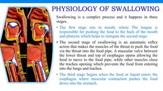
This document discusses dysphagia, or difficulty swallowing. It begins by defining dysphagia and describing the normal physiology of swallowing in three stages. It then discusses the various types, causes, signs and symptoms, diagnostic tests, complications and management approaches for dysphagia. Management may include dietary changes, swallowing exercises and techniques, botulinum toxin injections, dilation procedures, or surgeries like myotomy. Nurses play an important role in educating patients and monitoring for signs of aspiration during meals.
VIP Call Girls Mumbai Arpita 9910780858 Independent Escort Service Mumbai
Nursing Management of Dysphagia
1. ,
PhD Nursing, M.Sc Pediatric Nursing, B.Sc Nursing
Associate professor
EnamCollege of Nursing
Savar, Dhaka, Bangladesh
2. INTRODUCTION
The term dysphagia, is a medical terminology derived from
the Greek word “dys” (with difficulty) and “phagia” (to
eat), describes difficulty in the transfer of food or liquid
boluses from the mouth to the stomach. So Dysphagia
means difficulty in swallowing.
Dysphagia can be painful. In some cases, swallowing is
impossible.
3. PHYSIOLOGY OF SWALLOWING
Swallowing is a complex process and it happens in three
stages.
• The first stage sets in mouth, where The tongue is
responsible for pushing the food to the back of the mouth
and pharynx which helps to instigate the second stage.
• The second stage of swallowing is an automatic reflex
action that makes the muscles of the throat to push the food
via the throat into the food pipe. A muscular valve between
the lower throat and top of esophagus opens allowing the
food to move to the food pipe, while other muscles close
the trachea opening which prevents the food from entering
into the lungs and trachea.
• The third stage begins when the food or liquid enters the
esophagus where muscular contraction pushes the food
down into the stomach.
4. DEFINITION
Dysphagia is simply defined as a
swallowing disorder in which the
individual is having difficulty in
swallowing food or liquid via mouth
through esophageal region into the
stomach.
5. INCIDENCE
•The prevalence of dysphagia is
approximately 10% to 22% in
Americans aged 50 and over. The
prevalence increases with advances in
age, and it is approximately 40% in
people aged over 60..
6. TYPES OF DYSPHAGIA
Oropharyngeal Dysphagia
Difficulty moving food from
the mouth to the throat
Esophageal Dysphagia
Difficulty moving food
down the esophagus
Structural
(obstructive
) Dysphagia
Neurologica
l
(propulsive)
Dysphagia
Structural
(obstructive)
Dysphagia
Neurological
(propulsive)
Dysphagia
Internal obstruction
within the esophagus
External compression
of the esophagus
7. ETIOLOGY
OROPHARANGEAL STRUCTURAL OR OBSTRUCTIVE
DYSPHAGIA
Mouth cancer or throat cancer, such
as laryngeal cancer
Zenker’s diverticulum - A rare instance in
which a pouch-like structure forms between
the pharynx and the esophagus, making it
difficult to swallow.
Cleft lip and palate - It is a types of
abnormal developments of the face due to
incomplete fusing of bones in the head,
resulting in gaps (clefts) in the palate and lip
to nose area.
Peritonsillar abscess - is an area of pus-
filled tissue at the back of the mouth, next to
one of the tonsils. The abscess can be very
painful and can make it hard to open the
mouth.
Epiglottitis - Epiglottitis is characterized by
8. ETIOLOGY
The conditions that can prevent the muscles and nerves from working well enough to
pass food through the throat and esophagus include:
OROPHARANGEAL NEUROLOGICAL OR
PROPULSIVE DYSPHAGIA
•Stroke
•Brain or spine injury
•Nervous system
disorders
• Multiple sclerosis
• Myasthenia Gravis
or Gold flam Disease
• Post-polio syndrome
• Parkinson’s disease
• Muscular dystrophy
•Inflammation disorders
• Polymyositis
9. ETIOLOGY
Internal Compression
Foreign body impactions ( Sometimes food or another object can
partially block the throat or esophagus.)
Esophageal tumors
Esophageal rings or Schatzki's ring (is a ring of tissue that forms
inside the esophagus, the tube that carries food and liquid to your stomach.)
Esophageal webs (are thin membranes that grow across the inside of
the upper part of the esophagus and may cause dysphagia.)
Eosinophilic esophagitis ( is an inflammatory disorder in which the
wall of the esophagus becomes filled with large numbers of eosinophils)
Esophageal Strictures (A narrowed esophagus (stricture) can trap large
pieces of food. Tumors or scar tissue, often caused by gastroesophageal
reflux disease (GERD), can cause narrowing.)
External Compression
Mediastinal masses (Mediastinal tumors are growths that form in the
area of the chest that separates the lungs)
Goiter (A goiter is an enlargement of your thyroid gland.)
Enlarged Aorta or Heart
ESOPHAGEAL STRUCTURAL OR OBSTRUCTIVE DYSPHAGIA
10. ETIOLOGY
• Achalasia. When the lower esophageal muscle
(sphincter) doesn't relax properly to let food enter the
stomach, it can cause food to come back up into the
throat. Muscles in the wall of the esophagus might be
weak as well, a condition that tends to worsen over
time.
• GERD : Damage to esophageal tissues from stomach
acid backing up into the esophagus can lead to spasm
or scarring and narrowing of the lower esophagus.
• Scleroderma. Development of scar-like tissue,
causing stiffening and hardening of tissues, can weaken
the lower esophageal sphincter. As a result, acid backs
up into the esophagus and causes frequent heartburn.
• Nutcracker esophagus refers to having strong
spasms of your esophagus. It's also known as
jackhammer esophagus or hypercontractile esophagus.
ESOPHAGEAL NEUROLOGICAL OR PROPULSIVE DYSPHAGIA
12. DIAGNOSTIC EVALUATION
• History Collection: Collect history regarding duration of symptoms and
acuity of onset
• Physical examination : General factors such as body habitus, drooling,
and mental status should be noted
Voice quality (e.g. a wet sounding voice suggesting pooling of secretions),
Wheezing or labored breathing, and any cranial nerve weakness should be
noted
Gurgling noise in the neck or crepitus in the neck may indicate the presence
of Zenker’s diverticulum
Inspection or palpation of the tongue and tongue strength may unmask
fibrillation or fasciculation of one or both sides
The oropharynx should be inspected for palatal elevation and posterior
pharyngeal wall motion on phonation and Laryngeal examination is
important but can be made difficult by the presence of pooled secretions
• Fiberoptic endoscopic evaluation of swallowing (FEES): This
test involves passing a small camera into the client’s nose and feeding the
client by a dyed liquids and food to see how the client is swallowing. During
the test, the physician will also see check for aspiration.
13. DIAGNOSTIC EVALUATION
• X-ray with a contrast material (barium X-ray): To
see changes in the shape of esophagus and can assess the
muscular activity.
• Modified barium swallow: During this test ask the client to
chew and swallow barium covered foods and liquids , Barium
allows images of mouth, throat and esophagus to be seen on X-
ray images, the radiologist will take X-ray images while the
client swallow.
• Esophago-duodenoscopy (EGD) or upper endoscopy: This
test is used to look for any narrowing, tumors or Barrett’s
esophagus.
• Esophageal muscle test (manometry): In manometry , a small
tube is inserted into esophagus and connected to a pressure
recorder to measure the muscle contractions of esophagus
while swallowing.
15. MANAGEMENT
LIFE STYLE MODIFICATION :-
Avoidance of precipitating foods(fatty foods, alcohol, caffeine)
Oral hygiene
Smoking cessation
Limiting alcohol and caffeine. These can dry your mouth and throat, making swallowing more
difficult.
Weight reduction.
MANAGEMENT FOR OROPHARYNGEAL DYSPHAGIA
Disorders of oral and pharyngeal swallowing are usually amenable to rehabilitation, including
dietary modification and training in swallowing techniques and maneuvers.
Deep pharyngeal neurological stimulation is an intense stimulation program of the oral-pharyngeal
muscles
Thermal gustatory stimulation is a procedure performed by pressing a cold metal object against the
back of the throat to elicit a swallow response. Improvements in swallowing ability are generally
temporary.
16. MANAGEMENT
DIETARY MODIFICATION
Diet classifications The dysphagia diet can be classified according to
viscosity, as follows:
level I: Pudding, crushed potato, and ground meat
level II: Curd-type yogurt, orange juice (mixed with 3% thickener), cream
soup, and thin soup with starch
level III: Tomato juice, fluid-type yogurt, and thick, fluid rice
level IV: Water and orange juice
17. MANAGEMENT
PHARMACHOLOGICAL MANAGEMENT :-
Botulinum toxin type A (BoNT-A): Injected endoscopically into the gastroesophageal
sphincter and upper esophagus to decrease tone; this can be very useful in cricopharyngeal
spasms causing dysphagia or achalasia.
Nitrates: Including isosorbide dinitrate, which can especially be recommended in achalasia
Diltiazem: Can aid in esophageal contractions and motility, especially in the disorder known
as the nutcracker esophagus
Glucocorticoids and leukotriene antagonists represent currently accepted treatment for
Eosinophilic Esophagitis
Antibiotics and Antifungal for infection
Antacid - Acid ingestion Reflux esophagitis
18. MANAGEMENT
SURGICAL MANAGEMENT
• Esophageal dilation. The physician insert endoscope into the
esophagus and inflates an attached balloon to stretch it
(dilation). This treatment is used for achalasia, esophageal
stricture, Schatzki's ring, and motility disorders.
• Stent placement. The physician insert a metal or plastic tube
(stent) to prop open a narrowing or blocked esophagus. Some
stents are permanent, such as those for people with esophageal
cancer, while others are removed later.
• Laparoscopic Heller myotomy. This involves cutting the
muscle at the lower end of the esophagus (sphincter) when it
fails to open and release food into the stomach in people who
have achalasia.
• Peroral endoscopic myotomy (POEM) . The surgeon or
gastroenterologist uses an endoscope inserted through the
mouth and down the throat to create an incision in the inside
lining of the esophagus to treat achalasia Then, as in a Heller
myotomy, the surgeon or gastroenterologist cuts the muscle at
the lower end of the esophageal sphincter.
19. MANAGEMENT
SURGICAL
MANAGEMENT
Zenker’s diverticulum repair
• Open Cricomyotomy +
diverticulopexy/diverticulectomy
• Endoscopic diverticulotomy
(Dohlman's procedure) is a well-
established and effective
alternative to external
diverticulectomy in the treatment
of pharyngeal pouch
20. MANAGEMENT
NURSING MANAGEMENT
• Maintain the patient in high-Fowler’s position with the head flexed slightly forward during meals.
• Instruct the patient not to talk while eating. Provide verbal cueing as needed.
• Observe for uncoordinated chewing or swallowing; coughing shortly after eating or delayed coughing,
which may mean silent aspiration; pocketing of food; wet-sounding voice; sneezing when eating; delay
of more than 1 second in swallowing; or a variation in respiratory patterns. If any of these signs are
present, put on gloves, eliminate all food from oral cavity, end feedings, and consult with a speech and
language pathologist and a dysphagia team.
• Encourage high-calorie diet that involves all food groups, as appropriate. Avoid milk and milk
products.
• If the patient had a stroke, place food in the back of the mouth, on the unaffected side, and gently
massage the unaffected side of the throat.
• Discuss the importance of exercise to enhance the muscular strength of the face and tongue to enhance
swallowing.
• Educate patient, family, and all caregivers about rationales for food consistency and choices.