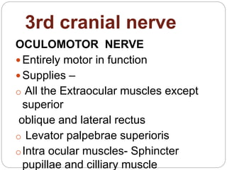
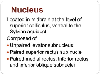
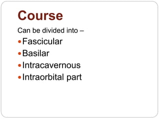
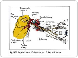
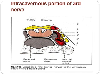
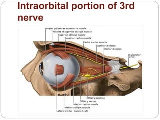
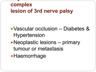
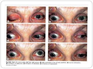
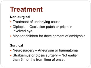
Dr Md Ferdous Islam
FCPS Part 2 Trainee
Dept of Ophthalmology
CMH, Dhaka
3rd Cranial Nerve Palsy
3rd cranial nerve
OCULOMOTOR NERVE
 Entirely motor in function
 Supplies –
o All the Extraocular muscles except
superior
oblique and lateral rectus
o Levator palpebrae superioris
oIntra ocular muscles- Sphincter
pupillae and cilliary muscle
Nucleus
Located in midbrain at the level of
superior colliculus, ventral to the
Sylvian aquiduct.
Composed of
 Unpaired levator subnucleus
 Paired superior rectus sub nuclei
 Paired medial rectus, inferior rectus
and inferior oblique subnuclei
Course
Can be divided into –
Fascicular
Basilar
Intracavernous
Intraorbital part
Course
Intracavernous portion of 3rd
nerve
Intraorbital portion of 3rd
nerve
complex
lesion of 3rd nerve palsy
Vascular occlusion – Diabetes &
Hypertension
Neoplastic lesions – primary
tumour or metastasis
Haemorrhage
Major causes of fascicular
lesion of
3rd nerve palsy
 Vascular occlusion – Diabetes &
Hypertension
 Neoplastic lesions – primary tumour or
metastasis
 Haemorrhage
 Demyelination
Syndromes of Fascicular
lesion
 Benedikt syndrome- Ipsilateral 3rd nerve palsy
and contralateral extrapyramidal signs
 Weber syndrome- Ipsilateral 3rd nerve palsy and
contralateral hemiparesis
 Nothnagel syndrome- Ipsilateral 3rd nerve palsy
and cerebellar ataxia
 Claude syndrome
Major causes of lesion in
Basilar
region
 The 3rd nerve traverses the basilar part
unaccompanied by any other cranial
nerves.
 Isolated 3rd nerve palsies are commonly
basilar.
The important causes are
 Aneurysm
 Head trauma-Extradural or subdural
Major causes of
Intracavernous
lesion
 Usually associated with involvement of
4th, 6th nerves & first division of 5th
nerve.
 Diabetes – causes pupil sparing 3rd
nerve palsy
 Pituitary apoplexy
 Others – Aneurysm, Meningeoma,
Carotid-cavernous fistula.
Intraorbital causes of 3rd
nerve
palsy
 Trauma
 Vascular
 Neoplasm
 Inflammation
Pupillomotor fibers
 Parasympathetic fibers
 Located superficially between the
brainstem and the cavernous sinus
 Blood supply derived from the pial blood
vessels
 Main trunk of 3rd nerve supplied by the
vasa nervorum
Causes of isolated 3rd nerve
palsy
 Idiopathic – about 25%
 Vascular – Hypertension & Diabetes
(commonly pupil sparing)
 Aneurysm – posterior communicating
artery at its junction with internal carotid
artery
 Trauma – subdural haematoma with
uncal herniation
 Miscellaneous
Clinical features of total 3rd
nerve
palsy
SYMPTOMS
 Drooping of eyelid
 Binocular double vision
 Pain (may be present)
SIGNS
 Ptosis
 Abduction of globe
 Intortion of the globe which increases on
attempted down gaze
 Limitation of adduction
 Limitation of elevation
 Limitation of depression
 Dilated pupil with defective accommodation
History of Patient
 Onset
 Duration
 Diplopia
 Trauma
 Associated systemic disorders
Examination
 Pupillary reactions
 Motility restrictions
 Ptosis
 Other cranial nerves
Investigations
Investigations
Treatment
Non-surgical
 Treatment of underlying cause
 Diplopia – Occlusion patch or prism in
involved eye
 Monitor children for development of amblyopia
Surgical
 Neurosurgery – Aneurysm or haematoma
 Strabismus or ptosis surgery – Not earlier
than 6 months from time of onset
Follow-up
 Pupil sparing – Observe daily for 5 days
for pupil involvement
 Recheck every 4 to 6 weeks
 If secondary to ischemia function usually
returns within 3 months
Thank
You

3rd nerve palsy

  • 1.
    Dr Md FerdousIslam FCPS Part 2 Trainee Dept of Ophthalmology CMH, Dhaka 3rd Cranial Nerve Palsy
  • 2.
    3rd cranial nerve OCULOMOTORNERVE  Entirely motor in function  Supplies – o All the Extraocular muscles except superior oblique and lateral rectus o Levator palpebrae superioris oIntra ocular muscles- Sphincter pupillae and cilliary muscle
  • 3.
    Nucleus Located in midbrainat the level of superior colliculus, ventral to the Sylvian aquiduct. Composed of  Unpaired levator subnucleus  Paired superior rectus sub nuclei  Paired medial rectus, inferior rectus and inferior oblique subnuclei
  • 4.
    Course Can be dividedinto – Fascicular Basilar Intracavernous Intraorbital part
  • 5.
  • 7.
  • 8.
  • 9.
    complex lesion of 3rdnerve palsy Vascular occlusion – Diabetes & Hypertension Neoplastic lesions – primary tumour or metastasis Haemorrhage
  • 10.
    Major causes offascicular lesion of 3rd nerve palsy  Vascular occlusion – Diabetes & Hypertension  Neoplastic lesions – primary tumour or metastasis  Haemorrhage  Demyelination
  • 11.
    Syndromes of Fascicular lesion Benedikt syndrome- Ipsilateral 3rd nerve palsy and contralateral extrapyramidal signs  Weber syndrome- Ipsilateral 3rd nerve palsy and contralateral hemiparesis  Nothnagel syndrome- Ipsilateral 3rd nerve palsy and cerebellar ataxia  Claude syndrome
  • 12.
    Major causes oflesion in Basilar region  The 3rd nerve traverses the basilar part unaccompanied by any other cranial nerves.  Isolated 3rd nerve palsies are commonly basilar. The important causes are  Aneurysm  Head trauma-Extradural or subdural
  • 15.
    Major causes of Intracavernous lesion Usually associated with involvement of 4th, 6th nerves & first division of 5th nerve.  Diabetes – causes pupil sparing 3rd nerve palsy  Pituitary apoplexy  Others – Aneurysm, Meningeoma, Carotid-cavernous fistula.
  • 16.
    Intraorbital causes of3rd nerve palsy  Trauma  Vascular  Neoplasm  Inflammation
  • 17.
    Pupillomotor fibers  Parasympatheticfibers  Located superficially between the brainstem and the cavernous sinus  Blood supply derived from the pial blood vessels  Main trunk of 3rd nerve supplied by the vasa nervorum
  • 19.
    Causes of isolated3rd nerve palsy  Idiopathic – about 25%  Vascular – Hypertension & Diabetes (commonly pupil sparing)  Aneurysm – posterior communicating artery at its junction with internal carotid artery  Trauma – subdural haematoma with uncal herniation  Miscellaneous
  • 20.
    Clinical features oftotal 3rd nerve palsy SYMPTOMS  Drooping of eyelid  Binocular double vision  Pain (may be present)
  • 21.
    SIGNS  Ptosis  Abductionof globe  Intortion of the globe which increases on attempted down gaze  Limitation of adduction  Limitation of elevation  Limitation of depression  Dilated pupil with defective accommodation
  • 23.
    History of Patient Onset  Duration  Diplopia  Trauma  Associated systemic disorders
  • 24.
    Examination  Pupillary reactions Motility restrictions  Ptosis  Other cranial nerves
  • 25.
  • 26.
  • 27.
    Treatment Non-surgical  Treatment ofunderlying cause  Diplopia – Occlusion patch or prism in involved eye  Monitor children for development of amblyopia Surgical  Neurosurgery – Aneurysm or haematoma  Strabismus or ptosis surgery – Not earlier than 6 months from time of onset
  • 28.
    Follow-up  Pupil sparing– Observe daily for 5 days for pupil involvement  Recheck every 4 to 6 weeks  If secondary to ischemia function usually returns within 3 months
  • 29.