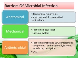
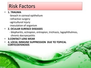
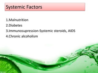
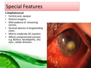
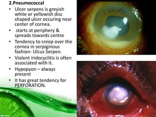
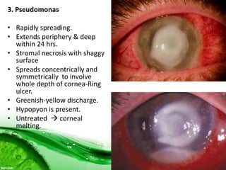
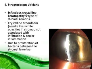
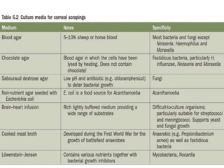
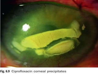
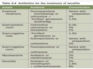
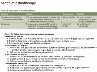
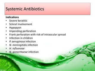
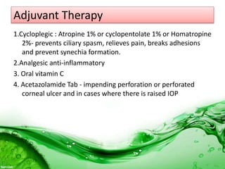
Dr Md Ferdous Islam
Introduction
• Bacterial keratitis usually develops when ocular defens
have been compromised.
•Host cellular and immunologic responses to offending agent which
may be bacterial, viral, fungal or protozoal organisms leads to
formation of ulcer.
•Sight threatening condition and should be considered as ocular
emergency.
Barriers Of Microbial Infection
• Bony orbital rim,eyelids,
• Intact corneal & conjunctival
epithelium
Anatomical
• Tear film-mucus layer
• Lacrimal systemMechanical
• Tear film constitutes-IgA, complement
components, and enzymes lysozyme,
lactoferrin, betalysins
• CALT
Antimicrobial
Risk Factors
• 1. TRAUMA
-breach in corneal epithelium
-refractive surgery
-agricultural injury
-inoculation of organism
• 2. OCULAR SURFACE DISEASES
- blepharitis, ectropion, entropion, trichiasis, lagophthalmos,
chronic dacryocystitis
• 3.CONTACT LENS WEAR
• 4. LOCAL IMMUNE SUPPRESSION DUE TO TOPICAL
CORTICOSTEROIDS
Systemic Factors
1.Malnutrition
2.Diabetes
3.Immunosupression-Systemic steroids, AIDS
4.Chronic alcoholism
Aetiology
• Caused by organisms which produce toxins causing tissue
death i.e. necrosis characterized by pus formation.
• Such purulent keratitis is usually exogenous due to infection
by pyogenic bacteria such as
-Pseudomonas,
-Staphylococcus,
-Streptococcus,
-N. gonorrhoeae and
-C. diphtheriae
Aetiology
• Most of the bacteria are capable of producing corneal ulcer
only when the epithelium is damaged.
• N. gonorrhoeae, C. diphtheriae, Haemophilus , N. meningitidis
can penetrate intact corneal epithelium.
Pathogenesis
Corneal abrasion Microbes adhere to epithelium, cloning ,invasion to stromal
lamellae,release toxins & lytic enzymes
Host response
PMNs at the site of ulcer from tears & limbal vessels release of cytokines &
interleukins  progressive invasion of cornea & increase in size of ulcer
Phagocytosis
Release of free radicals, proteolytic enymes Necrosis & sloughing of epithelium,
Bowman’s membrane & stroma
A saucer shaped defect with projecting walls above the normal surface due to
swelling of tissue resulting from fluid imbibition by corneal stroma with grey zone
of infiltration
Presentation
1. Diminution of vision, depending on location of
corneal ulcer
2. Watering due to reflex lacrimation
3. Photophobia
4. Pain due to exposed nerve endings
5. Mucopurulent / purulent discharge
Ocular Examination
1.Visual acuity-reduced
2.Slit lamp Biomicroscope
Lids - edema
Conjunctiva – Ciliary congestion
4. Cornea
-Location of the ulcer- central, paracentral peripheral,total.
-Size , shape, depth, margins & floor- depends on stage of
ulcer.
-Density and extent of stromal infiltration.
5. Anterior chamber
- Cells/flare, mobile Hypopyon.
Iris- muddy
Toxin induced iritis
Pupil – miotic
Other:
-Sac syringing
-corneal sensation
-Fluorescein staining
Special Features
1.Staphylococcal
• Central,oval, opaque
• Distinct margins.
• Mild oedema of remaining
cornea.
• Stromal abscess in longstanding
cases.
• Mild to moderate AC reaction.
• Affects compromised corneas
e.g. Bullous keratopathy , dry
eyes , atopic diseases.
2.Pneumococcal
• Ulcer serpens is greyish
white or yellowish disc
shaped ulcer occuring near
center of cornea.
• starts at periphery &
spreads towards centre
• Tendency to creep over the
cornea in serpiginous
fashion- Ulcus Serpen.
• Violent iridocyclitis is often
associated with it.
• Hypopyon – always
present
• It has great tendency for
PERFORATION.
3. Pseudomonas
• Rapidly spreading.
• Extends periphery & deep
within 24 hrs.
• Stromal necrosis with shaggy
surface
• Spreads concentrically and
symmetrically to involve
whole depth of cornea-Ring
ulcer.
• Greenish-yellow discharge.
• Hypopyon is present.
• Untreated  corneal
melting.
4. Streptococcus viridans
• Infectious crystalline
keratopathytype of
stromal keratitis.
• Crystalline arborifoem
(needle like) white
opacities in stroma , not
associated with
infiltration & ocular
inflammation
• Due to proliferation of
bacteria between the
stromal lamellae.
Complications Of Corneal Ulcer
1. Spread of ulcer horizontally and depth-wise, leading to thinning
of cornea
2. Descemetocele
3. Perforation of ulcer –
sudden exertion such as coughing, sneezing, straining at stool or
firm closure of eyes increase in intra-ocular pressure (IOP)
perforation
a) Peripheral perforation -iris prolapse through opening.
Exudation takes place on prolapsed tissue an  adherent
leucoma .
b) Central perforation  anterior chamber collapse
 lens comes in contact with corneal endothelial surface 
anterior capsular cataract  repeated healing and perforation
leading to corneal fistula formation
c) Sloughing of whole cornea: prolapse of iris  pupillary block
and exudation on iris  pseudocornea  anterior synechiae
 angle of anterior chamber is occluded leading to secondary
glaucoma  anterior staphyloma .
d) Intra-ocular purulent infection: due to perforation bacteria
enter in the eye and causes endophthalmitis /
panophthalmitis
Investigations
Specific – Corneal scraping
Gram stain, Culture &
Antibiotic sensitivity
Culture of contact lens & solution
Conjunctival Swab
Gen Consideration
• Hospitalization
• Discontinuation of contact lens wear
• Eye shield
LOCAL TREATMENT
Control of infection with appropriate antibiotic(s)
a. based on clinical judgment
b. based on finding of smear examination
c. based on culture and sensitivity report
• Antibiotic Monotherapy
-fluroquinolone
-Ciprofloxacin or Ofloxacin
-New generation fluroquinolone
•Antibiotic duotherapy
•Subconjunctival antibiotics
•Mydriatics
•Steroids
Systemic Antibiotics
Indications
• Severe keratitis
• Scleral involvement
• Hypopyon
• Impending perforation
• Frank perforation with risk of intraocular spread
• Infection in children
• P. aeruginosa infection
• N. meningitidis infection
• H. influenzae
• N. gonorrhoeae infection
Adjuvant Therapy
1.Cycloplegic : Atropine 1% or cyclopentolate 1% or Homatropine
2%- prevents ciliary spasm, relieves pain, breaks adhesions
and prevent synechia formation.
2.Analgesic anti-inflammatory
3. Oral vitamin C
4. Acetazolamide Tab - impending perforation or perforated
corneal ulcer and in cases where there is raised IOP
Treatment Of Impending Perforation
1. Straining should be avoided.
2. Pressure bandage
3. Lowering of IOP
4. Tissue adhesive glue (cynoacrylate)
5. Conjunctival flap
6. Soft contact lens Bandage
7. Penetrating keratoplasty
Treatment Of Perforated Corneal Ulcer
• Tissue adhesives
• Conjunctival flap
• Soft bandage
• Keratoplasty
• Modification of initial antimicrobial therapy:
-Should be based on clinical response not on culture sensitivity
• If pt is responding  no change in initial treatment
• If pt is not responding/ worsening drugs are changed
according to antimicrobial sensitivity
• SIGNS OF HEALING :
 -resolution of lid edema, congestion
 -decreased density of stromal infiltrate
 -reduction of corneal oedema
 -reduction in AC reaction/hypopyon
 -re-epithelization
 -corneal vascularization
• Antibiotic frequency-tapered to 4hrly after 72 hrs
• SIGNS OF NON-RESPONSE
- Increase in infiltration, epithelial defect, height of hypopyon,
Corneal thinning, perforation
Treatment
• Re-evaluate for
Drug toxicity,Non-infectious causes or Unusual organisms
• Modification of anti-microbial therapy according to
antimicrobial sensitivity
• Scraping of ulcer floor followed by cauterization with pure
(100%) carbolic acid or 10-20% trichloracetic acid.
• Therapeutic keratoplasty
Topical Corticosteroids
• Controversial in bacterial keratitis
• The rationale for using steroids - to decrease tissue destruction.
CRITERIA FOR TOPICAL STEROIDS IN ULCER --
1.Must not be used in presence of active infected corneal ulcer
2.If bacteria shows in-vitro sensitivity to the antibiotic being used
3.Patients compliance for follow-up
4. No other virulent organism is found
Surgical Treatment
• 1.Tissue adhesive-Cyanoacrylate glue- small perforations< 3mm
- descemetocele
• 2. Patch graft –perforation- 5mm in diameter
• 3 . Therapeutic keratoplasty
-large areas of perforation, necrosis
-Non-healing ulcer
Ferdous bacterial keratitis   copy

Ferdous bacterial keratitis copy

  • 1.
  • 2.
    Introduction • Bacterial keratitisusually develops when ocular defens have been compromised. •Host cellular and immunologic responses to offending agent which may be bacterial, viral, fungal or protozoal organisms leads to formation of ulcer. •Sight threatening condition and should be considered as ocular emergency.
  • 3.
    Barriers Of MicrobialInfection • Bony orbital rim,eyelids, • Intact corneal & conjunctival epithelium Anatomical • Tear film-mucus layer • Lacrimal systemMechanical • Tear film constitutes-IgA, complement components, and enzymes lysozyme, lactoferrin, betalysins • CALT Antimicrobial
  • 4.
    Risk Factors • 1.TRAUMA -breach in corneal epithelium -refractive surgery -agricultural injury -inoculation of organism • 2. OCULAR SURFACE DISEASES - blepharitis, ectropion, entropion, trichiasis, lagophthalmos, chronic dacryocystitis • 3.CONTACT LENS WEAR • 4. LOCAL IMMUNE SUPPRESSION DUE TO TOPICAL CORTICOSTEROIDS
  • 5.
  • 6.
    Aetiology • Caused byorganisms which produce toxins causing tissue death i.e. necrosis characterized by pus formation. • Such purulent keratitis is usually exogenous due to infection by pyogenic bacteria such as -Pseudomonas, -Staphylococcus, -Streptococcus, -N. gonorrhoeae and -C. diphtheriae
  • 7.
    Aetiology • Most ofthe bacteria are capable of producing corneal ulcer only when the epithelium is damaged. • N. gonorrhoeae, C. diphtheriae, Haemophilus , N. meningitidis can penetrate intact corneal epithelium.
  • 8.
    Pathogenesis Corneal abrasion Microbesadhere to epithelium, cloning ,invasion to stromal lamellae,release toxins & lytic enzymes Host response PMNs at the site of ulcer from tears & limbal vessels release of cytokines & interleukins  progressive invasion of cornea & increase in size of ulcer Phagocytosis Release of free radicals, proteolytic enymes Necrosis & sloughing of epithelium, Bowman’s membrane & stroma A saucer shaped defect with projecting walls above the normal surface due to swelling of tissue resulting from fluid imbibition by corneal stroma with grey zone of infiltration
  • 9.
    Presentation 1. Diminution ofvision, depending on location of corneal ulcer 2. Watering due to reflex lacrimation 3. Photophobia 4. Pain due to exposed nerve endings 5. Mucopurulent / purulent discharge
  • 10.
    Ocular Examination 1.Visual acuity-reduced 2.Slitlamp Biomicroscope Lids - edema Conjunctiva – Ciliary congestion
  • 11.
    4. Cornea -Location ofthe ulcer- central, paracentral peripheral,total. -Size , shape, depth, margins & floor- depends on stage of ulcer. -Density and extent of stromal infiltration. 5. Anterior chamber - Cells/flare, mobile Hypopyon.
  • 13.
    Iris- muddy Toxin inducediritis Pupil – miotic Other: -Sac syringing -corneal sensation -Fluorescein staining
  • 14.
    Special Features 1.Staphylococcal • Central,oval,opaque • Distinct margins. • Mild oedema of remaining cornea. • Stromal abscess in longstanding cases. • Mild to moderate AC reaction. • Affects compromised corneas e.g. Bullous keratopathy , dry eyes , atopic diseases.
  • 15.
    2.Pneumococcal • Ulcer serpensis greyish white or yellowish disc shaped ulcer occuring near center of cornea. • starts at periphery & spreads towards centre • Tendency to creep over the cornea in serpiginous fashion- Ulcus Serpen. • Violent iridocyclitis is often associated with it. • Hypopyon – always present • It has great tendency for PERFORATION.
  • 16.
    3. Pseudomonas • Rapidlyspreading. • Extends periphery & deep within 24 hrs. • Stromal necrosis with shaggy surface • Spreads concentrically and symmetrically to involve whole depth of cornea-Ring ulcer. • Greenish-yellow discharge. • Hypopyon is present. • Untreated  corneal melting.
  • 17.
    4. Streptococcus viridans •Infectious crystalline keratopathytype of stromal keratitis. • Crystalline arborifoem (needle like) white opacities in stroma , not associated with infiltration & ocular inflammation • Due to proliferation of bacteria between the stromal lamellae.
  • 18.
    Complications Of CornealUlcer 1. Spread of ulcer horizontally and depth-wise, leading to thinning of cornea 2. Descemetocele 3. Perforation of ulcer – sudden exertion such as coughing, sneezing, straining at stool or firm closure of eyes increase in intra-ocular pressure (IOP) perforation a) Peripheral perforation -iris prolapse through opening. Exudation takes place on prolapsed tissue an  adherent leucoma .
  • 19.
    b) Central perforation anterior chamber collapse  lens comes in contact with corneal endothelial surface  anterior capsular cataract  repeated healing and perforation leading to corneal fistula formation c) Sloughing of whole cornea: prolapse of iris  pupillary block and exudation on iris  pseudocornea  anterior synechiae  angle of anterior chamber is occluded leading to secondary glaucoma  anterior staphyloma . d) Intra-ocular purulent infection: due to perforation bacteria enter in the eye and causes endophthalmitis / panophthalmitis
  • 20.
    Investigations Specific – Cornealscraping Gram stain, Culture & Antibiotic sensitivity Culture of contact lens & solution Conjunctival Swab
  • 24.
    Gen Consideration • Hospitalization •Discontinuation of contact lens wear • Eye shield
  • 25.
    LOCAL TREATMENT Control ofinfection with appropriate antibiotic(s) a. based on clinical judgment b. based on finding of smear examination c. based on culture and sensitivity report • Antibiotic Monotherapy -fluroquinolone -Ciprofloxacin or Ofloxacin -New generation fluroquinolone
  • 28.
  • 29.
    Systemic Antibiotics Indications • Severekeratitis • Scleral involvement • Hypopyon • Impending perforation • Frank perforation with risk of intraocular spread • Infection in children • P. aeruginosa infection • N. meningitidis infection • H. influenzae • N. gonorrhoeae infection
  • 30.
    Adjuvant Therapy 1.Cycloplegic :Atropine 1% or cyclopentolate 1% or Homatropine 2%- prevents ciliary spasm, relieves pain, breaks adhesions and prevent synechia formation. 2.Analgesic anti-inflammatory 3. Oral vitamin C 4. Acetazolamide Tab - impending perforation or perforated corneal ulcer and in cases where there is raised IOP
  • 31.
    Treatment Of ImpendingPerforation 1. Straining should be avoided. 2. Pressure bandage 3. Lowering of IOP 4. Tissue adhesive glue (cynoacrylate) 5. Conjunctival flap 6. Soft contact lens Bandage 7. Penetrating keratoplasty
  • 32.
    Treatment Of PerforatedCorneal Ulcer • Tissue adhesives • Conjunctival flap • Soft bandage • Keratoplasty
  • 33.
    • Modification ofinitial antimicrobial therapy: -Should be based on clinical response not on culture sensitivity • If pt is responding  no change in initial treatment • If pt is not responding/ worsening drugs are changed according to antimicrobial sensitivity
  • 34.
    • SIGNS OFHEALING :  -resolution of lid edema, congestion  -decreased density of stromal infiltrate  -reduction of corneal oedema  -reduction in AC reaction/hypopyon  -re-epithelization  -corneal vascularization • Antibiotic frequency-tapered to 4hrly after 72 hrs
  • 35.
    • SIGNS OFNON-RESPONSE - Increase in infiltration, epithelial defect, height of hypopyon, Corneal thinning, perforation Treatment • Re-evaluate for Drug toxicity,Non-infectious causes or Unusual organisms • Modification of anti-microbial therapy according to antimicrobial sensitivity • Scraping of ulcer floor followed by cauterization with pure (100%) carbolic acid or 10-20% trichloracetic acid. • Therapeutic keratoplasty
  • 36.
    Topical Corticosteroids • Controversialin bacterial keratitis • The rationale for using steroids - to decrease tissue destruction. CRITERIA FOR TOPICAL STEROIDS IN ULCER -- 1.Must not be used in presence of active infected corneal ulcer 2.If bacteria shows in-vitro sensitivity to the antibiotic being used 3.Patients compliance for follow-up 4. No other virulent organism is found
  • 37.
    Surgical Treatment • 1.Tissueadhesive-Cyanoacrylate glue- small perforations< 3mm - descemetocele • 2. Patch graft –perforation- 5mm in diameter • 3 . Therapeutic keratoplasty -large areas of perforation, necrosis -Non-healing ulcer

Editor's Notes

  • #4 CALT- conjunctiva associated lymphoid tissue.