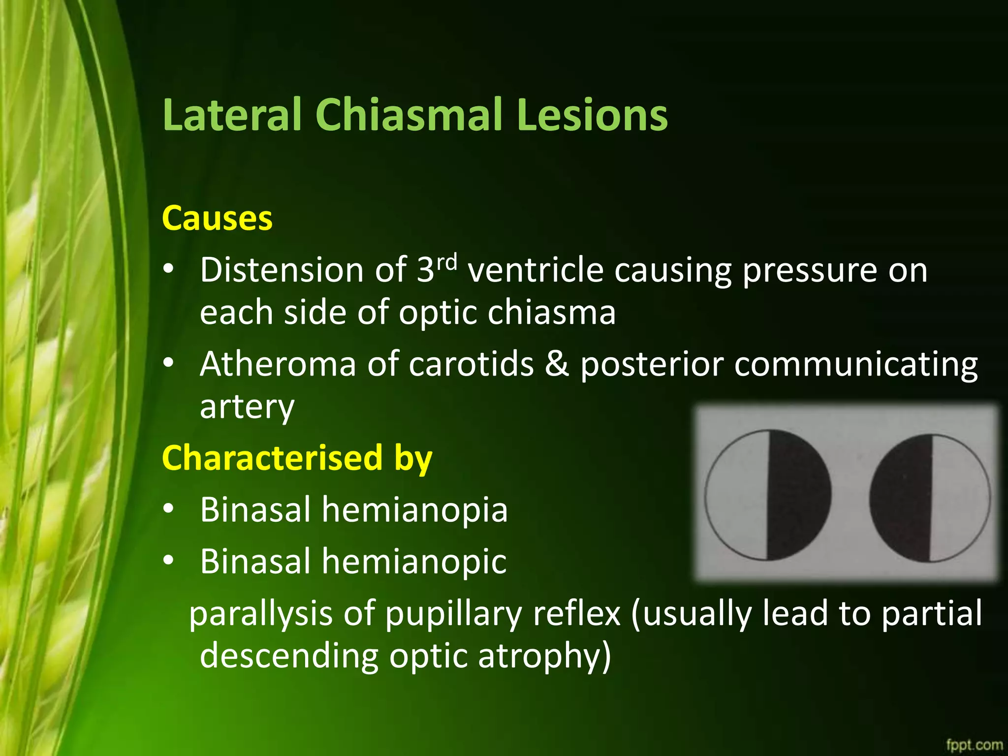
CHIASMA
Dr Md Ferdous Islam
Department of Ophthalmology
CMH,DHAKA
Visual Pathway
Optic Nerve
Optic Chiasma
Optic Tract
Lateral Geniculate Body
Optic Radiations
Visual cortex
Chiasma
•Flattened structure,12 mm horizontally & 8mm
anteroposteriorly, 4 mm thick
•Ensheathed by pia & surrounded by CSF
• Lies over diaphragma sellae so visual field defects
seen in patient with pituitary tumor having
suprasellar extension
•Posteriorly chiasma continous with the optic tracts
& form the anterior wall of 3rd ventricle
•Nerve fibres arising from nasal half of two retina
decussate at the chiasma
• Floor of the third ventricle
• 5-10 mm above the diphragma sella and the
hypophysis cerebri
• Important relations: 3rd ventricle, hypothalmus,
pituitary stalk, sella, dorsum sellam anterior and
posterior clinoid processes, cavernous sinus
• Nasal fibers cross ; temporal fibers do not (53:47)
• Wilband’s knee
Anatomical Variation
a) central : lies directly over sella,
expanding pituitary tumor involves
chiasma first
b) pre-fixed : lies more anteriorly
over tuberculum sellae,pituitary
tumor involves optic tract first.
c) post-fixed : lies more posterior
over dorsum sellae,pituitary tumor
damage optic nerve first
Relations Of Chiasma
• Anterior - anterior cerebral arteries & its
communicating arteries
• Posterior- tuber cinereum, infundibulum ,pitutary
body ,posterior perforated substance
• Superior- third ventricle
• Inferior- hypophysis
• Lateral- extra cavernous part of internal carotid
artery& anterior perforated substance
Arrangement Of Nerve Fibers
• Temporal fibers from retina
remains uncrossed and runs
backward in lateral part of
optic chaisma
• Nasal peripheral fibers
o ¾ of fibers
o Cross over to enter medial
part of opposite optic tract in
fol
 lower nasal fibers in optic
tract traverse chiasma low and
anteriorly
 Upper nasal fibers in optic
tract trasverse chiasma high
and posteriorly
Arrangement Of Nerve Fibers
Macular fibers
• Some fibers crossed n
runs backward in opposite
optic tract
• Some fibers uncrossed n
runs on same side in optic
tract
Blood Supply Of Optic Chiasma
Arterial
Venous
• B/o anterior cerebral & anterior
communicating arterySuperior aspect
• B/o internal carotid artery ,posterior
communicating artery ,anterior superior
hypophyseal artery
Inferior aspect
• Superior chiasmal vein drains into
anterior cerebral vein
Superior
aspect
• Pre-infundibular vein draining into
basilar veinInferior aspect
Central Lesions Of Chiasma (Sagittal)
 Causes
 suprasellar aneurysm
 tumors of pituitary gland
 craniopharyngioma
 suprasellar meningioma & glioma of 3rd ventricle
 third ventricular dilatation due to obstructive
hydrocephalus
 chronic chiasmal arachnoiditis
 Characterised by
 Bitemporal hemianopia
 Bitemporal hemianopic
paralysis of pupillary reflex (usually lead to partial
descending optic atrophy)
Lateral Chiasmal Lesions
Causes
• Distension of 3rd ventricle causing pressure on
each side of optic chiasma
• Atheroma of carotids & posterior communicating
artery
Characterised by
• Binasal hemianopia
• Binasal hemianopic
parallysis of pupillary reflex (usually lead to partial
descending optic atrophy)
Chiasma

Chiasma

  • 1.
    CHIASMA Dr Md FerdousIslam Department of Ophthalmology CMH,DHAKA
  • 2.
    Visual Pathway Optic Nerve OpticChiasma Optic Tract Lateral Geniculate Body Optic Radiations Visual cortex
  • 3.
    Chiasma •Flattened structure,12 mmhorizontally & 8mm anteroposteriorly, 4 mm thick •Ensheathed by pia & surrounded by CSF • Lies over diaphragma sellae so visual field defects seen in patient with pituitary tumor having suprasellar extension •Posteriorly chiasma continous with the optic tracts & form the anterior wall of 3rd ventricle •Nerve fibres arising from nasal half of two retina decussate at the chiasma
  • 4.
    • Floor ofthe third ventricle • 5-10 mm above the diphragma sella and the hypophysis cerebri • Important relations: 3rd ventricle, hypothalmus, pituitary stalk, sella, dorsum sellam anterior and posterior clinoid processes, cavernous sinus • Nasal fibers cross ; temporal fibers do not (53:47) • Wilband’s knee
  • 6.
    Anatomical Variation a) central: lies directly over sella, expanding pituitary tumor involves chiasma first b) pre-fixed : lies more anteriorly over tuberculum sellae,pituitary tumor involves optic tract first. c) post-fixed : lies more posterior over dorsum sellae,pituitary tumor damage optic nerve first
  • 8.
    Relations Of Chiasma •Anterior - anterior cerebral arteries & its communicating arteries • Posterior- tuber cinereum, infundibulum ,pitutary body ,posterior perforated substance • Superior- third ventricle • Inferior- hypophysis • Lateral- extra cavernous part of internal carotid artery& anterior perforated substance
  • 9.
    Arrangement Of NerveFibers • Temporal fibers from retina remains uncrossed and runs backward in lateral part of optic chaisma • Nasal peripheral fibers o ¾ of fibers o Cross over to enter medial part of opposite optic tract in fol  lower nasal fibers in optic tract traverse chiasma low and anteriorly  Upper nasal fibers in optic tract trasverse chiasma high and posteriorly
  • 10.
    Arrangement Of NerveFibers Macular fibers • Some fibers crossed n runs backward in opposite optic tract • Some fibers uncrossed n runs on same side in optic tract
  • 11.
    Blood Supply OfOptic Chiasma Arterial Venous • B/o anterior cerebral & anterior communicating arterySuperior aspect • B/o internal carotid artery ,posterior communicating artery ,anterior superior hypophyseal artery Inferior aspect • Superior chiasmal vein drains into anterior cerebral vein Superior aspect • Pre-infundibular vein draining into basilar veinInferior aspect
  • 12.
    Central Lesions OfChiasma (Sagittal)  Causes  suprasellar aneurysm  tumors of pituitary gland  craniopharyngioma  suprasellar meningioma & glioma of 3rd ventricle  third ventricular dilatation due to obstructive hydrocephalus  chronic chiasmal arachnoiditis  Characterised by  Bitemporal hemianopia  Bitemporal hemianopic paralysis of pupillary reflex (usually lead to partial descending optic atrophy)
  • 13.
    Lateral Chiasmal Lesions Causes •Distension of 3rd ventricle causing pressure on each side of optic chiasma • Atheroma of carotids & posterior communicating artery Characterised by • Binasal hemianopia • Binasal hemianopic parallysis of pupillary reflex (usually lead to partial descending optic atrophy)