Downloaded 138 times

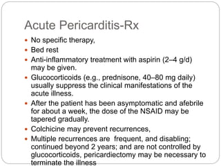


The document discusses the pericardium, a double-layered sac that protects the heart, its functions, and various pericardial diseases such as acute and chronic pericarditis. It describes clinical features, diagnostic methods, and treatment options including medications like aspirin and glucocorticoids, as well as pericardiocentesis for cardiac tamponade. The document also highlights the importance of recognizing cardiac tamponade in clinical settings due to its variable presentations.


