Download as PDF, PPTX
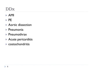
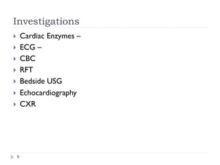
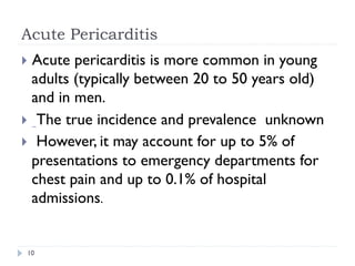
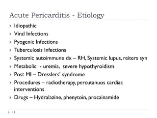


The document presents a case study on acute pericarditis, detailing the medical history, symptoms, and examination findings of a 38-year-old female patient. It outlines the etiology, classification, diagnostic tests, treatment options, and potential complications of pericarditis. The content is intended for educational purposes and emphasizes the importance of professional medical evaluation.


