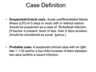
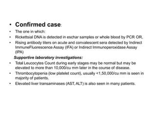
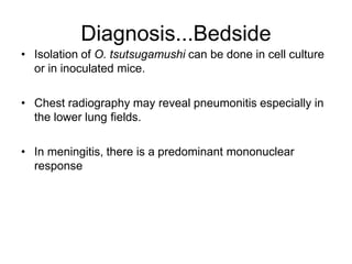
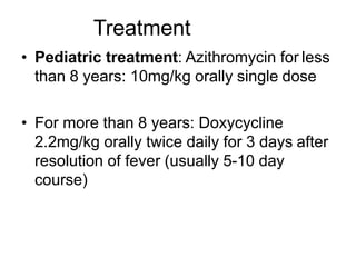
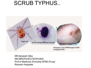
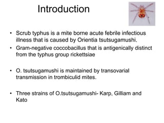
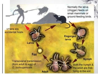
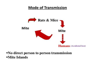
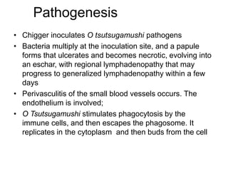
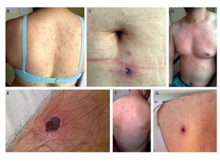
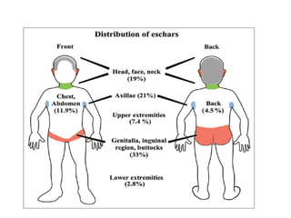
SCRUB TYPHUS..
DR Ashutosh Ojha
MD,MRCP(UK)I,FACP(USA)
Prof of Medicine (Formerly AFMC,Pune)
Ramesh Hospitals
Reported outbreaks of scrub typhus in India
(till 42nd week of 2018).
Introduction
Historical Perspective
Epidemiology & Indian Scenario
Pathogenesis
Clinical features & Complications
Approach to the Disease
Management
Prevention & Control
Scheme of Presentation
Introduction
• Scrub typhus is a mite borne acute febrile infectious
illness that is caused by Orientia tsutsugamushi.
• Gram-negative coccobacillus that is antigenically distinct
from the typhus group rickettsiae
• O. tsutsugamushi is maintained by transovarial
transmission in trombiculid mites.
• Three strains of O.tsutsugamushi- Karp, Gilliam and
Kato
Introduction....
• The mites are both the vector and reservoir of
the disease.
• The mite is very small (0.2 –0.4mm) and can be
seen through a microscope or magnifying glass.
• The infected larval mites (chiggers, the only
stage that feeds on a host) inoculate organisms
into the skin.
• There is no human to human transmission.
Historical perspective
• Historically, in 313 AD, a clinical manual by Hong
Ge called “Zhouhofang” had mentioned the
clinical description of disease and morphological
description of mites.
• 1596 AD, well-known Chinese physician Shizen
Li described the characteristics of the disease.
• Japanese researcher started research on this
disease in 1879.
• The name Rickettsia tsutsugamushi was
first used in 1930.
• The word “tsutsugamushi” is derived from
a Japanese word tsutsuga meaning
something small and dangerous,
mushi, meaning creature, so it was
known as small dangerous creature.
Historical perspective...
Why is it linked to
War ?
• Napoleon’s retreat from Moscow
was forced by rickettsial disease
breaking out among his troops.
• Lenin is said to have remarked, in
reference to rickettsial disease during
Russian revolution,“either socialism will
defeat the louse or the louse will defeat
the socialism”
Major Impact on research during
WW2..
• Its impact on immunologically naive Allied
troops between 1942 and 1945 resulted in
18 000 cases and 639 deaths (4.0%), as
well as an estimated 20 000 cases in
Japanese troops.
• First batch of scrub typhus vaccine used
to inoculate human subjects was
dispatched to India for use by the Allied
Land Forces, South-East Asia Command,
in June 1945.
• Leading cause of pyrexia of unknown
origin (PUOs) in forces of USA during the
VietNam conflict.
• Scrub typhus is endemic to a part of the world known as
the “tsutsugamushi triangle”
Indian Scenario
• In India, the disease had occurred among troops
during the Second World War in Assam andWest
Bengal, and in the 1965 Indo-Pak war.
• There was a resurgence of the disease in 1990 in a
unit of an army deployed at the Pakistan border of
India.
• Occurrence reported from several states in India
including Jammu and Kashmir, Himachal Pradesh,
Uttarakhand, Bihar, West Bengal, Meghalaya,
Rajasthan, Maharashtra, Karnataka, Tamil Nadu
and Kerala.
• Scrub typhus accounts for upto 50% of
undifferentiated fever presenting to hospitals.
• It remains a major underdiagnosed(suspected)
cause of undifferentiated fever.
Reserviour of infection at our door
Scrub typhus shpuld be considered Diffrential diagnosis
The study reveals rickettsial activity amongst rodents at Jammu,
Dehradun and Udhampur-Nagrota belt. The results correlate
well with the presence of vectors of scrub and tick typhus and
corroborate the occurrence of outbreaks of these diseases in
the respective areas.
um
• Infections are prevalent in these regions; in some areas,
>3% of the population is infected or re-infected each
month.
• Immunity wanes over 1–3 years, and the organism
exhibits remarkable antigenic diversity.
• Infected chiggers are particularly likely to be found in
areas of heavy scrub vegetation during the wet season,
when mites lay eggs
• Humans are accidental hosts
Pathogenesis
• Chigger inoculates O tsutsugamushi pathogens
• Bacteria multiply at the inoculation site, and a papule
forms that ulcerates and becomes necrotic, evolving into
an eschar, with regional lymphadenopathy that may
progress to generalized lymphadenopathy within a few
days
• Perivasculitis of the small blood vessels occurs. The
endothelium is involved;
• O Tsutsugamushi stimulates phagocytosis by the
immune cells, and then escapes the phagosome. It
replicates in the cytoplasm and then buds from the cell
Clinical Manifestations
COURSE OF ILLNESS
• Mild and self-limiting to fatal.
• incubation period of 6–21 days
• Scrub typhus lasts for 14 to 21 days without
treatment.
• Death may occur end of 2nd week due to
complications.
• Fever is high grade (>104 *F)
• Severe headache, Profuse sweating,
Conjunctival injection
• Myalgia, cough, and gastrointestinal symptoms
(Nausea, vomiting, and/or diarrhea are
prominent)
• Fever Lasts for long periods in untreated
patients .
Median 14.4 days, range 9 to 19 days
Clinical Manifestations
Seasonal deviation and habitat
diversity
• Scrub typhus was historically known to
occur in post monsoon season, however
reports of occurrence of outbreaks in
cooler months7 has widened the seasonal
window of scrub .
Mathai E, Rolain JM, Verghese GM, et al. Outbreak of scrub typhus in Southern
India during the cooler months. Ann N Y Acad Sci. 2003;990:359e364.
Symptoms and Signs
• The classic case description includes an eschar (fewer
than 50%)where the chigger has fed, regional
lymphadenopathy, and a transient maculopapular rash.
– fewer than 40% develop a rash (on day 4–6 of
illness).
– comprises 5 to 40 macular, then papular and
vesicular spots.
– Non-pruritic
– The rash typically begins on the abdomen and
spreads to the extremities. The face is also often
involved.
– Rarely, petechiae may develop.
• Eschar
– Painless papule often at the site of the
infecting chigger bite
– Subsequent central necrosis then occurs
forming eschar with black crust
Eschar.....Charecteristics
• Single
• Black necrosis ...hallmark
• 66-97% cases
• Missing should not exclude diagosis
• Scalp
• Groin
• Inguinal
• Perineal
• Axillary area
Eschar....Missed when
Eschar distribution
Signs
• Relative bradycardia
• Lymphadenopathy - Tender lymph node, usually
proximal to site of mite bite
• Hepatomegaly and splenomegaly can be observed.
• Respiratory-
– Cough
– Acute Respiratory Distress Syndrome
– Pathogenesis of ARDS in scrub typhus not known, thought to be
immunological response of the lung to the infection without direct
invasion of the organism and diffuse alveolar damage without
evidence of vasculitis.
• Neurological
– Involvement of blood vessels in the central nervous system may
produce meningitis
– Mental changes are usual and range from slight intellectual
blunting to coma or delirium
• In severe cases, evolution to a multiple-organ dysfunction syndrome
with hemorrhage can be observed
• Relapse is usually less severe than the first attack
Neurological findings may suggest meningo-
encephalitis.
Delirium, confusion, seizures
Multi-organ failure
Death may occur as a result of these complications
Spontaneous abortion may occur during pregnancy if
infected
Acute hearing loss or tinnitus
Complications
Complications
Overwhelming pneumonia with ARDS–likepresentation
Acute Kidney Injury
Atypical pneumonia,
Myocarditis, Congestive heart failure
Pulmonary edema
Circulatory collapse
Disseminated intravascular coagulation (DIC).
• Some patient recover spontaneously.
• Case-fatality rate for untreated classic cases is
70% (the fatality rate ranges from 0 to 30%.)
• Scrub typhus is not more severe in HIV-infected
patients, and surprisingly, HIV suppressive factors
appear to be produced during infection.
Complications
Indian Journal of Nephrology · November 2017 ,April to December 2016
Multivariate analysis of Scrub typhus revealed that the presence of
pneumonia, shock and ARDS predicted the development of AKI
Older age and high Serum Creatinine independent predictor of poor
outcome in patients with Scrum typhus admitted in ICUs
Study design influences the reported eschar rates in ST and MSF significantly.
NERD is likely to be a vastly underdiagnosed entity, and clinicians should
consider and test for the disease more often.
Differential Diagnosis
• The most common signs are similar to a variety of other
infectious diseases
• typhoid fever
• Malaria
• leptospirosis
• dengue fever
• Brucelosis
• Chickenguenia etc.
Lab Parameters
• Leucocytosis or leucopenia may be present, but
mostly normal WBC count .
• Lymphocyte count is decreased
• Liver enzyme levels are increased in 60% of cases.
• Thrombocytopenia may be sufficient to cause
bleeding.
• Hyperbilirubinemia and increased Creatinine
• The most common radiologic
abnormalities includes
– Bilateral reticular opacities (49%)
– Cardiomegaly (29%)
– Congestive heart failure (19%)
Lab Parameters
Table 3 e Characteristics of various Diagnostic tests for scrub typhus.
Test Principle Time taken Time of Positivity Sensitivity & specificity
1. Serologicala Weil Felix Based on antigenic similarity with OX-K
strain of Proteus mirabilis
Detects IgMFluorescent anti-human
antibody to detect antibody in patient
serumTests IgM & IgG Similar to IFA
Uses Light Microscope Uses outer
membrane proteins as Antigen Detects
both IgM & IgG Based on chromatography
Detects both IgM & IgG
Detects rickettsial DNA before antibody
response Eschar/Lymph node biopsy/ Skin
Biopsy as sample Recombinant 56 kDa protein
used as antigen Uses amplification and detection
of DNA
Cell culture of organisms in monolayer
cells
Can use ELISA or
Immunochromatography to detect
antibodiesLAMP detects antigen
6e18 h 5e10 daysUsually
second week
Poor sensitivity Good
Specificity
b Immunofluorescent assay (IFA) 2 h 3e4 days Highly sensitive and
specificGOLD STANDARD
Indirect immuno- peroxidase Assay
(IIP)Enzyme linked immunosorbent assay
(ELISA)
2 h 30 min 3e4 days 7 days for IgM,
End of 2nd week for IgG
Highly sensitive and
specific IgM capture assays
most sensitive
C.Immunochromato graphic Test(ICT) 30 min More than 5 days after
onset
Good sensitivity and
specificity
2. Moleculara Polymerase chain
reaction (PCR)
Few hours Samples on day 1 can
test positive
Highly sensitive and
specific
bRapid flow assay 15 min Positive on Day 1 Highly sensitive and
specific
c Loop-mediated isothermal
amplification assays (LAMP)
2e3 h Positive after day 2 of
fever up to 10 days
Sensitive and specific
3. CultureIn vitro
27 days Day 1 (for sample) Most sensitive and specific
4. Rapid tests
Few min2e3 h
After 7 days
Day2 of onset
Sensitive and specific(???)
Diagnosis
• Serologic assays
– indirect fluorescent antibody (gold standard)
– indirect immunoperoxidase
– enzyme immunoassays
– Serological methods are most reliable when a four-fold rise in
antibody titre is looked for.
– When a single measurement is performed, the most common
cut off titre is 1:50
• PCR amplification of Orientia genes from eschars, lymphnodes
and blood
• O.tsutsugamushi specific gene (56-kDa protein-encoding gene)
WEIL FELIX TEST
• The Weil-Felix test detects cross-reacting antibodies to
Proteus mirabilis OX-K. The Weil- Felix test is still used
because of its low cost.
– notoriously unreliable and no longer advised.
– Fifty per cent of patients have a positive test result
during the second week
• Weil felix test is based on cross reactions which occur
between antibodies produced in acute rickettsial
infections with antigens of OX (OX19, OX 2 and OX K )
strains of proteus.
– Typhus group (R.prowazekii, R typhi) react with P.vulgaris OX 19
– Scrub typhus reacts with P.mirabilis OX K
– Spotted fever group react with OX2 and OX19.
Diagnosis
• Biopsy of an eschar or generalised rash
– Pathological hallmark- lymphohistiocytic vasculitis
– Endothelial injury causes loss of vascular integrity.
Egress of plasma and plasma proteins and
microscopic and macroscopic hemorrhages.
– Histologic change in biopsies of eschars shows focal
areas of cutaneous necrosis surrounded by a zone of
intense vasculitis with perivascular collection of
lymphocytes and macrophages
Case Definition
• Suspected/clinical case: Acute undifferentiated febrile
illness (UFI) of 5 days or more with or without eschar
should be suspected as a case of Rickettsial infection.
(If eschar is present, fever of less than 5 days duration
should be considered as scrub typhus.)
• Probable case: A suspected clinical case with an IgM
titer > 1:32 and/or a four-fold increase of titers between
two sera confirm a recent infection.
• Confirmed case:
• The one in which:
• Rickettsial DNA is detected in eschar samples or whole blood by PCR OR,
• Rising antibody titers on acute and convalescent sera detected by Indirect
ImmuneFluorescence Assay (IFA) or Indirect Immunoperoxidase Assay
(IPA)
Supportive laboratory investigations:
• Total Leucocytes Count during early stages may be normal but may be
elevated to more than 10,000/cu mm later in the course of disease.
• Thrombocytopenia (low platelet count), usually <1,50,000/cu mm is seen in
majority of patients.
• Elevated liver transaminases (AST, ALT) is also seen in many patients.
Diagnosis...Bedside
• Isolation of O. tsutsugamushi can be done in cell culture
or in inoculated mice.
• Chest radiography may reveal pneumonitis especially in
the lower lung fields.
• In meningitis, there is a predominant mononuclear
response
Treatment
• Pediatric treatment: Azithromycin for less
than 8 years: 10mg/kg orally single dose
• For more than 8 years: Doxycycline
2.2mg/kg orally twice daily for 3 days after
resolution of fever (usually 5-10 day
course)
Adult treatment:
• Doxycycline (100 mg bid orally for 7–15 days),
– but can also be given in a single dose or for short periods (3 to 7
days), although relapse can occur .
• Azithromycin (500 mg orally for 3 days) especially for the
pregnant patients.
• Alternatives:
• Ciprofloxacin 10 mg/kg twice daily for 5-10 days
• Chloramphenicol 25 mg/kg/dose 6 hourly for 5-10 days
• Rifampicin
Treatment
• Rifampin (600 to 900 mg/day) may be used especially in
doxycycline resistant areas and a combination therapy is
recommended.
– Known to have a good CNS penetration, hence
valuable in Scrub Meningitis.
• Rapid defervescence after antibiotic treatment is so
characteristic that it is used as a diagnostic test for O.
tsutsugamushi infection ( resolution of fever expected
within 24-36 hours.)
• Failure of defervescence should lead to suspicion of
other diseases like Malaria.
Treatment
• Prophylaxis:
• Single oral dose of chloramphenicol or
tetracycline given every five days for a
total of 35 days, with 5- day non-treatment
intervals (for endemic regions).
• No vaccine is available for scrub typhus.
• Prevention/Control/Precautions:
• Early case detection by healthcare workers is needed.
• Other strategies are to make public aware and give preventive information
like:
• Wear protective clothing including boots
• Insect repellents containing benzyl benzoate, DEET, permethrin, diethyl
toluamide, during travel in rural areas of endemic countries.
• Do not sit or lie on bare ground or grass; use a suitable ground sheet or
other ground cover
• Clear vegetation spray insecticides on the soil to break up the cycle of
transmission
Conclusion
• Common disease
• Changes in presentation , disease
distribution
• Acute Encephalitis syndrome
• Complications
• Poor prognostic markers
• Good index of suspicion can save life
References
1. Harrison’s principles of Internal Medicine 19E
2. Goldman’s Cecil Medicine 24E
3. Mandell, Douglas, Bennett’s Principles and Practice of Infectious
Diseases 7th Edition
4. UptoDate© , Medscape
5. WHO SEAR on Scrub typhus
6. (http://www.searo.who.int/entity/emerging_diseases/CDS_faq_Scrub_Typhus.pdf)
7. Descriptive Epidemiology of Scrub Typhus in Nepal, 2017
8. EDCD interim guideline on prevention and control of scrub typhus
9. Laskar A, Suri S, Acharya AS. Scrub typhus. Re emerging public
health problem in India. J Commun Dis. 2015;47(3):19e25.
10.Koh GC, Maude RJ, Paris DH, Newton PN, Blacksell SD. Diagnosis
of scrub typhus. Am J Trop Med Hyg. 2010;82:368e370.
11.Rahi M, Gupte MD, Bhargava A, Varghese GM, Arora R. DHR- ICMR
Guidelines for diagnosis & management of Rickettsial diseases in
India. Indian J Med Res. 2015;141(4):417e422.
Thank You

Scrub Typhus Recent advances

  • 1.
    SCRUB TYPHUS.. DR AshutoshOjha MD,MRCP(UK)I,FACP(USA) Prof of Medicine (Formerly AFMC,Pune) Ramesh Hospitals
  • 3.
    Reported outbreaks ofscrub typhus in India (till 42nd week of 2018).
  • 4.
    Introduction Historical Perspective Epidemiology &Indian Scenario Pathogenesis Clinical features & Complications Approach to the Disease Management Prevention & Control Scheme of Presentation
  • 5.
    Introduction • Scrub typhusis a mite borne acute febrile infectious illness that is caused by Orientia tsutsugamushi. • Gram-negative coccobacillus that is antigenically distinct from the typhus group rickettsiae • O. tsutsugamushi is maintained by transovarial transmission in trombiculid mites. • Three strains of O.tsutsugamushi- Karp, Gilliam and Kato
  • 6.
    Introduction.... • The mitesare both the vector and reservoir of the disease. • The mite is very small (0.2 –0.4mm) and can be seen through a microscope or magnifying glass. • The infected larval mites (chiggers, the only stage that feeds on a host) inoculate organisms into the skin. • There is no human to human transmission.
  • 7.
    Historical perspective • Historically,in 313 AD, a clinical manual by Hong Ge called “Zhouhofang” had mentioned the clinical description of disease and morphological description of mites. • 1596 AD, well-known Chinese physician Shizen Li described the characteristics of the disease. • Japanese researcher started research on this disease in 1879.
  • 8.
    • The nameRickettsia tsutsugamushi was first used in 1930. • The word “tsutsugamushi” is derived from a Japanese word tsutsuga meaning something small and dangerous, mushi, meaning creature, so it was known as small dangerous creature. Historical perspective...
  • 9.
    Why is itlinked to War ? • Napoleon’s retreat from Moscow was forced by rickettsial disease breaking out among his troops. • Lenin is said to have remarked, in reference to rickettsial disease during Russian revolution,“either socialism will defeat the louse or the louse will defeat the socialism”
  • 10.
    Major Impact onresearch during WW2.. • Its impact on immunologically naive Allied troops between 1942 and 1945 resulted in 18 000 cases and 639 deaths (4.0%), as well as an estimated 20 000 cases in Japanese troops. • First batch of scrub typhus vaccine used to inoculate human subjects was dispatched to India for use by the Allied Land Forces, South-East Asia Command, in June 1945. • Leading cause of pyrexia of unknown origin (PUOs) in forces of USA during the VietNam conflict.
  • 11.
    • Scrub typhusis endemic to a part of the world known as the “tsutsugamushi triangle”
  • 12.
    Indian Scenario • InIndia, the disease had occurred among troops during the Second World War in Assam andWest Bengal, and in the 1965 Indo-Pak war. • There was a resurgence of the disease in 1990 in a unit of an army deployed at the Pakistan border of India. • Occurrence reported from several states in India including Jammu and Kashmir, Himachal Pradesh, Uttarakhand, Bihar, West Bengal, Meghalaya, Rajasthan, Maharashtra, Karnataka, Tamil Nadu and Kerala. • Scrub typhus accounts for upto 50% of undifferentiated fever presenting to hospitals. • It remains a major underdiagnosed(suspected) cause of undifferentiated fever.
  • 13.
  • 14.
    Scrub typhus shpuldbe considered Diffrential diagnosis
  • 15.
    The study revealsrickettsial activity amongst rodents at Jammu, Dehradun and Udhampur-Nagrota belt. The results correlate well with the presence of vectors of scrub and tick typhus and corroborate the occurrence of outbreaks of these diseases in the respective areas.
  • 17.
  • 18.
    • Infections areprevalent in these regions; in some areas, >3% of the population is infected or re-infected each month. • Immunity wanes over 1–3 years, and the organism exhibits remarkable antigenic diversity.
  • 19.
    • Infected chiggersare particularly likely to be found in areas of heavy scrub vegetation during the wet season, when mites lay eggs • Humans are accidental hosts
  • 24.
    Pathogenesis • Chigger inoculatesO tsutsugamushi pathogens • Bacteria multiply at the inoculation site, and a papule forms that ulcerates and becomes necrotic, evolving into an eschar, with regional lymphadenopathy that may progress to generalized lymphadenopathy within a few days • Perivasculitis of the small blood vessels occurs. The endothelium is involved; • O Tsutsugamushi stimulates phagocytosis by the immune cells, and then escapes the phagosome. It replicates in the cytoplasm and then buds from the cell
  • 25.
    Clinical Manifestations COURSE OFILLNESS • Mild and self-limiting to fatal. • incubation period of 6–21 days • Scrub typhus lasts for 14 to 21 days without treatment. • Death may occur end of 2nd week due to complications.
  • 26.
    • Fever ishigh grade (>104 *F) • Severe headache, Profuse sweating, Conjunctival injection • Myalgia, cough, and gastrointestinal symptoms (Nausea, vomiting, and/or diarrhea are prominent) • Fever Lasts for long periods in untreated patients . Median 14.4 days, range 9 to 19 days Clinical Manifestations
  • 27.
    Seasonal deviation andhabitat diversity • Scrub typhus was historically known to occur in post monsoon season, however reports of occurrence of outbreaks in cooler months7 has widened the seasonal window of scrub . Mathai E, Rolain JM, Verghese GM, et al. Outbreak of scrub typhus in Southern India during the cooler months. Ann N Y Acad Sci. 2003;990:359e364.
  • 29.
    Symptoms and Signs •The classic case description includes an eschar (fewer than 50%)where the chigger has fed, regional lymphadenopathy, and a transient maculopapular rash. – fewer than 40% develop a rash (on day 4–6 of illness). – comprises 5 to 40 macular, then papular and vesicular spots. – Non-pruritic – The rash typically begins on the abdomen and spreads to the extremities. The face is also often involved. – Rarely, petechiae may develop.
  • 30.
    • Eschar – Painlesspapule often at the site of the infecting chigger bite – Subsequent central necrosis then occurs forming eschar with black crust
  • 31.
    Eschar.....Charecteristics • Single • Blacknecrosis ...hallmark • 66-97% cases • Missing should not exclude diagosis
  • 32.
    • Scalp • Groin •Inguinal • Perineal • Axillary area Eschar....Missed when
  • 34.
  • 36.
    Signs • Relative bradycardia •Lymphadenopathy - Tender lymph node, usually proximal to site of mite bite • Hepatomegaly and splenomegaly can be observed.
  • 37.
    • Respiratory- – Cough –Acute Respiratory Distress Syndrome – Pathogenesis of ARDS in scrub typhus not known, thought to be immunological response of the lung to the infection without direct invasion of the organism and diffuse alveolar damage without evidence of vasculitis. • Neurological – Involvement of blood vessels in the central nervous system may produce meningitis – Mental changes are usual and range from slight intellectual blunting to coma or delirium • In severe cases, evolution to a multiple-organ dysfunction syndrome with hemorrhage can be observed • Relapse is usually less severe than the first attack
  • 38.
    Neurological findings maysuggest meningo- encephalitis. Delirium, confusion, seizures Multi-organ failure Death may occur as a result of these complications Spontaneous abortion may occur during pregnancy if infected Acute hearing loss or tinnitus Complications
  • 41.
    Complications Overwhelming pneumonia withARDS–likepresentation Acute Kidney Injury Atypical pneumonia, Myocarditis, Congestive heart failure Pulmonary edema Circulatory collapse Disseminated intravascular coagulation (DIC).
  • 42.
    • Some patientrecover spontaneously. • Case-fatality rate for untreated classic cases is 70% (the fatality rate ranges from 0 to 30%.) • Scrub typhus is not more severe in HIV-infected patients, and surprisingly, HIV suppressive factors appear to be produced during infection. Complications
  • 43.
    Indian Journal ofNephrology · November 2017 ,April to December 2016 Multivariate analysis of Scrub typhus revealed that the presence of pneumonia, shock and ARDS predicted the development of AKI
  • 44.
    Older age andhigh Serum Creatinine independent predictor of poor outcome in patients with Scrum typhus admitted in ICUs
  • 45.
    Study design influencesthe reported eschar rates in ST and MSF significantly. NERD is likely to be a vastly underdiagnosed entity, and clinicians should consider and test for the disease more often.
  • 46.
    Differential Diagnosis • Themost common signs are similar to a variety of other infectious diseases • typhoid fever • Malaria • leptospirosis • dengue fever • Brucelosis • Chickenguenia etc.
  • 47.
    Lab Parameters • Leucocytosisor leucopenia may be present, but mostly normal WBC count . • Lymphocyte count is decreased • Liver enzyme levels are increased in 60% of cases. • Thrombocytopenia may be sufficient to cause bleeding. • Hyperbilirubinemia and increased Creatinine
  • 48.
    • The mostcommon radiologic abnormalities includes – Bilateral reticular opacities (49%) – Cardiomegaly (29%) – Congestive heart failure (19%) Lab Parameters
  • 49.
    Table 3 eCharacteristics of various Diagnostic tests for scrub typhus. Test Principle Time taken Time of Positivity Sensitivity & specificity 1. Serologicala Weil Felix Based on antigenic similarity with OX-K strain of Proteus mirabilis Detects IgMFluorescent anti-human antibody to detect antibody in patient serumTests IgM & IgG Similar to IFA Uses Light Microscope Uses outer membrane proteins as Antigen Detects both IgM & IgG Based on chromatography Detects both IgM & IgG Detects rickettsial DNA before antibody response Eschar/Lymph node biopsy/ Skin Biopsy as sample Recombinant 56 kDa protein used as antigen Uses amplification and detection of DNA Cell culture of organisms in monolayer cells Can use ELISA or Immunochromatography to detect antibodiesLAMP detects antigen 6e18 h 5e10 daysUsually second week Poor sensitivity Good Specificity b Immunofluorescent assay (IFA) 2 h 3e4 days Highly sensitive and specificGOLD STANDARD Indirect immuno- peroxidase Assay (IIP)Enzyme linked immunosorbent assay (ELISA) 2 h 30 min 3e4 days 7 days for IgM, End of 2nd week for IgG Highly sensitive and specific IgM capture assays most sensitive C.Immunochromato graphic Test(ICT) 30 min More than 5 days after onset Good sensitivity and specificity 2. Moleculara Polymerase chain reaction (PCR) Few hours Samples on day 1 can test positive Highly sensitive and specific bRapid flow assay 15 min Positive on Day 1 Highly sensitive and specific c Loop-mediated isothermal amplification assays (LAMP) 2e3 h Positive after day 2 of fever up to 10 days Sensitive and specific 3. CultureIn vitro 27 days Day 1 (for sample) Most sensitive and specific 4. Rapid tests Few min2e3 h After 7 days Day2 of onset Sensitive and specific(???)
  • 50.
    Diagnosis • Serologic assays –indirect fluorescent antibody (gold standard) – indirect immunoperoxidase – enzyme immunoassays – Serological methods are most reliable when a four-fold rise in antibody titre is looked for. – When a single measurement is performed, the most common cut off titre is 1:50 • PCR amplification of Orientia genes from eschars, lymphnodes and blood • O.tsutsugamushi specific gene (56-kDa protein-encoding gene)
  • 51.
    WEIL FELIX TEST •The Weil-Felix test detects cross-reacting antibodies to Proteus mirabilis OX-K. The Weil- Felix test is still used because of its low cost. – notoriously unreliable and no longer advised. – Fifty per cent of patients have a positive test result during the second week • Weil felix test is based on cross reactions which occur between antibodies produced in acute rickettsial infections with antigens of OX (OX19, OX 2 and OX K ) strains of proteus. – Typhus group (R.prowazekii, R typhi) react with P.vulgaris OX 19 – Scrub typhus reacts with P.mirabilis OX K – Spotted fever group react with OX2 and OX19.
  • 52.
    Diagnosis • Biopsy ofan eschar or generalised rash – Pathological hallmark- lymphohistiocytic vasculitis – Endothelial injury causes loss of vascular integrity. Egress of plasma and plasma proteins and microscopic and macroscopic hemorrhages. – Histologic change in biopsies of eschars shows focal areas of cutaneous necrosis surrounded by a zone of intense vasculitis with perivascular collection of lymphocytes and macrophages
  • 53.
    Case Definition • Suspected/clinicalcase: Acute undifferentiated febrile illness (UFI) of 5 days or more with or without eschar should be suspected as a case of Rickettsial infection. (If eschar is present, fever of less than 5 days duration should be considered as scrub typhus.) • Probable case: A suspected clinical case with an IgM titer > 1:32 and/or a four-fold increase of titers between two sera confirm a recent infection.
  • 54.
    • Confirmed case: •The one in which: • Rickettsial DNA is detected in eschar samples or whole blood by PCR OR, • Rising antibody titers on acute and convalescent sera detected by Indirect ImmuneFluorescence Assay (IFA) or Indirect Immunoperoxidase Assay (IPA) Supportive laboratory investigations: • Total Leucocytes Count during early stages may be normal but may be elevated to more than 10,000/cu mm later in the course of disease. • Thrombocytopenia (low platelet count), usually <1,50,000/cu mm is seen in majority of patients. • Elevated liver transaminases (AST, ALT) is also seen in many patients.
  • 55.
    Diagnosis...Bedside • Isolation ofO. tsutsugamushi can be done in cell culture or in inoculated mice. • Chest radiography may reveal pneumonitis especially in the lower lung fields. • In meningitis, there is a predominant mononuclear response
  • 56.
    Treatment • Pediatric treatment:Azithromycin for less than 8 years: 10mg/kg orally single dose • For more than 8 years: Doxycycline 2.2mg/kg orally twice daily for 3 days after resolution of fever (usually 5-10 day course)
  • 57.
    Adult treatment: • Doxycycline(100 mg bid orally for 7–15 days), – but can also be given in a single dose or for short periods (3 to 7 days), although relapse can occur . • Azithromycin (500 mg orally for 3 days) especially for the pregnant patients. • Alternatives: • Ciprofloxacin 10 mg/kg twice daily for 5-10 days • Chloramphenicol 25 mg/kg/dose 6 hourly for 5-10 days • Rifampicin
  • 58.
    Treatment • Rifampin (600to 900 mg/day) may be used especially in doxycycline resistant areas and a combination therapy is recommended. – Known to have a good CNS penetration, hence valuable in Scrub Meningitis.
  • 59.
    • Rapid defervescenceafter antibiotic treatment is so characteristic that it is used as a diagnostic test for O. tsutsugamushi infection ( resolution of fever expected within 24-36 hours.) • Failure of defervescence should lead to suspicion of other diseases like Malaria. Treatment
  • 60.
    • Prophylaxis: • Singleoral dose of chloramphenicol or tetracycline given every five days for a total of 35 days, with 5- day non-treatment intervals (for endemic regions). • No vaccine is available for scrub typhus.
  • 61.
    • Prevention/Control/Precautions: • Earlycase detection by healthcare workers is needed. • Other strategies are to make public aware and give preventive information like: • Wear protective clothing including boots • Insect repellents containing benzyl benzoate, DEET, permethrin, diethyl toluamide, during travel in rural areas of endemic countries. • Do not sit or lie on bare ground or grass; use a suitable ground sheet or other ground cover • Clear vegetation spray insecticides on the soil to break up the cycle of transmission
  • 62.
    Conclusion • Common disease •Changes in presentation , disease distribution • Acute Encephalitis syndrome • Complications • Poor prognostic markers • Good index of suspicion can save life
  • 63.
    References 1. Harrison’s principlesof Internal Medicine 19E 2. Goldman’s Cecil Medicine 24E 3. Mandell, Douglas, Bennett’s Principles and Practice of Infectious Diseases 7th Edition 4. UptoDate© , Medscape 5. WHO SEAR on Scrub typhus 6. (http://www.searo.who.int/entity/emerging_diseases/CDS_faq_Scrub_Typhus.pdf) 7. Descriptive Epidemiology of Scrub Typhus in Nepal, 2017 8. EDCD interim guideline on prevention and control of scrub typhus 9. Laskar A, Suri S, Acharya AS. Scrub typhus. Re emerging public health problem in India. J Commun Dis. 2015;47(3):19e25. 10.Koh GC, Maude RJ, Paris DH, Newton PN, Blacksell SD. Diagnosis of scrub typhus. Am J Trop Med Hyg. 2010;82:368e370. 11.Rahi M, Gupte MD, Bhargava A, Varghese GM, Arora R. DHR- ICMR Guidelines for diagnosis & management of Rickettsial diseases in India. Indian J Med Res. 2015;141(4):417e422.
  • 64.