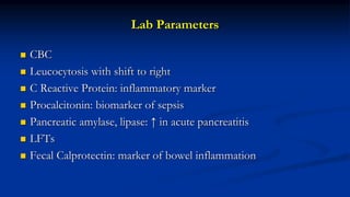
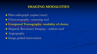
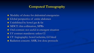
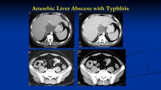
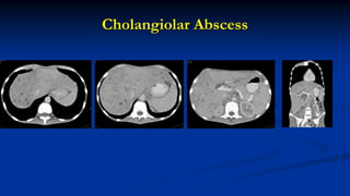
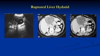
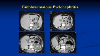
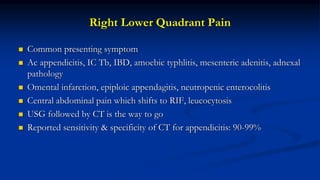
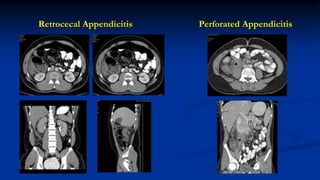
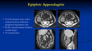
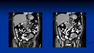
Clinical & Imaging Inputs in Acute Abdominal
Inflammatory Conditions
Prof. Raju Sharma
All India Institute of Medical Sciences, New Delhi
Clinical Inputs
History
 Pain: location, type, duration,
 Fever
 Vomiting
 Diarrhoea
 Distension
Examination
 Tachycardia
 Guarding
 Rigidity
 Lump
Lab Parameters
 CBC
 Leucocytosis with shift to right
 C Reactive Protein: inflammatory marker
 Procalcitonin: biomarker of sepsis
 Pancreatic amylase, lipase: ↑ in acute pancreatitis
 LFTs
 Fecal Calprotectin: marker of bowel inflammation
Spectrum
 Acute hepatitis
 Liver abscess
 Acute cholecystitis
 Cholangitis
 Acute pancreatitis
 Acute pyelonephritis
 Acute gynae disease: PID, TO
abscess
 Acute gastroenteritis
 Appendicitis
 Bowel obstruction
 Inflammatory Bowel Disease
 Enterocolitis: Infectious
 Diverticulitis
IMAGING MODALITIES
 Plain radiograph (supine/erect)
 Ultrasonography- screening tool
 Computed Tomography- modality of choice
 Magnetic Resonance Imaging – seldom used
 Angiography
 Image guided intervention
Computed Tomography
 Modality of choice for abdominal emergencies
 Global perspective of entire abdomen
 Uninhibited by bowel gas & fat
 MDCT: thin collimation, MPRs
 Oral contrast: not useful in emergent situation
 I.V contrast mandatory unless CI
 CT Angiography: bowel ischemia, GI bleed
 Radiation concern: ASIR, low dose protocols
Bowel Obstruction
 Small bowel obstruction is more common than large
 Common causes: adhesions, hernia, volvulus, inflammatory strictures,
intussusception, ischemic
 Accounts for 20% of acute abdominal surgical conditions
 Simple/Strangulated
 Complete/Incomplete
 Open/Closed loop
Acute Abdomen: Quadrant Approach
 Helps choose modality
 Narrows differential diagnosis
 ACR appropriateness criteria
 RUQ: USG (9) MRI & CT (6)
 LUQ
 RLQ: CT (8) USG (6)
 LLQ: CT (9) USG (4)
 Non-localized pain – CT (8) USG (4)
Beta HCG testing should be done in women of reproductive age group
Acute Abdomen
Bowel Obstruction
Perforation
Right Upper Quadrant Pain
Biliary pathology
Lower Quadrant Pain
Plain Radiographs
CT
USG
Bowel Ischemia
GI Bleed
CT Angiography
MRCP/CT DSA/Intervention
USG
CT
Right Upper Quadrant Pain
 Among the commonest reasons for ED visit
 USG is the modality of choice, occ CT or MRI are required
 Liver: Ac hepatitis, abscess, hydatid
 Gall Bladder: calculi, ac. cholecystitis & its complications,
Mirrizi syndrome
 Acute pancreatitis
 Peptic ulcer disease
Complications of Cholelithiasis
 Acute cholecystitis
 Perforation, peri-cholecystic abscess
 Pancreatitis
 Gall stone ileus
 Biliary fistula
 Mirrizi’s syndrome
Acute Cholecystitis
 Commonest cause: gall stones
 Sonography: modality of choice
 Hallmarks: thickened GB wall (>3mm), distended GB,
pericholecystic edema, sonographic Murphy’s sign (92% sensitivity)
 Acalculus cholecystitis – seen in sick ICU patients
 Gangrenous, Emphysematous, Xanthogranulomatous cholecystitis
Acute Cholecystitis
Gangrenous Cholecystitis
Striated thickening of wall, irregularity, intraluminal membrane, Murphy’s sign may be absent
Emphysematous Cholecystitis
• More common in males & diabetics
• Stones seen only in 50%
• Small vessel disease may be
responsible
• Murphy’s sign is often absent
• Organism: Cl. welchii, E coli
• Mortality 15%
Ruptured GB
Gall stone eroding & causing Pseudoaneurysm
Gall Stone Ileus
 Accounts for 1-5% of non-malignant small bowel
obstruction
 GB perforation with fistula formation with
duodenum
 Gall stone impaction occurs in ileum(54-65%),
jejunum (27%) and rarely duodenum (1-3%)
 Bouveret syndrome
 Riggler triad: bowel obstruction, pneumobilia,
obstr. Gall stone – seen in 30-40% patients
Mirrizi’s Syndrome
• CHD/ CBD obstruction due to impacted
calculus in GB neck/ cystic duct
• May be associated with cholecystocholedocal
fistula
• Uncommon, 2% of pts operated for
symptomatic gall stones
• Preop diagnosis is important: USG, MRI
• Standard technique of Sx: increased risk of
injury to CBD due to dense adhesions in hepato-
duodenal ligament
Liver Abscess
Amoebic Liver Abscess with Typhlitis
Cholangiolar Abscess
Ruptured Liver Hydatid
32yr old: pain, jaundice fever
Oriental Cholangiohepatitits (OCH)
• Common in south east Asia
• Rec episodes of RUQ pain, fever,
jaundice
• Multiple intra and extrahepatic
strictures
• Intraductal calculi specially in absence
of gall stones
• Segmental atrophy specially of lateral
segment of left lobe
Acute Pancreatitis
 Causes: gall stones, alcohol, abdominal surgery
 CT is the modality of choice
 Balthazar grading system and CT scoring system
 Ideal for detecting pancreatic necrosis
 Can diagnose complications of acute pancreatitis
Types
Interstitial Pancreatitis (80-90%) Necrotizing Pancreatitis (5-10%)
Necrosis
Pancreatic Peri-pancreatic Combined
Infected Walled off Necrosis
Complications Of Acute Pancreatitis
 Pseudocyst
 Infected walled off necrosis
 Pseudoaneurysms
 Vascular thrombosis
Left Upper Quadrant Pain
 Less common than other sites
 Splenic pathology: infarct, abscess, spontaneous rupture
 Gastritis
 Pancreatitis & its complications
 Left pyelonephritis
 Splenic flexure colitis, ischemia
 Sub-phrenic abscess
Splenic Infarction
 Causes: pancreatitis, portal
hypertension, sickle cell anemia,
splenomegaly
 Presents with left upper quadrant pain
 Peripherally located wedge shaped
hypodense, non-enhancing lesion
Splenic Abscess with Rupture
Emphysematous Pyelonephritis
Right Lower Quadrant Pain
 Common presenting symptom
 Ac appendicitis, IC Tb, IBD, amoebic typhlitis, mesenteric adenitis, adnexal
pathology
 Omental infarction, epiploic appendagitis, neutropenic enterocolitis
 Central abdominal pain which shifts to RIF, leucocytosis
 USG followed by CT is the way to go
 Reported sensitivity & specificity of CT for appendicitis: 90-99%
Retrocecal Appendicitis Perforated Appendicitis
Crohn’s Disease
Epiploic Appendagitis
• Oval fat density mass with a
central dot & an inflamed
peripheral hyperdense rim
• D/D: omental infarct which is
usually larger
• Tt: conservative
Omental Infarct
 Sudden onset of right lower
quadrant pain
 Fat density with stranding
 Usually > 5cm
 Primary/ secondary
 Usually self limiting
 Conservative Tt
Neutropenic Enterocolitis
• Oedema & inflammation of cecum, asc colon
& occ small bowel
• Combination of chemoRx induced mucosal
injury - facilitating infection, altered immune
response
• Should be considered in any neutropenic
patient (<500 cells/cumm) presenting with
fever, pain RIF, GI bleed &/or diarrhoea
Left Lower Quadrant Pain
 Causes: colitis, diverticulitis, urolithiasis, epiploic appendagitis, gynae causes
 CT is considered the modality of choice
 Colitis: ischemic, infective, pseudomembranous, UC, Crohn’s
 An empty colon sign favours infective colitis over ischemic & inflammatory
etiology
 Crohn’s & pseudomembranous colitis: greater wall thickening than in
ischemic & UC
 Adnexal pathology
Crohn’s Colitis
Ischemic Colitis
Diverticulitis
• Bowel wall thickening, peri-colonic
inflammation, diverticulosis
• Inflamed diverticuli may be
hyperdense on CT
• Sigmoid colon: most often involved
• Hinchey’s classification
• Perforated malignancy may mimic
perforated diverticulitis
Intra-Abdominal Abscess
 Post-operative patient
 Complication of appendicitis, bowel perforation
 Inflammatory Bowel Disease
 Diverticulitis
 On USG: fluid with debris
 Fluid attenuating lesion with thick rim enhancement
 On MRI: fluid signal with restricted diffusion
Sub-Phrenic Abscess
Post-Op Patient
Acute Abdomen in Pregnancy &
Women in Reproductive Age Group
 As far as possible avoid ionizing radiation
 Sonography first line
 MRI (no Gadolinium) is very useful
 CT only if benefit outweighs the risk
 Non-pregnancy related: biliary, acute appendicitis
 Pregnancy related: ectopic, abruption, HELLP
 Gynae causes: PID, TO abscess, ovarian hgic cyst, torsion
Conclusion
 Wide spectrum of diseases
 Clinical findings: often non-specific & frequently overlap
 Judicious use of imaging modalities: narrow D/d & triage patients
 Diagnostic laparotomy: thing of the past
 USG & CT: mainstay of imaging
 Niche indications for MRI
 Quality of imaging & interpretation must be high
Thank you for your attention!

Dr.raju sharma 1

  • 1.
    Clinical & ImagingInputs in Acute Abdominal Inflammatory Conditions Prof. Raju Sharma All India Institute of Medical Sciences, New Delhi
  • 2.
    Clinical Inputs History  Pain:location, type, duration,  Fever  Vomiting  Diarrhoea  Distension Examination  Tachycardia  Guarding  Rigidity  Lump
  • 3.
    Lab Parameters  CBC Leucocytosis with shift to right  C Reactive Protein: inflammatory marker  Procalcitonin: biomarker of sepsis  Pancreatic amylase, lipase: ↑ in acute pancreatitis  LFTs  Fecal Calprotectin: marker of bowel inflammation
  • 4.
    Spectrum  Acute hepatitis Liver abscess  Acute cholecystitis  Cholangitis  Acute pancreatitis  Acute pyelonephritis  Acute gynae disease: PID, TO abscess  Acute gastroenteritis  Appendicitis  Bowel obstruction  Inflammatory Bowel Disease  Enterocolitis: Infectious  Diverticulitis
  • 5.
    IMAGING MODALITIES  Plainradiograph (supine/erect)  Ultrasonography- screening tool  Computed Tomography- modality of choice  Magnetic Resonance Imaging – seldom used  Angiography  Image guided intervention
  • 6.
    Computed Tomography  Modalityof choice for abdominal emergencies  Global perspective of entire abdomen  Uninhibited by bowel gas & fat  MDCT: thin collimation, MPRs  Oral contrast: not useful in emergent situation  I.V contrast mandatory unless CI  CT Angiography: bowel ischemia, GI bleed  Radiation concern: ASIR, low dose protocols
  • 7.
    Bowel Obstruction  Smallbowel obstruction is more common than large  Common causes: adhesions, hernia, volvulus, inflammatory strictures, intussusception, ischemic  Accounts for 20% of acute abdominal surgical conditions  Simple/Strangulated  Complete/Incomplete  Open/Closed loop
  • 8.
    Acute Abdomen: QuadrantApproach  Helps choose modality  Narrows differential diagnosis  ACR appropriateness criteria  RUQ: USG (9) MRI & CT (6)  LUQ  RLQ: CT (8) USG (6)  LLQ: CT (9) USG (4)  Non-localized pain – CT (8) USG (4) Beta HCG testing should be done in women of reproductive age group
  • 9.
    Acute Abdomen Bowel Obstruction Perforation RightUpper Quadrant Pain Biliary pathology Lower Quadrant Pain Plain Radiographs CT USG Bowel Ischemia GI Bleed CT Angiography MRCP/CT DSA/Intervention USG CT
  • 10.
    Right Upper QuadrantPain  Among the commonest reasons for ED visit  USG is the modality of choice, occ CT or MRI are required  Liver: Ac hepatitis, abscess, hydatid  Gall Bladder: calculi, ac. cholecystitis & its complications, Mirrizi syndrome  Acute pancreatitis  Peptic ulcer disease
  • 11.
    Complications of Cholelithiasis Acute cholecystitis  Perforation, peri-cholecystic abscess  Pancreatitis  Gall stone ileus  Biliary fistula  Mirrizi’s syndrome
  • 12.
    Acute Cholecystitis  Commonestcause: gall stones  Sonography: modality of choice  Hallmarks: thickened GB wall (>3mm), distended GB, pericholecystic edema, sonographic Murphy’s sign (92% sensitivity)  Acalculus cholecystitis – seen in sick ICU patients  Gangrenous, Emphysematous, Xanthogranulomatous cholecystitis
  • 13.
  • 14.
    Gangrenous Cholecystitis Striated thickeningof wall, irregularity, intraluminal membrane, Murphy’s sign may be absent
  • 15.
    Emphysematous Cholecystitis • Morecommon in males & diabetics • Stones seen only in 50% • Small vessel disease may be responsible • Murphy’s sign is often absent • Organism: Cl. welchii, E coli • Mortality 15%
  • 16.
  • 17.
    Gall stone eroding& causing Pseudoaneurysm
  • 18.
    Gall Stone Ileus Accounts for 1-5% of non-malignant small bowel obstruction  GB perforation with fistula formation with duodenum  Gall stone impaction occurs in ileum(54-65%), jejunum (27%) and rarely duodenum (1-3%)  Bouveret syndrome  Riggler triad: bowel obstruction, pneumobilia, obstr. Gall stone – seen in 30-40% patients
  • 19.
    Mirrizi’s Syndrome • CHD/CBD obstruction due to impacted calculus in GB neck/ cystic duct • May be associated with cholecystocholedocal fistula • Uncommon, 2% of pts operated for symptomatic gall stones • Preop diagnosis is important: USG, MRI • Standard technique of Sx: increased risk of injury to CBD due to dense adhesions in hepato- duodenal ligament
  • 20.
  • 21.
    Amoebic Liver Abscesswith Typhlitis
  • 22.
  • 23.
  • 24.
    32yr old: pain,jaundice fever
  • 25.
    Oriental Cholangiohepatitits (OCH) •Common in south east Asia • Rec episodes of RUQ pain, fever, jaundice • Multiple intra and extrahepatic strictures • Intraductal calculi specially in absence of gall stones • Segmental atrophy specially of lateral segment of left lobe
  • 26.
    Acute Pancreatitis  Causes:gall stones, alcohol, abdominal surgery  CT is the modality of choice  Balthazar grading system and CT scoring system  Ideal for detecting pancreatic necrosis  Can diagnose complications of acute pancreatitis
  • 27.
    Types Interstitial Pancreatitis (80-90%)Necrotizing Pancreatitis (5-10%)
  • 28.
  • 30.
  • 31.
    Complications Of AcutePancreatitis  Pseudocyst  Infected walled off necrosis  Pseudoaneurysms  Vascular thrombosis
  • 32.
    Left Upper QuadrantPain  Less common than other sites  Splenic pathology: infarct, abscess, spontaneous rupture  Gastritis  Pancreatitis & its complications  Left pyelonephritis  Splenic flexure colitis, ischemia  Sub-phrenic abscess
  • 33.
    Splenic Infarction  Causes:pancreatitis, portal hypertension, sickle cell anemia, splenomegaly  Presents with left upper quadrant pain  Peripherally located wedge shaped hypodense, non-enhancing lesion
  • 34.
  • 35.
  • 36.
    Right Lower QuadrantPain  Common presenting symptom  Ac appendicitis, IC Tb, IBD, amoebic typhlitis, mesenteric adenitis, adnexal pathology  Omental infarction, epiploic appendagitis, neutropenic enterocolitis  Central abdominal pain which shifts to RIF, leucocytosis  USG followed by CT is the way to go  Reported sensitivity & specificity of CT for appendicitis: 90-99%
  • 38.
  • 39.
  • 40.
    Epiploic Appendagitis • Ovalfat density mass with a central dot & an inflamed peripheral hyperdense rim • D/D: omental infarct which is usually larger • Tt: conservative
  • 41.
    Omental Infarct  Suddenonset of right lower quadrant pain  Fat density with stranding  Usually > 5cm  Primary/ secondary  Usually self limiting  Conservative Tt
  • 42.
    Neutropenic Enterocolitis • Oedema& inflammation of cecum, asc colon & occ small bowel • Combination of chemoRx induced mucosal injury - facilitating infection, altered immune response • Should be considered in any neutropenic patient (<500 cells/cumm) presenting with fever, pain RIF, GI bleed &/or diarrhoea
  • 43.
    Left Lower QuadrantPain  Causes: colitis, diverticulitis, urolithiasis, epiploic appendagitis, gynae causes  CT is considered the modality of choice  Colitis: ischemic, infective, pseudomembranous, UC, Crohn’s  An empty colon sign favours infective colitis over ischemic & inflammatory etiology  Crohn’s & pseudomembranous colitis: greater wall thickening than in ischemic & UC  Adnexal pathology
  • 44.
  • 45.
  • 46.
    Diverticulitis • Bowel wallthickening, peri-colonic inflammation, diverticulosis • Inflamed diverticuli may be hyperdense on CT • Sigmoid colon: most often involved • Hinchey’s classification • Perforated malignancy may mimic perforated diverticulitis
  • 48.
    Intra-Abdominal Abscess  Post-operativepatient  Complication of appendicitis, bowel perforation  Inflammatory Bowel Disease  Diverticulitis  On USG: fluid with debris  Fluid attenuating lesion with thick rim enhancement  On MRI: fluid signal with restricted diffusion
  • 49.
  • 50.
  • 51.
    Acute Abdomen inPregnancy & Women in Reproductive Age Group  As far as possible avoid ionizing radiation  Sonography first line  MRI (no Gadolinium) is very useful  CT only if benefit outweighs the risk  Non-pregnancy related: biliary, acute appendicitis  Pregnancy related: ectopic, abruption, HELLP  Gynae causes: PID, TO abscess, ovarian hgic cyst, torsion
  • 52.
    Conclusion  Wide spectrumof diseases  Clinical findings: often non-specific & frequently overlap  Judicious use of imaging modalities: narrow D/d & triage patients  Diagnostic laparotomy: thing of the past  USG & CT: mainstay of imaging  Niche indications for MRI  Quality of imaging & interpretation must be high
  • 53.
    Thank you foryour attention!