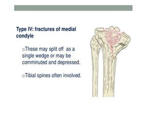
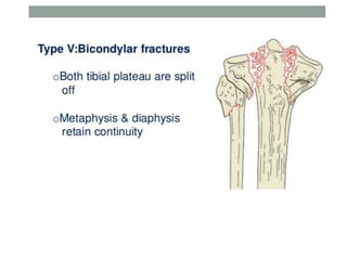
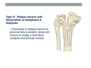
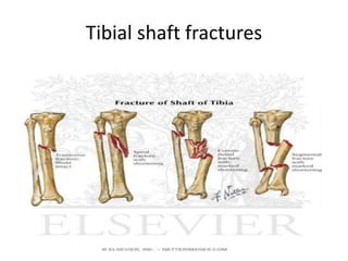
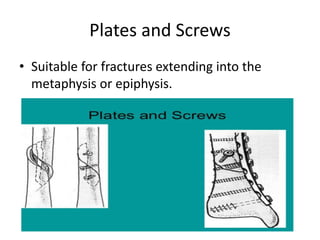
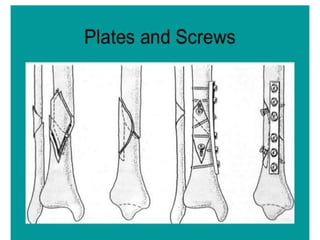
TIBIA FRACTURES
Presented by:
Bimal Pokharel
MBBS 4TH BATCH
INTERN
Tibial Plateau
o The tibial plateau is the proximal end of the
tibia including the metaphyseal and
epiphyseal regions as well as articular surfaces
made up of hyaline cartilage.
o It is one of the most critical load bearing
areas in human body ; fractures of the plateau
affect knee alignment, stability and motion.
TIBIAL PLATEAU FRACTURES
Tibial plateau fractures comprises 1% of all
fractures and 8% of all fractures of elderly
Most of these fractures are associated
with:
1) Neurological and vascular injuries
2) Compartment syndrome
3) Contusion and crush injuries of soft
tissue
Mechanism of injury
• Caused by valgus or varus forces combined
with axial loading
• Fall from a height in which the knee is forced
into valgus and varus. The tibial condyle is
crushed or split by the opposing femoral
condyle, which remains intact
Classification of tibial plateau fractures
Associated injuries
• 90% of these fractures are associated with soft
tissue injuries
• Meniscal injuries occurs in 50% of these
fractures
• Associated ligamentous injuries ( cruciate or
collateral ligaments) occurs in 30% of these
fractures
• Others: common peroneal nerve injuries
popliteal artery injury
Evaluation of injury
• Clinical evaluation:
1) Neurovascular examination to rule out any
neurological or vascular injury
2) Assessment for any ligament injury
3) Assessment for compartment syndrome
4) Assessment for haemarthrosis
Radiographic evaluation
• X-Ray:
a) AP view
b) Lateral view
c) 40 degree internal rotation view ( lateral plateau)
d) 40 degree external rotation view ( medial plateau)
• CT: provides information on:
 Location of main fracture lines
 The site and size of the portion of condyle that is depressed
 Position of major parts of articular surface that have been
displaced
• MRI:
Useful for evaluating the injuries of menisci
and cruciate and collateral ligaments injuries
Treatment
• Type I fractures:
 Undisplaced type 1 fractures can be treated
conservatively.
 Haemarthrosis is aspirated and compression bandage is
applied.
 As soon as acute pain and swelling subsides, a hinged-cast
brace is applied; however weight bearing is not allowed for
another 3 weeks.
 Thereafter partial weightbearing is permitted but full
weight bearing is delayed until the bone has fully healed ( 8
weeks)
 Displaced fractures are treated by ORIF method. Two lag
screws or buttress plate are usually sufficient for fixation.
• Type II fractures
 If the depression is slight ( less than 5 mm)
and the knee is stable or if the patient is old,
the fracture is treated by skeletal traction.
 After 3 to 4 weeks, traction pin is removed
and a hinged- cast brace is applied and patient
is allowed to move on crutches. Full weight
bearing is restricted for another 6 weeks
If the depression is more ( more than 5 mm),
in younger patients and with central
depression, ORIF is preferred
Tibial shaft fractures
TIBIAL SHAFT FRACTURES
• Mechanism of injury:
Direct injury: RTA are the commonest cause of
these fractures, mostly due to direct violence.
Frequently the object causing the fracture lacerates
the skin over it, resulting in open fracture.
Indirect injury: A bending or torsional force on the
tibia may result in an oblique or spiral fracture. The
sharp edge of fracture fragment may pierce the
skin from within, resulting in an open fracture.
Patho-anatomy
• May be closed or open
• May occur at various levels ( upper, middle or
lower thirds )
• Displacements may be sideways, angulatory or
rotational.
Imaging
• Diagnosis usually cofirmed by x- rays
• CT
• MRI
• Arteriography or venography
Acceptable Fracture Reduction
• Less than 5 degrees of valgus/ varus
angulation is recommended
• Less than 10 degrees of anterior/ posterior
angulation is recommended ( < 5 degrees
preferred)
• Less than 10 degrees of rotational deformity is
and less than 1 cm of shortening.
• More than 50% of cortical contact is
recommended.
NON-OPERATIVE MANAGEMENT
• Fracture reduction followed by application of
a long leg cast with progressive weight bearing
can be used for fractures with minimal
displacement
• Cast with the knee in 0 to 5 degrees of flexon
to allow for weight bearing with crutches as
soon as tolerated by patient with full weight
bearing by 2nd to 4th week
Operative treatment
Intramedullary ( IM) nailing:
 Method of choice for internal fixation
 IM nailing carries the advantage of preservation of
periosteal blood supply and limited soft tissue damage.
 Fracture is reduced under x ray control, proximal end of
tibia is exposed ; a guide wire is passed down the medullary
canal and the canal is reamed
 A nail of appropriate size and shape is then introduced
from the proximal end across the fracture site.
 Transverse locking screws are inserted at the proximal and
distal ends
 Postoperatively, partial weightbearing is started early.
• Plate Fixation:
Plating is suitable for metaphyseal fractures
that are not suitable for nailing.
• External Fixation:
Primarily used to treat severe open fractures, it
can also be used in closed fractures complicated
by compartment syndrome.
Union rates : 90% with about 4-6 months for
union.
Plates and Screws
• Suitable for fractures extending into the
metaphysis or epiphysis.
Complications
1) Delayed union and non union
2) Malunion
3) Infections
4) Compartment syndrome
5) Injury to major vessels and nerves
Fracture of the malleolus
• Fractures and fracture dislocations of the
ankle is common.

Tibia fractures

  • 1.
    TIBIA FRACTURES Presented by: BimalPokharel MBBS 4TH BATCH INTERN
  • 2.
    Tibial Plateau o Thetibial plateau is the proximal end of the tibia including the metaphyseal and epiphyseal regions as well as articular surfaces made up of hyaline cartilage. o It is one of the most critical load bearing areas in human body ; fractures of the plateau affect knee alignment, stability and motion.
  • 3.
    TIBIAL PLATEAU FRACTURES Tibialplateau fractures comprises 1% of all fractures and 8% of all fractures of elderly Most of these fractures are associated with: 1) Neurological and vascular injuries 2) Compartment syndrome 3) Contusion and crush injuries of soft tissue
  • 4.
    Mechanism of injury •Caused by valgus or varus forces combined with axial loading • Fall from a height in which the knee is forced into valgus and varus. The tibial condyle is crushed or split by the opposing femoral condyle, which remains intact
  • 5.
    Classification of tibialplateau fractures
  • 11.
    Associated injuries • 90%of these fractures are associated with soft tissue injuries • Meniscal injuries occurs in 50% of these fractures • Associated ligamentous injuries ( cruciate or collateral ligaments) occurs in 30% of these fractures • Others: common peroneal nerve injuries popliteal artery injury
  • 12.
    Evaluation of injury •Clinical evaluation: 1) Neurovascular examination to rule out any neurological or vascular injury 2) Assessment for any ligament injury 3) Assessment for compartment syndrome 4) Assessment for haemarthrosis
  • 13.
    Radiographic evaluation • X-Ray: a)AP view b) Lateral view c) 40 degree internal rotation view ( lateral plateau) d) 40 degree external rotation view ( medial plateau) • CT: provides information on:  Location of main fracture lines  The site and size of the portion of condyle that is depressed  Position of major parts of articular surface that have been displaced
  • 14.
    • MRI: Useful forevaluating the injuries of menisci and cruciate and collateral ligaments injuries
  • 15.
    Treatment • Type Ifractures:  Undisplaced type 1 fractures can be treated conservatively.  Haemarthrosis is aspirated and compression bandage is applied.  As soon as acute pain and swelling subsides, a hinged-cast brace is applied; however weight bearing is not allowed for another 3 weeks.  Thereafter partial weightbearing is permitted but full weight bearing is delayed until the bone has fully healed ( 8 weeks)  Displaced fractures are treated by ORIF method. Two lag screws or buttress plate are usually sufficient for fixation.
  • 16.
    • Type IIfractures  If the depression is slight ( less than 5 mm) and the knee is stable or if the patient is old, the fracture is treated by skeletal traction.  After 3 to 4 weeks, traction pin is removed and a hinged- cast brace is applied and patient is allowed to move on crutches. Full weight bearing is restricted for another 6 weeks If the depression is more ( more than 5 mm), in younger patients and with central depression, ORIF is preferred
  • 22.
  • 23.
    TIBIAL SHAFT FRACTURES •Mechanism of injury: Direct injury: RTA are the commonest cause of these fractures, mostly due to direct violence. Frequently the object causing the fracture lacerates the skin over it, resulting in open fracture. Indirect injury: A bending or torsional force on the tibia may result in an oblique or spiral fracture. The sharp edge of fracture fragment may pierce the skin from within, resulting in an open fracture.
  • 24.
    Patho-anatomy • May beclosed or open • May occur at various levels ( upper, middle or lower thirds ) • Displacements may be sideways, angulatory or rotational.
  • 25.
    Imaging • Diagnosis usuallycofirmed by x- rays • CT • MRI • Arteriography or venography
  • 26.
    Acceptable Fracture Reduction •Less than 5 degrees of valgus/ varus angulation is recommended • Less than 10 degrees of anterior/ posterior angulation is recommended ( < 5 degrees preferred) • Less than 10 degrees of rotational deformity is and less than 1 cm of shortening. • More than 50% of cortical contact is recommended.
  • 27.
    NON-OPERATIVE MANAGEMENT • Fracturereduction followed by application of a long leg cast with progressive weight bearing can be used for fractures with minimal displacement • Cast with the knee in 0 to 5 degrees of flexon to allow for weight bearing with crutches as soon as tolerated by patient with full weight bearing by 2nd to 4th week
  • 28.
    Operative treatment Intramedullary (IM) nailing:  Method of choice for internal fixation  IM nailing carries the advantage of preservation of periosteal blood supply and limited soft tissue damage.  Fracture is reduced under x ray control, proximal end of tibia is exposed ; a guide wire is passed down the medullary canal and the canal is reamed  A nail of appropriate size and shape is then introduced from the proximal end across the fracture site.  Transverse locking screws are inserted at the proximal and distal ends  Postoperatively, partial weightbearing is started early.
  • 30.
    • Plate Fixation: Platingis suitable for metaphyseal fractures that are not suitable for nailing. • External Fixation: Primarily used to treat severe open fractures, it can also be used in closed fractures complicated by compartment syndrome. Union rates : 90% with about 4-6 months for union.
  • 32.
    Plates and Screws •Suitable for fractures extending into the metaphysis or epiphysis.
  • 34.
    Complications 1) Delayed unionand non union 2) Malunion 3) Infections 4) Compartment syndrome 5) Injury to major vessels and nerves
  • 35.
    Fracture of themalleolus • Fractures and fracture dislocations of the ankle is common.