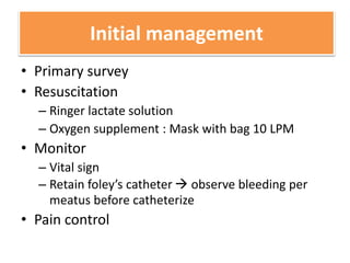
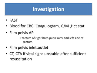
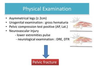
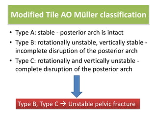
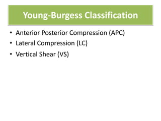
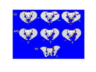
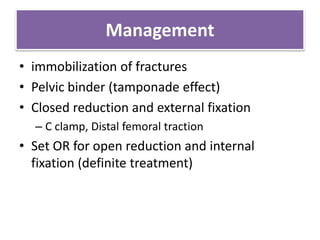
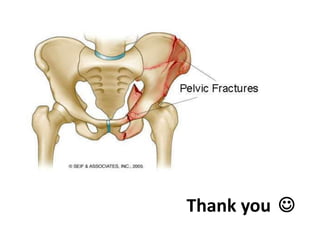
Case discussion
Emergency conditions in orthopaedic
Group 1
Rotate 503-504
Faculty of Medicine Siriraj Hospital,Mahidol University
Pelvic injury with hypovolumic shock
ผู้ป่วยชายไทยอายุ 30 ปี ประสบอุบัติเหตุจราจร 15 นาที ก่อนมา
โรงพยาบาล ไม่รู้สึกตัว ตรวจร่างกาย BP 80/50 mmHg pulse 120/min
pelvic compression test +ve ขาขวาสั้นกว่าอีกข้าง 3 ซม.
Initial management
• Primary survey
• Resuscitation
– Ringer lactate solution
– Oxygen supplement : Mask with bag 10 LPM
• Monitor
– Vital sign
– Retain foley’s catheter  observe bleeding per
meatus before catheterize
• Pain control
Investigation
• FAST
• Blood for CBC, Coagulogram, G/M ,Hct stat
• Film pelvis AP
Fracture of right both pubic rami and left side of
sacrum
• Film pelvis inlet,outlet
• CT, CTA if vital signs unstable after sufficient
resuscitation
Physical Examination
• Asymmetrical legs (≥ 2cm)
• Urogenital examination : gross hematuria
• Pelvic compression test positive (AP, Lat.)
• Neurovascular injury
- lower extremities pulse
- neurological examination : DRE, DTR
Pelvic fracture
Modified Tile AO Müller classification
• Type A: stable - posterior arch is intact
• Type B: rotationally unstable, vertically stable -
incomplete disruption of the posterior arch
• Type C: rotationally and vertically unstable -
complete disruption of the posterior arch
Type B, Type C  Unstable pelvic fracture
Young-Burgess Classification
• Anterior Posterior Compression (APC)
• Lateral Compression (LC)
• Vertical Shear (VS)
Management
• immobilization of fractures
• Pelvic binder (tamponade effect)
• Closed reduction and external fixation
– C clamp, Distal femoral traction
• Set OR for open reduction and internal
fixation (definite treatment)
Thank you 

Orthopedic pelvic fracture

  • 1.
    Case discussion Emergency conditionsin orthopaedic Group 1 Rotate 503-504 Faculty of Medicine Siriraj Hospital,Mahidol University
  • 2.
    Pelvic injury withhypovolumic shock ผู้ป่วยชายไทยอายุ 30 ปี ประสบอุบัติเหตุจราจร 15 นาที ก่อนมา โรงพยาบาล ไม่รู้สึกตัว ตรวจร่างกาย BP 80/50 mmHg pulse 120/min pelvic compression test +ve ขาขวาสั้นกว่าอีกข้าง 3 ซม.
  • 3.
    Initial management • Primarysurvey • Resuscitation – Ringer lactate solution – Oxygen supplement : Mask with bag 10 LPM • Monitor – Vital sign – Retain foley’s catheter  observe bleeding per meatus before catheterize • Pain control
  • 4.
    Investigation • FAST • Bloodfor CBC, Coagulogram, G/M ,Hct stat • Film pelvis AP Fracture of right both pubic rami and left side of sacrum • Film pelvis inlet,outlet • CT, CTA if vital signs unstable after sufficient resuscitation
  • 5.
    Physical Examination • Asymmetricallegs (≥ 2cm) • Urogenital examination : gross hematuria • Pelvic compression test positive (AP, Lat.) • Neurovascular injury - lower extremities pulse - neurological examination : DRE, DTR Pelvic fracture
  • 6.
    Modified Tile AOMüller classification • Type A: stable - posterior arch is intact • Type B: rotationally unstable, vertically stable - incomplete disruption of the posterior arch • Type C: rotationally and vertically unstable - complete disruption of the posterior arch Type B, Type C  Unstable pelvic fracture
  • 7.
    Young-Burgess Classification • AnteriorPosterior Compression (APC) • Lateral Compression (LC) • Vertical Shear (VS)
  • 9.
    Management • immobilization offractures • Pelvic binder (tamponade effect) • Closed reduction and external fixation – C clamp, Distal femoral traction • Set OR for open reduction and internal fixation (definite treatment)
  • 10.

Editor's Notes

  • #6 True length วัดจาก asis ไป lat malleolus Apparent วัดจากสะดือไป lat malleolus
  • #7 Type Description Type A: stable - posterior arch is intact A1: fracture does not involve the pelvic ring (avulsion fracture or fracture of the iliac wing) - A1.1: iliac spine - A1.2: iliac crest - A1.3: ischial tuberosity A2: stable or minimally displaced fracture of the pelvic ring - A2.1: iliac wing fractures - A2.2: unilateral fracture of anterior arch - A2.3: bifocal fracture of anterior arch A3: transverse fracture of the sacrum - A3.1: sacrococcygeal dislocation - A3.2: sacrum undisplaced - A3.3: sacrum displaced Type B: rotationally unstable, vertically stable - incomplete disruption of the posterior arch B1: open book injury (external rotation) - B1.1: sacroiliac joint, anterior disruption - B1.2: sacral fracture B2: lateral compression injury (internal rotation) - B2.1: anterior compression fracture, sacrum - B2.2: partial sacroiliac joint fracture, subluxation - B2.3: incomplete posterior iliac fracture B3: bilateral type B fracture - B3.1: bilateral open book fracture - B3.2: open book fracture and lateral compression - B3.3: bilateral lateral compression Type C: rotationally and vertically unstable - complete disruption of the posterior arch   C1: unilateral fracture - C1.1: fracture of the iliac bone - C1.2: sacroiliac dislocation and/or fracture dislocation - C1.3: sacral fracture C2: bilateral fracture with one side type B fracture (rotationally unstable) and one side type C fracture (vertically unstable) C3: bilateral fracture with both sides type C fracture (both sides completely unstable)
  • #8 APC I Symphysis widening < 2.5 cm Non-operative. Protected weight bearing   APC II Symphysis widening > 2.5 cm. Anterior SI joint diastasis  . Posterior SI ligaments intact. Disruption of sacrospinous and sacrotuberous ligaments.  Anterior symphyseal plate or external fixator +/- posterior fixation APC III Disruption of anterior and posterior SI ligaments (SI dislocation). Disruption of sacrospinous and sacrotuberous ligaments. APCIII associated with vascular injury   Anterior symphyseal multi-hole plate or external fixator and posterior stabilization with SI screws or plate/screws Lateral Compression (LC) LC Type I Oblique or transverse ramus fracture and ipsilateral anterior sacral ala compression fracture.  Non-operative. Protected weight bearing (complete, comminuted sacral component. Weight bearing as tolerated (simple, incomplete sacral fracture).  LC Type II Rami fracture and ipsilateral posterior ilium fracture dislocation (crescent fracture).  Open reduction and internal fixation of ilium   LC Type III Ipsilateral lateral compression and contralateral APC (windswept pelvis).  Common mechanism is rollover vehicle accident or pedestrian vs auto.  Posterior stabilization with plate or SI screws as needed. Percutaneous or open based on injury pattern and surgeon preference.    Vertical Shear (VS) Vertical shear Posterior and superior directed force.  Associated with the highest risk of hypovolemic shock (63%); mortality rate up to 25% Posterior stabilization with plate or SI screws as needed. Percutaneous or open based on injury pattern and surgeon preference.   http://www.orthobullets.com/trauma/1030/pelvic-ring-fractures