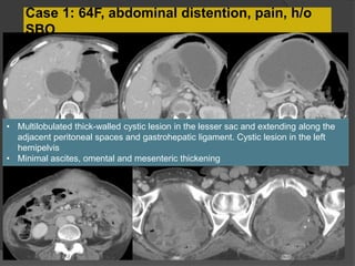
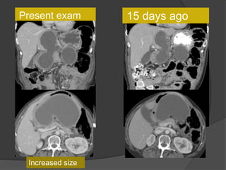
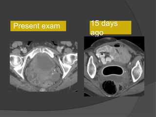
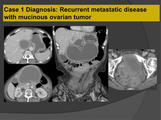
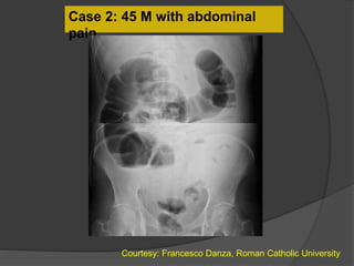
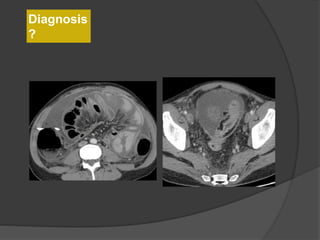
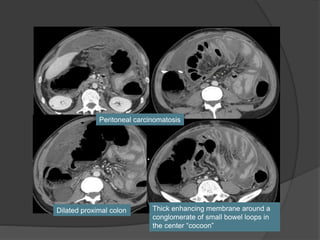
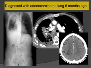
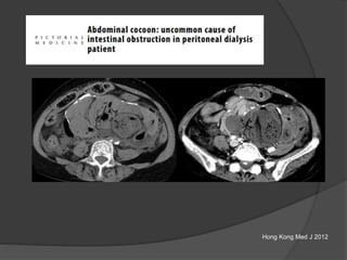
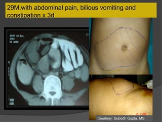
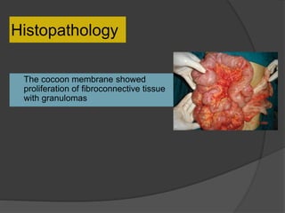
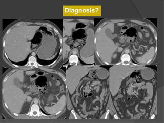
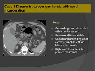
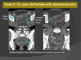
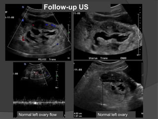
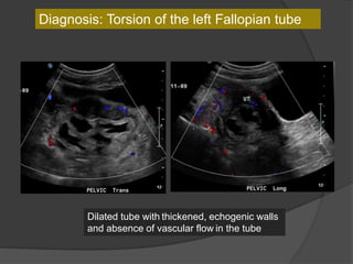
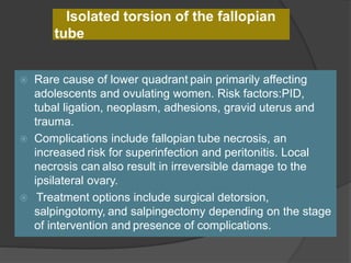
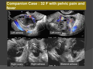
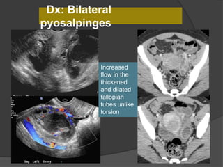
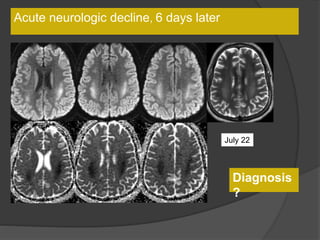
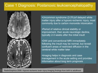
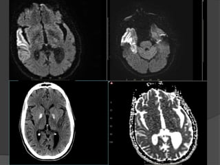
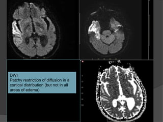
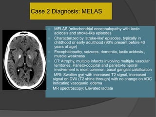
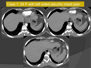
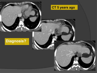
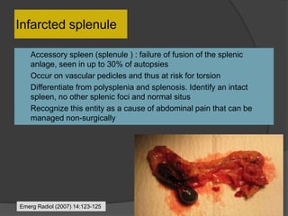
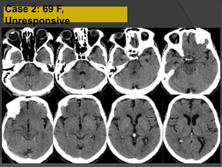
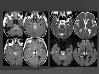
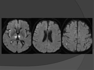
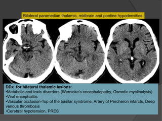
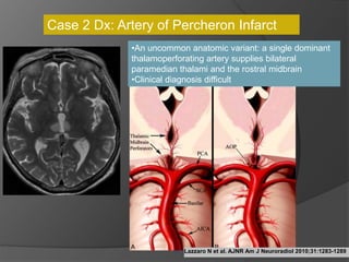
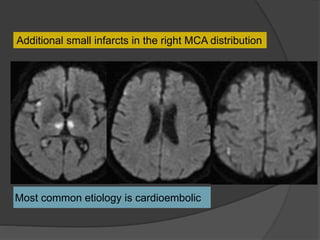
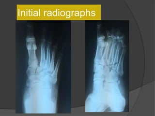
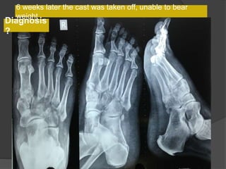
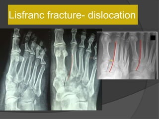
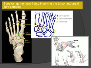
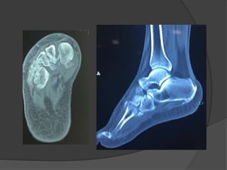
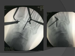
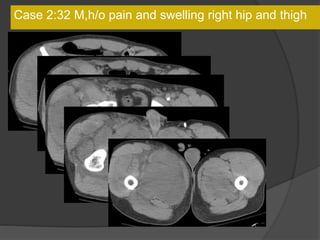
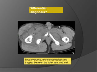
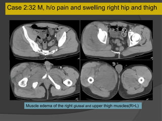
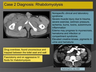
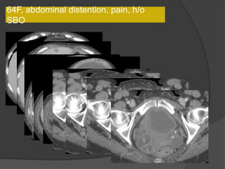
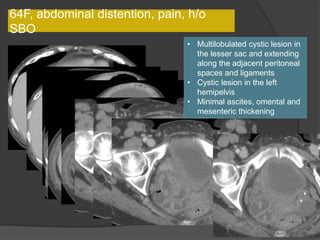
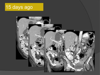
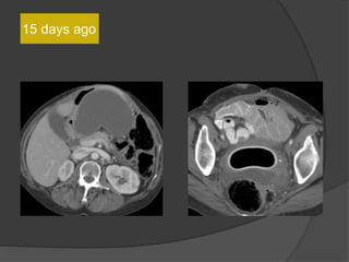
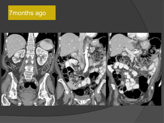
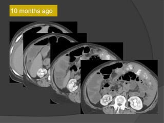
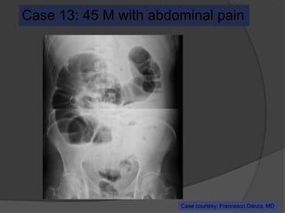
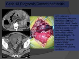
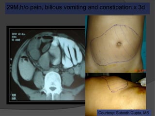
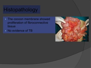
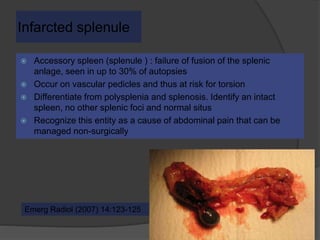
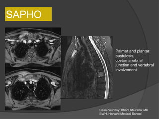
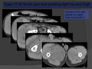
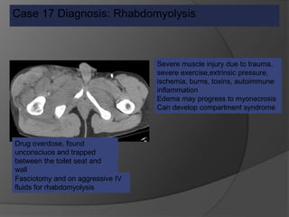
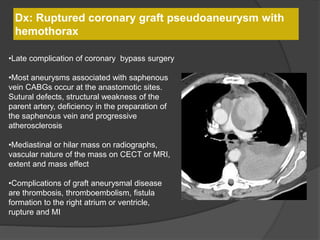
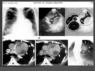
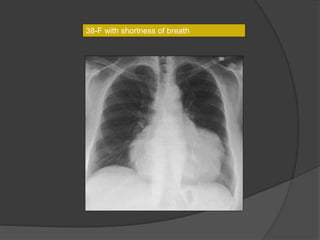
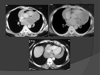
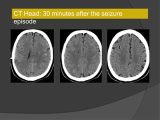
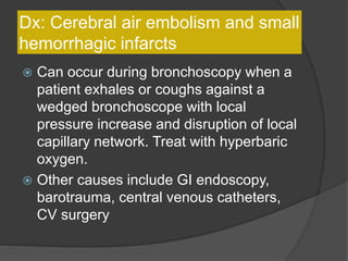
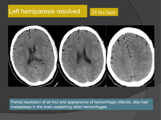
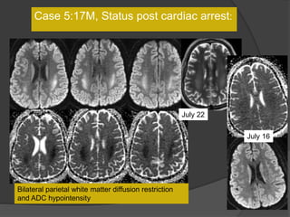
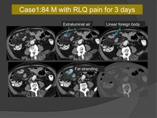
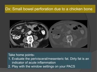
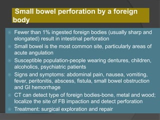
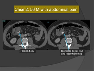
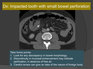
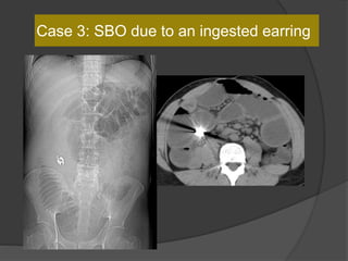
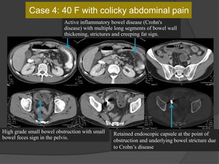
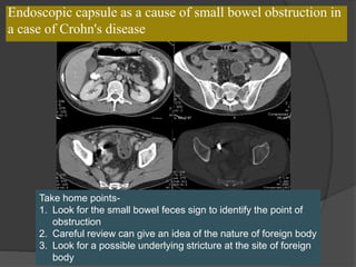
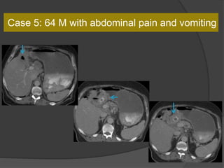
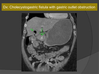
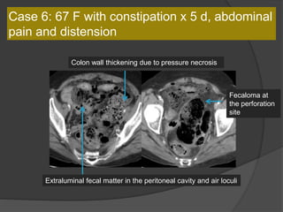
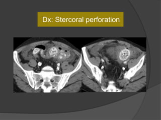
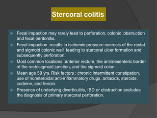
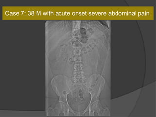
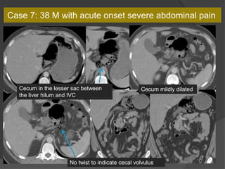
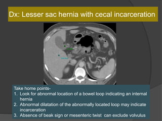
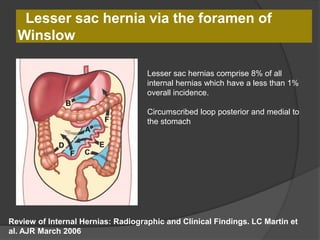
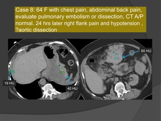
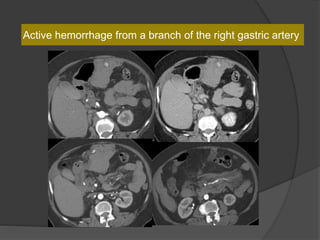
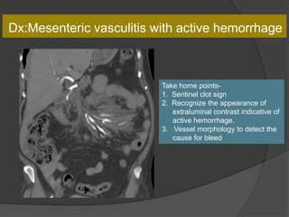
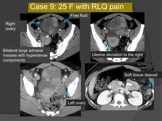
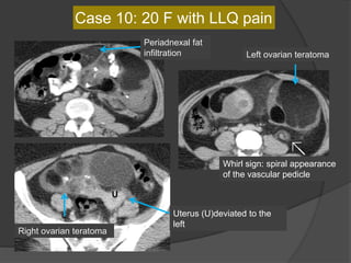
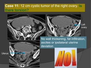
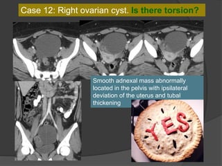
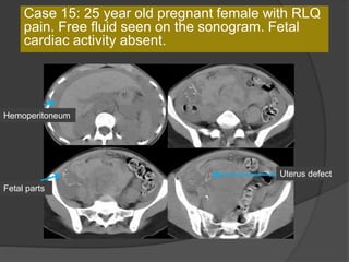
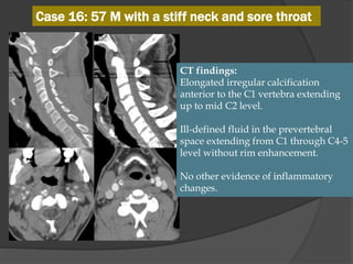
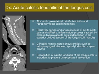
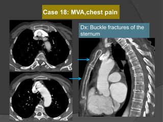
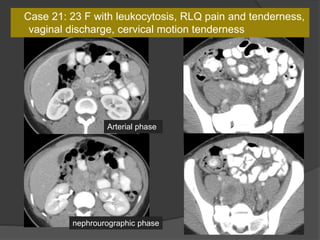
ER potpourri-
Film reading panel
Anjali Agrawal, MD
Consultant,Teleradiology Solutions
SER 2016, Bangalore
Panelists
 Dr Raju Sharma
 Dr Shanmuganathan
 Dr Dinesh Varma
 Dr Rathachai Kaewlai
 Dr Adnan Sheikh
MBBS: Maulana Azad Medical College, New Delhi
MD: AIIMS, New Delhi
Fellowship in GI Radiology: Massachusetts General Hospital, Boston
Joined as Assistant Professor, AIIMS in 1993
Professor in Dept of Radiology AIIMS, New Delhi since 2008
Area of Interest: Abdominal Imaging
RAJU SHARMA, MD, MAMS
Case 1: 64F, abdominal distention, pain, h/o
SBO
Axial CT images
Coronal images
15 days ago
15d ago
This lesion is larger compared to the CT 15 days
ago
Differential
Diagnosis?
7months ago
10 months ago
Case 1: 64F, abdominal distention, pain, h/o
SBO
• Multilobulated thick-walled cystic lesion in the lesser sac and extending along the
adjacent peritoneal spaces and gastrohepatic ligament. Cystic lesion in the left
hemipelvis
• Minimal ascites, omental and mesenteric thickening
Present exam 15 days ago
Increased size
15 days
ago
Present exam
Case 1 Diagnosis: Recurrent metastatic disease
with mucinous ovarian tumor
Clinical clues are useful
Case 2: 45 M with abdominal
pain
Courtesy: Francesco Danza, Roman Catholic University
6 months ago
Diagnosis
?
Peritoneal carcinomatosis
Thick enhancing membrane around a
conglomerate of small bowel loops in
the center “cocoon”
Dilated proximal colon
Diagnosed with adenocarcinoma lung 6 months ago
Case 2 Diagnosis: “Cocoon
peritonitis”
•AKA sclerosing
encapsulating peritonitis
•Rare cause of bowel
obstruction due to fibrotic
encapsulation of the bowel
forming a sac or cocoon
•May be idiopathic or
secondary to chronic
peritoneal dialysis, TB,
sarcoidosis, GI malignancy,
fibrogenic foreign material
•Treatment –Surgical
removal of the covering
membrane
Hong Kong Med J 2012
29M,with abdominal pain, bilious vomiting and
constipation x 3d
Courtesy: Subodh Gupta, MS
Histopathology
 The cocoon membrane showed
proliferation of fibroconnective tissue
with granulomas
RATHACHAI KAEWLAI, MD
 Ramathibodi Hospital, Faculty of
Medicine, Mahidol University,
Bangkok,Thailand
 Subspecialties: Emergency radiology and
body imaging
 Training:
 MD – Siriraj Hospital, BKK
 Residency – Ramathibodi, BKK
 American Board (Diagnostic Radiology) –
MGH, Boston, USA
 Clinical Fellowships – MGH (Boston) and
NEOUCOM (Ohio)
Case 1: 38 M with acute onset severe abdominal
pain
Cecum in the lesser sac
Diagnosis?
Cecum in the lesser sac
Cecum mildly dilatedCecum in the lesser sac between
the liver hilum and IVC
No twist to indicate cecal volvulus
Lesser Sac
Memorangapp.com
Case 1 Diagnosis: Lesser sac hernia with cecal
incarceration
 Cecum large and distended
within the lesser sac
 Cecum and bowel viable
 Cecum and ascending colon
extremely mobile with no
lateral attachments
 Right colectomy done to
prevent recurrence
Surgery:
Lesser sac hernia via the foramen of
Winslow
Review of Internal Hernias: Radiographic and Clinical Findings. LC Martin et
al. AJR March 2006
Lesser sac hernias comprise 8% of all
internal hernias which have a less than 1%
overall incidence.
Circumscribed loop posterior and medial to
the stomach
Case 2: 12 F with abdominal pain
Follow-up US
Diagnosis
?
Case 2: 12 year old female with abdominal pain
Dilated fallopian tube with thickened and enhancing tubal wall
Right
ovary
Complex
tubular
mass
Follow-up US
Normal left ovaryNormal left ovary flow
Diagnosis: Torsion of the left Fallopian tube
Dilated tube with thickened, echogenic walls
and absence of vascular flow in the tube
Isolated torsion of the fallopian
tube
 Rare cause of lower quadrant pain primarily affecting
adolescents and ovulating women. Risk factors:PID,
tubal ligation, neoplasm, adhesions, gravid uterus and
trauma.
 Complications include fallopian tube necrosis, an
increased risk for superinfection and peritonitis. Local
necrosis can also result in irreversible damage to the
ipsilateral ovary.
 Treatment options include surgical detorsion,
salpingotomy, and salpingectomy depending on the stage
of intervention and presence of complications.
Companion Case : 32 F with pelvic pain and
fever
Left ovaryLeft adnexa
Right ovary Right adnexa Bilateral adnexa
Dx: Bilateral
pyosalpinges
Increased
flow in the
thickened
and dilated
fallopian
tubes unlike
torsion
Acting Director Radiology; Head of
Emergency/Trauma Radiology
The Alfred Hospital, Melbourne, Australia
Areas of Interest:Emergency / Trauma
Radiology
Past President RANZCR
Chairman :ANZERG
President Elect: AOSR
DINESH VARMA, MBBS,
FRANZCR
Case 1:17M, Status post cardiac
arrest
July 16
Acute neurologic decline, 6 days later
July 22
Diagnosis
?
Case 1:17M, Status post cardiac arrest:
July 22
July 16
Bilateral parietal white matter diffusion restriction
and ADC hypointensity
Case 1 Diagnosis: Postanoxic leukoencephalopathy
•Uncommon syndrome (2-3%)of delayed white
matter injury after a hypoxic-ischemic injury, most
commonly due to carbon monoxide intoxication
•Period of relative clinical stability or
improvement, then acute neurologic decline,
typically 2-3 weeks after the initial insult
•DWI and conventional MRI immediately
following the insult may be normal, but reveal
confluent areas of restricted diffusion in the
cerebral white matter later
•Imaging helps in diagnosis and case
management in the acute setting and provides
information about long term prognosis
RadioGraphics 2008. Hypoxic-ischemic brain injury:Imaging findings from birth to adulthood
June 2014
Case 2: 41M, AMS, s/p
seizure
Courtesy: Matt Fox, MD
Feb 2014June 2014
DWI FLAIR
T2 T1
DWI FLAIR
T2 ADC
DWI Flair
ADC
DWI Flair
ADC
DWI
Patchy restriction of diffusion in a
cortical distribution (but not in all
areas of edema)
Case 2 Diagnosis: MELAS
 MELAS (mitochondrial encephalopathy with lactic
acidosis and stroke-like episodes
 Characterized by 'stroke-like' episodes, typically in
childhood or early adulthood (90% present before 40
years of age)
 Encephalopathy, seizures, dementia, lactic acidosis ,
muscle weakness
 CT: Atrophy, multiple infarcts involving multiple vascular
territories. Parieto-occipital and parieto-temporal
involvement is most common, basal ganglial calcification
• MRI: Swollen gyri with increased T2 signal, increased
signal on DWI (T2 shine through) with no change on ADC
indicating vasogenic edema
• MR spectroscopy: Elevated lactate
K.
Shanmuganathan
1979-MD University of Sri Lanka
Radiology-St, Bartholomew’s Hospital,
London
1991-Present, University of Maryland
School of Medicine, Baltimore
Professor Diagnostic Radiology, Shock
Trauma Center, University of Maryland
School of Medicine
120 publications, textbooks and
chapters, 200 invited lectures
Case 1: 24 F with left sided pleuritic chest pain
CT 5 years ago
Diagnosis?
CT 5 years agoCurrent CT
Case 1 Diagnosis: Infarcted splenule
Infarcted splenule
 Accessory spleen (splenule ) : failure of fusion of the splenic
anlage, seen in up to 30% of autopsies
 Occur on vascular pedicles and thus at risk for torsion
 Differentiate from polysplenia and splenosis. Identify an intact
spleen, no other splenic foci and normal situs
 Recognize this entity as a cause of abdominal pain that can be
managed non-surgically
Emerg Radiol (2007) 14:123-125
Case 2: 69 F,
Unresponsive
Bilateral paramedian thalamic, midbrain and pontine hypodensities
DDx for bilateral thalamic lesions:
•Metabolic and toxic disorders (Wernicke’s encephalopathy, Osmotic myelinolysis)
•Viral encephalitis
•Vascular occlusion-Top of the basilar syndrome, Artery of Percheron infarcts, Deep
venous thrombosis
•Cerebral hypotension, PRES
Acute infarcts in the pons, midbrain and bilateral thalami
“V sign”
Lazzaro N et al. AJNR Am J Neuroradiol 2010;31:1283-1289
•An uncommon anatomic variant: a single dominant
thalamoperforating artery supplies bilateral
paramedian thalami and the rostral midbrain
•Clinical diagnosis difficult
Case 2 Dx: Artery of Percheron Infarct
Most common etiology is cardioembolic
Additional small infarcts in the right MCA distribution
ADNAN SHEIKH, MD
MD – JJMMC, Davangere, India
Musculoskeletal fellowship – Vancouver General
Hospital
Emergency trauma fellowship – Vancouver
General Hospital
Head, ER /Trauma radiology, The Ottawa
Hospital.
Fellowship director, ER/ Trauma radiology, The
Ottawa Hospital.
Medical Director , 3D printing lab , The Ottawa
Hospital
Case 1: A“healthy” 50 year old
Fell off a 3 ft high parapet
c/o pain, inability to bear weight on the right
foot
Initial radiographs
6 weeks later the cast was taken off, unable to bear
weight
Diagnosis
?
Lisfranc fracture- dislocation
Bony or ligamentous injury involving the tarsometatarsal
joint complex
Case 2:32 M,h/o pain and swelling right hip and thigh
Drug overdose, found unconscious and
trapped between the toilet seat and wall
Differential
Diagnosis?
Case 2:32 M, h/o pain and swelling right hip and thigh
Muscle edema of the right gluteal and upper thigh muscles(R>L)
Case 2 Diagnosis: Rhabdomyolysis
Nonspecific clinical and laboratory
syndrome
Severe muscle injury due to trauma,
severe exercise, extrinsic pressure,
ischemia, burns, toxins, autoimmune
inflammation
Edema may progress to myonecrosis,
hematoma and infection or
compartment syndrome.
Elevated creatine kinase, pigments in
urine and hematuria
Fasciotomy and on aggressive IV
fluids for rhabdomyolysis
Drug overdose, found unconscious and
trapped between the toilet seat and wall
RadioGraphics July 2004
ER potpourri-
Film Reading
Panel
anjali.agrawal@telradsol.com
64F, abdominal distention, pain, h/o
SBO
64F, abdominal distention, pain, h/o
SBO
• Multilobulated cystic lesion in
the lesser sac and extending
along the adjacent peritoneal
spaces and ligaments
• Cystic lesion in the left
hemipelvis
• Minimal ascites, omental and
mesenteric thickening
15 days ago
15 days ago
7months ago
10 months ago
Diagnosis: Recurrent metastatic disease
with mucinous tumor
Case 13: 45 M with abdominal pain
Case courtesy: Francesco Danza, MD
Peritoneal carcinomatosis Thick enhancing membrane
around a conglomerate of
small bowel loops in the
center “cocoon”
Diagnosed with adenocarcinoma lung 6 months ago
Case 13 Diagnosis:Cocoon peritonitis
•AKA sclerosing
encapsulating peritonitis
•Rare cause of bowel
obstruction due to fibrotic
encapsulation of the bowel
forming a sac or cocoon
•May be idiopathic or
secondary to chronic
peritoneal dialysis, TB,
sarcoidosis, GI
malignancy, fibrogenic
foreign material
•Treatment –Surgical
removal of the covering
membrane
Hong Kong Med J 2012
29M,h/o pain, bilious vomiting and constipation x 3d
Courtesy: Subodh Gupta, MS
Histopathology
 The cocoon membrane showed
proliferation of fibroconnective
tissue
 No evidence of TB
Case 14: 24 F with left sided pleuritic chest pain
CT 5 years ago
CT 5 years agoCurrent CT
Case 14 Diagnosis: Infarcted splenule
Infarcted splenule
 Accessory spleen (splenule ) : failure of fusion of the splenic
anlage, seen in up to 30% of autopsies
 Occur on vascular pedicles and thus at risk for torsion
 Differentiate from polysplenia and splenosis. Identify an intact
spleen, no other splenic foci and normal situs
 Recognize this entity as a cause of abdominal pain that can be
managed non-surgically
Emerg Radiol (2007) 14:123-125
8 M with ankle and hip pain for a few weeks and
fatigue
Follow-up radiographs 3 weeks
later
CBC, DLC, ESR, CRP-Normal
Increased IgA level
DDx: Rheumatic condition, infection, neoplasm
Uptake in the left ankle,
greater trochanter apophysis
Uptake in a right rib
Biopsy: Osteomyelitis
Organism:Propionibacterium acnes
Treated with Clindamycin and steroids
Diagnosis: CRMO
SAPHO
Palmar and plantar
pustulosis,
costomanubrial
junction and vertebral
involvement
Case courtesy: Bharti Khurana, MD
BWH, Harvard Medical School
Costomanubrial involvement
and clavicular osteitis
SAPHO
Case 17:32 M,h/o pain and swelling right hip and thigh
Myositis of the right
gluteal and upper
thigh muscles(R>L)
Case 17 Diagnosis: Rhabdomyolysis
Severe muscle injury due to trauma,
severe exercise,extrinsic pressure,
ischemia, burns, toxins, autoimmune
inflammation
Edema may progress to myonecrosis
Can develop compartment syndrome
Fasciotomy and on aggressive IV
fluids for rhabdomyolysis
Drug overdose, found
unconsciuos and trapped
between the toilet seat and
wall
Case A 3:
73 year old male , R/O mass,
heart attack
Dx: Ruptured coronary graft pseudoaneurysm with
hemothorax
•Late complication of coronary bypass surgery
•Most aneurysms associated with saphenous
vein CABGs occur at the anastomotic sites.
Sutural defects, structural weakness of the
parent artery, deficiency in the preparation of
the saphenous vein and progressive
atherosclerosis
•Mediastinal or hilar mass on radiographs,
vascular nature of the mass on CECT or MRI,
extent and mass effect
•Complications of graft aneurysmal disease
are thrombosis, thromboembolism, fistula
formation to the right atrium or ventricle,
rupture and MI
38-F with shortness of breath
Pericardial Hydatid
 Rare -may be mistaken for tubercular pericarditis
 Non specific symptoms
 Imaging
CT- cystic nature, daughter cysts & membranes
- pericardial effusion +/-
MR- highly specific
- characteristic T2 hypointense wall of the cyst
Singhal M et al. Isolated pericardial hydatid cyst.
Postgraduate Medical Journal 2011; 87: 790.
Case 1:
5 M
Had a CT chest for worsening cough. 2.6 x 2.5
cm nodule in the RUL and COPD
Underwent flexible trans-bronchial biopsy
using fenestrated forceps.
Within a few seconds, developed generalized
tonic seizure and left hemiplegia
CT Head: 30 minutes after the seizure
episode
24 hrs laterLeft hemiparesis resolved
Partial resolution of air foci and appearance of hemorrhagic infarcts, also had
metastases in the brain explaining other hemorrhages
Dx: Cerebral air embolism and small
hemorrhagic infarcts
 Can occur during bronchoscopy when a
patient exhales or coughs against a
wedged bronchoscope with local
pressure increase and disruption of local
capillary network. Treat with hyperbaric
oxygen.
 Other causes include GI endoscopy,
barotrauma, central venous catheters,
CV surgery
24 hrs laterLeft hemiparesis resolved
Partial resolution of air foci and appearance of hemorrhagic infarcts, also had
metastases in the brain explaining other hemorrhages
Case 5:17M, Status post cardiac arrest:
July 22
July 16
Bilateral parietal white matter diffusion restriction
and ADC hypointensity
Case 5 Diagnosis: Postanoxic leukoencephalopathy
•Uncommon syndrome (2-3%)of delayed white
matter injury after a hypoxic-ischemic injury, most
commonly due to carbon monoxide intoxication
•Period of relative clinical stability or
improvement, then acute neurologic decline,
typically 2-3 weeks after the initial insult
•DWI and conventional MRI immediately
following the insult may be normal, but reveal
confluent areas of restricted diffusion in the
cerebral white matter later
•Imaging helps in diagnosis and case
management in the acute setting and provides
information about long term prognosis
RadioGraphics 2008. Hypoxic-ischemic brain injury:Imaging findings from birth to adulthood
Case1:84 M with RLQ pain for 3 days
Linear foreign bodyExtraluminal air
Fat stranding
Dx: Small bowel perforation due to a chicken bone
Take home points-
1. Evaluate the perivisceral/mesenteric fat. Dirty fat is an
indicator of acute inflammation
2. Play with the window settings on your PACS
Small bowel perforation by a foreign
body
 Fewer than 1% ingested foreign bodies (usually sharp and
elongated) result in intestinal perforation
 Small bowel is the most common site, particularly areas of
acute angulation
 Susceptible population-people wearing dentures, children,
alcoholics, psychiatric patients
 Signs and symptoms: abdominal pain, nausea, vomiting,
fever, peritonitis, abscess, fistula, small bowel obstruction
and GI hemorrhage
 CT can detect type of foreign bodies-bone, metal and wood;
localize the site of FB impaction and detect perforation
 Treatment: surgical exploration and repair
Don’t trust cows that write !
Case 2: 56 M with abdominal pain
Disrupted bowel wall
and focal thickening
Foreign body
Dx: Impacted tooth with small bowel perforation
Take home points-
1. Look for any discrepancy in bowel morphology.
2. Discontinuity in mucosal enhancement may indicate
perforation, in absence of free air.
3. Careful review can give an idea of the nature of foreign body
Case 3: SBO due to an ingested earring
Case 4: 40 F with colicky abdominal pain
High grade small bowel obstruction with small
bowel feces sign in the pelvis.
Retained endoscopic capsule at the point of
obstruction and underlying bowel stricture due
to Crohn’s disease
Active inflammatory bowel disease (Crohn's
disease) with multiple long segments of bowel wall
thickening, strictures and creeping fat sign.
Endoscopic capsule as a cause of small bowel obstruction in
a case of Crohn's disease
Take home points-
1. Look for the small bowel feces sign to identify the point of
obstruction
2. Careful review can give an idea of the nature of foreign body
3. Look for a possible underlying stricture at the site of foreign
body
Case 5: 64 M with abdominal pain and vomiting
Dx: Cholecystogastric fistula with gastric outlet obstruction
Gastric outlet obstruction caused by a large gallstone
passing into the duodenal bulb through a biliogastric or
bilioduodenal fistula.
What is it called?
Bouveret's syndrome
Extraluminal fecal matter in the peritoneal cavity and air loculi
Fecaloma at
the perforation
site
Colon wall thickening due to pressure necrosis
Case 6: 67 F with constipation x 5 d, abdominal
pain and distension
Dx: Stercoral perforation
Stercoral colitis
 Fecal impaction may rarely lead to perforation, colonic obstruction
and fecal peritonitis.
 Fecal impaction results in ischemic pressure necrosis of the rectal
and sigmoid colonic wall leading to stercoral ulcer formation and
subsequently perforation.
 Most common locations :anterior rectum, the antimesenteric border
of the rectosigmoid junction, and the sigmoid colon.
 Mean age 59 yrs. Risk factors : chronic intermittent constipation,
use of nonsteroidal anti-inflammatory drugs, antacids, steroids,
codeine, and heroin.
 Presence of underlying diverticulitis, IBD or obstruction excludes
the diagnosis of primary stercoral perforation.
Does it stink in your ER?
Case 7: 38 M with acute onset severe abdominal pain
Cecum in the lesser sac
Cecum mildly dilatedCecum in the lesser sac between
the liver hilum and IVC
No twist to indicate cecal volvulus
Case 7: 38 M with acute onset severe abdominal pain
Dx: Lesser sac hernia with cecal incarceration
Take home points-
1. Look for abnormal location of a bowel loop indicating an internal
hernia
2. Abnormal dilatation of the abnormally located loop may indicate
incarceration
3. Absence of beak sign or mesenteric twist can exclude volvulus
Lesser sac hernia via the foramen of
Winslow
Review of Internal Hernias: Radiographic and Clinical Findings. LC Martin et
al. AJR March 2006
Lesser sac hernias comprise 8% of all
internal hernias which have a less than 1%
overall incidence.
Circumscribed loop posterior and medial to
the stomach
Case 8: 64 F with chest pain, abdominal back pain,
evaluate pulmonary embolism or dissection, CT A/P
normal. 24 hrs later right flank pain and hypotension ,
?aortic dissection
19 HU
40 HU
60 HU
Active hemorrhage from a branch of the right gastric artery
Dx:Mesenteric vasculitis with active hemorrhage
Take home points-
1. Sentinel clot sign
2. Recognize the appearance of
extraluminal contrast indicative of
active hemorrhage.
3. Vessel morphology to detect the
cause for bleed
Bilateral large adnexal
masses with hyperdense
components.
Right
ovary
Left ovary
Free fluid
Uterine deviation to the right
Case 9: 25 F with RLQ pain
Soft tissue deposit
Dx: Bilateral struma ovarii with torsion of
the right ovary and benign strumosis
Take home points-
1. Consider the possibility of torsion in presence of a large adnexal
mass and appropriate clinical setting
2. Ascites , abnormal location of the ovary, ipsilateral deviation of the
uterus indicate adnexal torsion
Struma ovarii
 Struma ovarii is composed predominantly of thyroid
tissue. It accounts for approximately 3% of all mature
cystic teratomas
 US and CT demonstrate its complex appearance
with multiple cystic and solid areas. When struma
ovarii is not associated with hyperthyroidism, the
differential diagnosis should include mature cystic
teratoma without fatty tissue, cystadenoma or
cystadenocarcinoma, endometriosis, tuboovarian
abscess, and metastatic tumor
 Malignant transformation of thyroid tissue in struma
ovarii and metastasis are extremely uncommon
 In rare cases, benign thyroid tissue may spread to
the peritoneal cavity. This condition is termed
"peritoneal strumosis."
Case 10: 20 F with LLQ pain
Whirl sign: spiral appearance
of the vascular pedicle
Periadnexal fat
infiltration
Uterus (U)deviated to the
left
U
Right ovarian teratoma
Left ovarian teratoma
Dx: Bilateral ovarian dermoid cysts with
torsion on the left
Take home point-
Look for the whirl sign in adnexal torsion
Case 11: 12 cm cystic tumor of the right ovary. Is
there torsion?
No wall thickening, fat infiltration,
ascites or ipsilateral uterine
deviation
Left
ovary
Uterus
Case 12: Right ovarian cyst. Is there torsion?
Smooth adnexal mass abnormally
located in the pelvis with ipsilateral
deviation of the uterus and tubal
thickening
Case 15: 25 year old pregnant female with RLQ
pain. Free fluid seen on the sonogram. Fetal
cardiac activity absent.
Hemoperitoneum
Fetal parts
Uterus defect
Dx: Ruptured uterine pregnancy
 Uterine rupture in pregnancy is a rare (0.07%)and
catastrophic complication with high incidence of fetal
and maternal morbidity.
 Signs and symptoms nonspecific resulting in a delayed
diagnosis.
 Unlike uterine scar dehiscence, uterine rupture is a full-
thickness separation of the uterine wall and overlying
serosa. Associated with massive bleeding, fetal
distress and expulsion or protrusion of fetus, placenta
or both into the abdominal cavity.
 Risk factors:Scarred uterus, placenta accreta/percreta,
multiple gestation, molar pregnancy, obstructed labor
Case 16: 57 M with a stiff neck and sore throat
CT findings:
Elongated irregular calcification
anterior to the C1 vertebra extending
up to mid C2 level.
Ill-defined fluid in the prevertebral
space extending from C1 through C4-5
level without rim enhancement.
No other evidence of inflammatory
changes.
Dx: Acute calcific tendinitis of the longus colli
 Aka acute prevertebral calcific tendinitis and
retropharyngeal calcific tendinitis
 Relatively benign and unusual cause of acute neck
pain and stiffness. Inflammatory process caused by
calcium hydroxyapatite crystal deposition in the
superior oblique tendon of the longus colli muscles
 Clinically mimics more serious entities such as
retropharyngeal abscess, spondylodiscitis or spine
trauma
 Recognition of calcific tendinitis of the longus colli is
important to prevent unnecessary intervention
Case 18: MVA,chest pain
Dx: Buckle fractures of the
sternum
Case 21: 23 F with leukocytosis, RLQ pain and tenderness,
vaginal discharge, cervical motion tenderness
Arterial phase
nephrourographic phase
Dx: TOA with perihepatitis (Fitz-Hugh Curtis Syndrome)
 Characterized by right sided abdominal
pain and perihepatitis associated with
pelvic inflammatory disease (gonococcal
or chlamydial)
 Localized RUQ peritonitis (hepatic
capsular/pericapsular enhancement on
the arterial phase) with PID (mild pelvic
edema, thickened fallopian tubes,
enlarged ovary, abnormal endometrial
enhancement and fluid, frank
tuboovarian abscess) suggest the
diagnosis
ER potpourri-
An interactive case review
Anjali Agrawal, MD
Teleradiology Solutions
anjali.agrawal@telradsol.com
SAARC2012

Anjali agrawal case discussion by experts

  • 1.
    ER potpourri- Film readingpanel Anjali Agrawal, MD Consultant,Teleradiology Solutions SER 2016, Bangalore
  • 2.
    Panelists  Dr RajuSharma  Dr Shanmuganathan  Dr Dinesh Varma  Dr Rathachai Kaewlai  Dr Adnan Sheikh
  • 3.
    MBBS: Maulana AzadMedical College, New Delhi MD: AIIMS, New Delhi Fellowship in GI Radiology: Massachusetts General Hospital, Boston Joined as Assistant Professor, AIIMS in 1993 Professor in Dept of Radiology AIIMS, New Delhi since 2008 Area of Interest: Abdominal Imaging RAJU SHARMA, MD, MAMS
  • 5.
    Case 1: 64F,abdominal distention, pain, h/o SBO Axial CT images
  • 6.
  • 7.
  • 8.
    15d ago This lesionis larger compared to the CT 15 days ago Differential Diagnosis?
  • 9.
  • 10.
  • 11.
    Case 1: 64F,abdominal distention, pain, h/o SBO • Multilobulated thick-walled cystic lesion in the lesser sac and extending along the adjacent peritoneal spaces and gastrohepatic ligament. Cystic lesion in the left hemipelvis • Minimal ascites, omental and mesenteric thickening
  • 12.
    Present exam 15days ago Increased size
  • 13.
  • 14.
    Case 1 Diagnosis:Recurrent metastatic disease with mucinous ovarian tumor
  • 15.
  • 16.
    Case 2: 45M with abdominal pain Courtesy: Francesco Danza, Roman Catholic University
  • 18.
  • 19.
  • 20.
    Peritoneal carcinomatosis Thick enhancingmembrane around a conglomerate of small bowel loops in the center “cocoon” Dilated proximal colon
  • 21.
  • 22.
    Case 2 Diagnosis:“Cocoon peritonitis” •AKA sclerosing encapsulating peritonitis •Rare cause of bowel obstruction due to fibrotic encapsulation of the bowel forming a sac or cocoon •May be idiopathic or secondary to chronic peritoneal dialysis, TB, sarcoidosis, GI malignancy, fibrogenic foreign material •Treatment –Surgical removal of the covering membrane
  • 24.
  • 25.
    29M,with abdominal pain,bilious vomiting and constipation x 3d Courtesy: Subodh Gupta, MS
  • 26.
    Histopathology  The cocoonmembrane showed proliferation of fibroconnective tissue with granulomas
  • 27.
    RATHACHAI KAEWLAI, MD Ramathibodi Hospital, Faculty of Medicine, Mahidol University, Bangkok,Thailand  Subspecialties: Emergency radiology and body imaging  Training:  MD – Siriraj Hospital, BKK  Residency – Ramathibodi, BKK  American Board (Diagnostic Radiology) – MGH, Boston, USA  Clinical Fellowships – MGH (Boston) and NEOUCOM (Ohio)
  • 28.
    Case 1: 38M with acute onset severe abdominal pain
  • 29.
    Cecum in thelesser sac Diagnosis?
  • 30.
    Cecum in thelesser sac Cecum mildly dilatedCecum in the lesser sac between the liver hilum and IVC No twist to indicate cecal volvulus
  • 31.
  • 32.
    Case 1 Diagnosis:Lesser sac hernia with cecal incarceration  Cecum large and distended within the lesser sac  Cecum and bowel viable  Cecum and ascending colon extremely mobile with no lateral attachments  Right colectomy done to prevent recurrence Surgery:
  • 33.
    Lesser sac herniavia the foramen of Winslow Review of Internal Hernias: Radiographic and Clinical Findings. LC Martin et al. AJR March 2006 Lesser sac hernias comprise 8% of all internal hernias which have a less than 1% overall incidence. Circumscribed loop posterior and medial to the stomach
  • 34.
    Case 2: 12F with abdominal pain
  • 35.
  • 36.
  • 37.
    Case 2: 12year old female with abdominal pain Dilated fallopian tube with thickened and enhancing tubal wall Right ovary Complex tubular mass
  • 38.
    Follow-up US Normal leftovaryNormal left ovary flow
  • 39.
    Diagnosis: Torsion ofthe left Fallopian tube Dilated tube with thickened, echogenic walls and absence of vascular flow in the tube
  • 40.
    Isolated torsion ofthe fallopian tube  Rare cause of lower quadrant pain primarily affecting adolescents and ovulating women. Risk factors:PID, tubal ligation, neoplasm, adhesions, gravid uterus and trauma.  Complications include fallopian tube necrosis, an increased risk for superinfection and peritonitis. Local necrosis can also result in irreversible damage to the ipsilateral ovary.  Treatment options include surgical detorsion, salpingotomy, and salpingectomy depending on the stage of intervention and presence of complications.
  • 41.
    Companion Case :32 F with pelvic pain and fever Left ovaryLeft adnexa Right ovary Right adnexa Bilateral adnexa
  • 42.
    Dx: Bilateral pyosalpinges Increased flow inthe thickened and dilated fallopian tubes unlike torsion
  • 43.
    Acting Director Radiology;Head of Emergency/Trauma Radiology The Alfred Hospital, Melbourne, Australia Areas of Interest:Emergency / Trauma Radiology Past President RANZCR Chairman :ANZERG President Elect: AOSR DINESH VARMA, MBBS, FRANZCR
  • 45.
    Case 1:17M, Statuspost cardiac arrest July 16
  • 46.
    Acute neurologic decline,6 days later July 22 Diagnosis ?
  • 47.
    Case 1:17M, Statuspost cardiac arrest: July 22 July 16 Bilateral parietal white matter diffusion restriction and ADC hypointensity
  • 48.
    Case 1 Diagnosis:Postanoxic leukoencephalopathy •Uncommon syndrome (2-3%)of delayed white matter injury after a hypoxic-ischemic injury, most commonly due to carbon monoxide intoxication •Period of relative clinical stability or improvement, then acute neurologic decline, typically 2-3 weeks after the initial insult •DWI and conventional MRI immediately following the insult may be normal, but reveal confluent areas of restricted diffusion in the cerebral white matter later •Imaging helps in diagnosis and case management in the acute setting and provides information about long term prognosis RadioGraphics 2008. Hypoxic-ischemic brain injury:Imaging findings from birth to adulthood
  • 49.
    June 2014 Case 2:41M, AMS, s/p seizure Courtesy: Matt Fox, MD
  • 50.
  • 51.
  • 52.
  • 53.
  • 54.
    DWI Flair ADC DWI Patchy restrictionof diffusion in a cortical distribution (but not in all areas of edema)
  • 55.
    Case 2 Diagnosis:MELAS  MELAS (mitochondrial encephalopathy with lactic acidosis and stroke-like episodes  Characterized by 'stroke-like' episodes, typically in childhood or early adulthood (90% present before 40 years of age)  Encephalopathy, seizures, dementia, lactic acidosis , muscle weakness  CT: Atrophy, multiple infarcts involving multiple vascular territories. Parieto-occipital and parieto-temporal involvement is most common, basal ganglial calcification • MRI: Swollen gyri with increased T2 signal, increased signal on DWI (T2 shine through) with no change on ADC indicating vasogenic edema • MR spectroscopy: Elevated lactate
  • 56.
    K. Shanmuganathan 1979-MD University ofSri Lanka Radiology-St, Bartholomew’s Hospital, London 1991-Present, University of Maryland School of Medicine, Baltimore Professor Diagnostic Radiology, Shock Trauma Center, University of Maryland School of Medicine 120 publications, textbooks and chapters, 200 invited lectures
  • 57.
    Case 1: 24F with left sided pleuritic chest pain
  • 58.
    CT 5 yearsago Diagnosis?
  • 59.
    CT 5 yearsagoCurrent CT Case 1 Diagnosis: Infarcted splenule
  • 60.
    Infarcted splenule  Accessoryspleen (splenule ) : failure of fusion of the splenic anlage, seen in up to 30% of autopsies  Occur on vascular pedicles and thus at risk for torsion  Differentiate from polysplenia and splenosis. Identify an intact spleen, no other splenic foci and normal situs  Recognize this entity as a cause of abdominal pain that can be managed non-surgically Emerg Radiol (2007) 14:123-125
  • 61.
    Case 2: 69F, Unresponsive
  • 65.
    Bilateral paramedian thalamic,midbrain and pontine hypodensities DDx for bilateral thalamic lesions: •Metabolic and toxic disorders (Wernicke’s encephalopathy, Osmotic myelinolysis) •Viral encephalitis •Vascular occlusion-Top of the basilar syndrome, Artery of Percheron infarcts, Deep venous thrombosis •Cerebral hypotension, PRES
  • 66.
    Acute infarcts inthe pons, midbrain and bilateral thalami “V sign” Lazzaro N et al. AJNR Am J Neuroradiol 2010;31:1283-1289
  • 67.
    Lazzaro N etal. AJNR Am J Neuroradiol 2010;31:1283-1289 •An uncommon anatomic variant: a single dominant thalamoperforating artery supplies bilateral paramedian thalami and the rostral midbrain •Clinical diagnosis difficult Case 2 Dx: Artery of Percheron Infarct
  • 68.
    Most common etiologyis cardioembolic Additional small infarcts in the right MCA distribution
  • 69.
    ADNAN SHEIKH, MD MD– JJMMC, Davangere, India Musculoskeletal fellowship – Vancouver General Hospital Emergency trauma fellowship – Vancouver General Hospital Head, ER /Trauma radiology, The Ottawa Hospital. Fellowship director, ER/ Trauma radiology, The Ottawa Hospital. Medical Director , 3D printing lab , The Ottawa Hospital
  • 70.
    Case 1: A“healthy”50 year old Fell off a 3 ft high parapet c/o pain, inability to bear weight on the right foot
  • 71.
  • 72.
    6 weeks laterthe cast was taken off, unable to bear weight Diagnosis ?
  • 73.
  • 74.
    Bony or ligamentousinjury involving the tarsometatarsal joint complex
  • 77.
    Case 2:32 M,h/opain and swelling right hip and thigh
  • 78.
    Drug overdose, foundunconscious and trapped between the toilet seat and wall Differential Diagnosis?
  • 79.
    Case 2:32 M,h/o pain and swelling right hip and thigh Muscle edema of the right gluteal and upper thigh muscles(R>L)
  • 80.
    Case 2 Diagnosis:Rhabdomyolysis Nonspecific clinical and laboratory syndrome Severe muscle injury due to trauma, severe exercise, extrinsic pressure, ischemia, burns, toxins, autoimmune inflammation Edema may progress to myonecrosis, hematoma and infection or compartment syndrome. Elevated creatine kinase, pigments in urine and hematuria Fasciotomy and on aggressive IV fluids for rhabdomyolysis Drug overdose, found unconscious and trapped between the toilet seat and wall RadioGraphics July 2004
  • 81.
  • 83.
  • 84.
    64F, abdominal distention,pain, h/o SBO • Multilobulated cystic lesion in the lesser sac and extending along the adjacent peritoneal spaces and ligaments • Cystic lesion in the left hemipelvis • Minimal ascites, omental and mesenteric thickening
  • 85.
  • 86.
  • 87.
  • 88.
  • 89.
    Diagnosis: Recurrent metastaticdisease with mucinous tumor
  • 90.
    Case 13: 45M with abdominal pain Case courtesy: Francesco Danza, MD
  • 91.
    Peritoneal carcinomatosis Thickenhancing membrane around a conglomerate of small bowel loops in the center “cocoon”
  • 92.
  • 93.
    Case 13 Diagnosis:Cocoonperitonitis •AKA sclerosing encapsulating peritonitis •Rare cause of bowel obstruction due to fibrotic encapsulation of the bowel forming a sac or cocoon •May be idiopathic or secondary to chronic peritoneal dialysis, TB, sarcoidosis, GI malignancy, fibrogenic foreign material •Treatment –Surgical removal of the covering membrane
  • 95.
  • 96.
    29M,h/o pain, biliousvomiting and constipation x 3d Courtesy: Subodh Gupta, MS
  • 97.
    Histopathology  The cocoonmembrane showed proliferation of fibroconnective tissue  No evidence of TB
  • 98.
    Case 14: 24F with left sided pleuritic chest pain
  • 99.
  • 100.
    CT 5 yearsagoCurrent CT Case 14 Diagnosis: Infarcted splenule
  • 101.
    Infarcted splenule  Accessoryspleen (splenule ) : failure of fusion of the splenic anlage, seen in up to 30% of autopsies  Occur on vascular pedicles and thus at risk for torsion  Differentiate from polysplenia and splenosis. Identify an intact spleen, no other splenic foci and normal situs  Recognize this entity as a cause of abdominal pain that can be managed non-surgically Emerg Radiol (2007) 14:123-125
  • 102.
    8 M withankle and hip pain for a few weeks and fatigue
  • 104.
    Follow-up radiographs 3weeks later CBC, DLC, ESR, CRP-Normal Increased IgA level DDx: Rheumatic condition, infection, neoplasm
  • 105.
    Uptake in theleft ankle, greater trochanter apophysis Uptake in a right rib
  • 107.
    Biopsy: Osteomyelitis Organism:Propionibacterium acnes Treatedwith Clindamycin and steroids Diagnosis: CRMO
  • 108.
    SAPHO Palmar and plantar pustulosis, costomanubrial junctionand vertebral involvement Case courtesy: Bharti Khurana, MD BWH, Harvard Medical School
  • 109.
  • 110.
    Case 17:32 M,h/opain and swelling right hip and thigh Myositis of the right gluteal and upper thigh muscles(R>L)
  • 111.
    Case 17 Diagnosis:Rhabdomyolysis Severe muscle injury due to trauma, severe exercise,extrinsic pressure, ischemia, burns, toxins, autoimmune inflammation Edema may progress to myonecrosis Can develop compartment syndrome Fasciotomy and on aggressive IV fluids for rhabdomyolysis Drug overdose, found unconsciuos and trapped between the toilet seat and wall
  • 112.
    Case A 3: 73year old male , R/O mass, heart attack
  • 114.
    Dx: Ruptured coronarygraft pseudoaneurysm with hemothorax •Late complication of coronary bypass surgery •Most aneurysms associated with saphenous vein CABGs occur at the anastomotic sites. Sutural defects, structural weakness of the parent artery, deficiency in the preparation of the saphenous vein and progressive atherosclerosis •Mediastinal or hilar mass on radiographs, vascular nature of the mass on CECT or MRI, extent and mass effect •Complications of graft aneurysmal disease are thrombosis, thromboembolism, fistula formation to the right atrium or ventricle, rupture and MI
  • 116.
  • 120.
    Pericardial Hydatid  Rare-may be mistaken for tubercular pericarditis  Non specific symptoms  Imaging CT- cystic nature, daughter cysts & membranes - pericardial effusion +/- MR- highly specific - characteristic T2 hypointense wall of the cyst Singhal M et al. Isolated pericardial hydatid cyst. Postgraduate Medical Journal 2011; 87: 790.
  • 121.
    Case 1: 5 M Hada CT chest for worsening cough. 2.6 x 2.5 cm nodule in the RUL and COPD Underwent flexible trans-bronchial biopsy using fenestrated forceps. Within a few seconds, developed generalized tonic seizure and left hemiplegia
  • 122.
    CT Head: 30minutes after the seizure episode
  • 124.
    24 hrs laterLefthemiparesis resolved Partial resolution of air foci and appearance of hemorrhagic infarcts, also had metastases in the brain explaining other hemorrhages
  • 125.
    Dx: Cerebral airembolism and small hemorrhagic infarcts  Can occur during bronchoscopy when a patient exhales or coughs against a wedged bronchoscope with local pressure increase and disruption of local capillary network. Treat with hyperbaric oxygen.  Other causes include GI endoscopy, barotrauma, central venous catheters, CV surgery
  • 126.
    24 hrs laterLefthemiparesis resolved Partial resolution of air foci and appearance of hemorrhagic infarcts, also had metastases in the brain explaining other hemorrhages
  • 127.
    Case 5:17M, Statuspost cardiac arrest: July 22 July 16 Bilateral parietal white matter diffusion restriction and ADC hypointensity
  • 128.
    Case 5 Diagnosis:Postanoxic leukoencephalopathy •Uncommon syndrome (2-3%)of delayed white matter injury after a hypoxic-ischemic injury, most commonly due to carbon monoxide intoxication •Period of relative clinical stability or improvement, then acute neurologic decline, typically 2-3 weeks after the initial insult •DWI and conventional MRI immediately following the insult may be normal, but reveal confluent areas of restricted diffusion in the cerebral white matter later •Imaging helps in diagnosis and case management in the acute setting and provides information about long term prognosis RadioGraphics 2008. Hypoxic-ischemic brain injury:Imaging findings from birth to adulthood
  • 130.
    Case1:84 M withRLQ pain for 3 days Linear foreign bodyExtraluminal air Fat stranding
  • 131.
    Dx: Small bowelperforation due to a chicken bone Take home points- 1. Evaluate the perivisceral/mesenteric fat. Dirty fat is an indicator of acute inflammation 2. Play with the window settings on your PACS
  • 132.
    Small bowel perforationby a foreign body  Fewer than 1% ingested foreign bodies (usually sharp and elongated) result in intestinal perforation  Small bowel is the most common site, particularly areas of acute angulation  Susceptible population-people wearing dentures, children, alcoholics, psychiatric patients  Signs and symptoms: abdominal pain, nausea, vomiting, fever, peritonitis, abscess, fistula, small bowel obstruction and GI hemorrhage  CT can detect type of foreign bodies-bone, metal and wood; localize the site of FB impaction and detect perforation  Treatment: surgical exploration and repair
  • 133.
    Don’t trust cowsthat write !
  • 134.
    Case 2: 56M with abdominal pain Disrupted bowel wall and focal thickening Foreign body
  • 135.
    Dx: Impacted toothwith small bowel perforation Take home points- 1. Look for any discrepancy in bowel morphology. 2. Discontinuity in mucosal enhancement may indicate perforation, in absence of free air. 3. Careful review can give an idea of the nature of foreign body
  • 136.
    Case 3: SBOdue to an ingested earring
  • 137.
    Case 4: 40F with colicky abdominal pain High grade small bowel obstruction with small bowel feces sign in the pelvis. Retained endoscopic capsule at the point of obstruction and underlying bowel stricture due to Crohn’s disease Active inflammatory bowel disease (Crohn's disease) with multiple long segments of bowel wall thickening, strictures and creeping fat sign.
  • 138.
    Endoscopic capsule asa cause of small bowel obstruction in a case of Crohn's disease Take home points- 1. Look for the small bowel feces sign to identify the point of obstruction 2. Careful review can give an idea of the nature of foreign body 3. Look for a possible underlying stricture at the site of foreign body
  • 139.
    Case 5: 64M with abdominal pain and vomiting
  • 140.
    Dx: Cholecystogastric fistulawith gastric outlet obstruction
  • 141.
    Gastric outlet obstructioncaused by a large gallstone passing into the duodenal bulb through a biliogastric or bilioduodenal fistula. What is it called? Bouveret's syndrome
  • 142.
    Extraluminal fecal matterin the peritoneal cavity and air loculi Fecaloma at the perforation site Colon wall thickening due to pressure necrosis Case 6: 67 F with constipation x 5 d, abdominal pain and distension
  • 143.
  • 144.
    Stercoral colitis  Fecalimpaction may rarely lead to perforation, colonic obstruction and fecal peritonitis.  Fecal impaction results in ischemic pressure necrosis of the rectal and sigmoid colonic wall leading to stercoral ulcer formation and subsequently perforation.  Most common locations :anterior rectum, the antimesenteric border of the rectosigmoid junction, and the sigmoid colon.  Mean age 59 yrs. Risk factors : chronic intermittent constipation, use of nonsteroidal anti-inflammatory drugs, antacids, steroids, codeine, and heroin.  Presence of underlying diverticulitis, IBD or obstruction excludes the diagnosis of primary stercoral perforation.
  • 145.
    Does it stinkin your ER?
  • 146.
    Case 7: 38M with acute onset severe abdominal pain
  • 147.
    Cecum in thelesser sac Cecum mildly dilatedCecum in the lesser sac between the liver hilum and IVC No twist to indicate cecal volvulus Case 7: 38 M with acute onset severe abdominal pain
  • 148.
    Dx: Lesser sachernia with cecal incarceration Take home points- 1. Look for abnormal location of a bowel loop indicating an internal hernia 2. Abnormal dilatation of the abnormally located loop may indicate incarceration 3. Absence of beak sign or mesenteric twist can exclude volvulus
  • 149.
    Lesser sac herniavia the foramen of Winslow Review of Internal Hernias: Radiographic and Clinical Findings. LC Martin et al. AJR March 2006 Lesser sac hernias comprise 8% of all internal hernias which have a less than 1% overall incidence. Circumscribed loop posterior and medial to the stomach
  • 150.
    Case 8: 64F with chest pain, abdominal back pain, evaluate pulmonary embolism or dissection, CT A/P normal. 24 hrs later right flank pain and hypotension , ?aortic dissection 19 HU 40 HU 60 HU
  • 151.
    Active hemorrhage froma branch of the right gastric artery
  • 152.
    Dx:Mesenteric vasculitis withactive hemorrhage Take home points- 1. Sentinel clot sign 2. Recognize the appearance of extraluminal contrast indicative of active hemorrhage. 3. Vessel morphology to detect the cause for bleed
  • 153.
    Bilateral large adnexal masseswith hyperdense components. Right ovary Left ovary Free fluid Uterine deviation to the right Case 9: 25 F with RLQ pain Soft tissue deposit
  • 154.
    Dx: Bilateral strumaovarii with torsion of the right ovary and benign strumosis Take home points- 1. Consider the possibility of torsion in presence of a large adnexal mass and appropriate clinical setting 2. Ascites , abnormal location of the ovary, ipsilateral deviation of the uterus indicate adnexal torsion
  • 155.
    Struma ovarii  Strumaovarii is composed predominantly of thyroid tissue. It accounts for approximately 3% of all mature cystic teratomas  US and CT demonstrate its complex appearance with multiple cystic and solid areas. When struma ovarii is not associated with hyperthyroidism, the differential diagnosis should include mature cystic teratoma without fatty tissue, cystadenoma or cystadenocarcinoma, endometriosis, tuboovarian abscess, and metastatic tumor  Malignant transformation of thyroid tissue in struma ovarii and metastasis are extremely uncommon  In rare cases, benign thyroid tissue may spread to the peritoneal cavity. This condition is termed "peritoneal strumosis."
  • 156.
    Case 10: 20F with LLQ pain Whirl sign: spiral appearance of the vascular pedicle Periadnexal fat infiltration Uterus (U)deviated to the left U Right ovarian teratoma Left ovarian teratoma
  • 157.
    Dx: Bilateral ovariandermoid cysts with torsion on the left Take home point- Look for the whirl sign in adnexal torsion
  • 158.
    Case 11: 12cm cystic tumor of the right ovary. Is there torsion? No wall thickening, fat infiltration, ascites or ipsilateral uterine deviation Left ovary Uterus
  • 159.
    Case 12: Rightovarian cyst. Is there torsion? Smooth adnexal mass abnormally located in the pelvis with ipsilateral deviation of the uterus and tubal thickening
  • 160.
    Case 15: 25year old pregnant female with RLQ pain. Free fluid seen on the sonogram. Fetal cardiac activity absent. Hemoperitoneum Fetal parts Uterus defect
  • 161.
    Dx: Ruptured uterinepregnancy  Uterine rupture in pregnancy is a rare (0.07%)and catastrophic complication with high incidence of fetal and maternal morbidity.  Signs and symptoms nonspecific resulting in a delayed diagnosis.  Unlike uterine scar dehiscence, uterine rupture is a full- thickness separation of the uterine wall and overlying serosa. Associated with massive bleeding, fetal distress and expulsion or protrusion of fetus, placenta or both into the abdominal cavity.  Risk factors:Scarred uterus, placenta accreta/percreta, multiple gestation, molar pregnancy, obstructed labor
  • 162.
    Case 16: 57M with a stiff neck and sore throat CT findings: Elongated irregular calcification anterior to the C1 vertebra extending up to mid C2 level. Ill-defined fluid in the prevertebral space extending from C1 through C4-5 level without rim enhancement. No other evidence of inflammatory changes.
  • 163.
    Dx: Acute calcifictendinitis of the longus colli  Aka acute prevertebral calcific tendinitis and retropharyngeal calcific tendinitis  Relatively benign and unusual cause of acute neck pain and stiffness. Inflammatory process caused by calcium hydroxyapatite crystal deposition in the superior oblique tendon of the longus colli muscles  Clinically mimics more serious entities such as retropharyngeal abscess, spondylodiscitis or spine trauma  Recognition of calcific tendinitis of the longus colli is important to prevent unnecessary intervention
  • 164.
    Case 18: MVA,chestpain Dx: Buckle fractures of the sternum
  • 165.
    Case 21: 23F with leukocytosis, RLQ pain and tenderness, vaginal discharge, cervical motion tenderness Arterial phase nephrourographic phase
  • 166.
    Dx: TOA withperihepatitis (Fitz-Hugh Curtis Syndrome)  Characterized by right sided abdominal pain and perihepatitis associated with pelvic inflammatory disease (gonococcal or chlamydial)  Localized RUQ peritonitis (hepatic capsular/pericapsular enhancement on the arterial phase) with PID (mild pelvic edema, thickened fallopian tubes, enlarged ovary, abnormal endometrial enhancement and fluid, frank tuboovarian abscess) suggest the diagnosis
  • 167.
    ER potpourri- An interactivecase review Anjali Agrawal, MD Teleradiology Solutions anjali.agrawal@telradsol.com SAARC2012