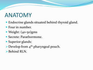
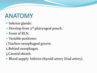
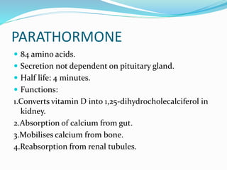
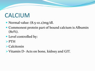
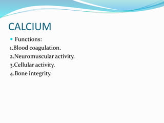
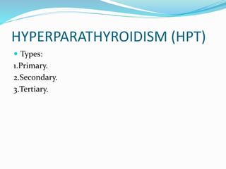
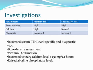
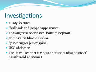
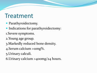
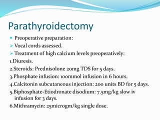
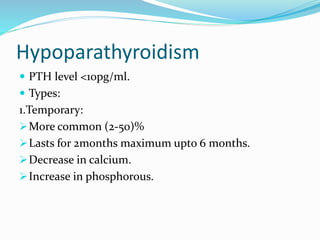
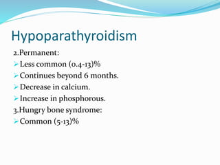
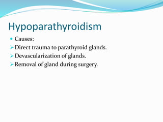
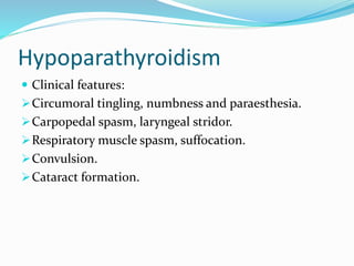
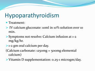
Dr. Ritesh Dhanbhar
ANATOMY
 Endocrine glands situated behind thyroid gland.
 Four in number.
 Weight: (40-50)gms
 Secrete: Parathormone.
 Superior glands:
Develop from 4th pharyngeal pouch.
Behind RLN.
ANATOMY
 Inferior glands:
Develop from 3rd pharyngeal pouch.
Front of RLN.
Variable positions:
1.Tracheo-oesophageal groove.
2.Behind oesophagus.
3.Carotid sheath.
 Blood supply: Inferior thyroid artery (End artery).
PARATHORMONE
 84 amino acids.
 Secretion not dependent on pituitary gland.
 Half life: 4 minutes.
 Functions:
1.Converts vitamin D into 1,25-dihydrocholecalciferol in
kidney.
2.Absorption of calcium from gut.
3.Mobilises calcium from bone.
4.Reabsorption from renal tubules.
CALCIUM
 Normal value: (8.5-10.2)mg/dl.
 Commonest protein part of bound calcium is Albumin
(80%).
 Level controlled by:
PTH
Calcitonin
Vitamin D- Acts on bone, kidney and GIT.
CALCIUM
 Functions:
1.Blood coagulation.
2.Neuromuscular activity.
3.Cellular activity.
4.Bone integrity.
HYPERPARATHYROIDISM (HPT)
 Types:
1.Primary.
2.Secondary.
3.Tertiary.
PRIMARY HPT
 3rd most common endocrine disease.
 Causes hypercalcaemia.
 Etiology:
Parathyroid adenoma.
Familial/ genetic causes.
MEN I syndrome.
Therapeutic ionizing radiation.
Lithium: no bone or renal problems.
CLINICAL FEATURES
 Clinical vignette: “Bones, stones, abdominal organs
and psychiatric moans”.
 Middle aged women (3:1).
 Incidence: 1: 1000.
 Asymptomatic >50% cases.
CLINICAL FEATURES
 Bones:
Raised PTH
Increased osteoclastic activity
Extensive decalcification of bone
Bone pain, subperiosteal erosions.
Osteitis fibrosa cystica: Single/ multiple cysts/
pseudotumours in the jaw, skull or phalanges.
Osteopenia, osteoporosis and pathalogical fractures.
CLINICAL FEATURES
 Stones:
Renal stones in 25% patients.
Recurrent stones.
Calcium phosphate and oxalate type.
Metastatic calcification, nephrocalcinosis, renal
failure.
Calcification in renal vessels: renal hypertension.
CLINICAL FEATURES
 Abdominal organs:
Stimulates gastrin release: peptic ulceration.
Precipitate acute pancreatitis.
Increases gall stone disease (Calcium bicarbonate).
 Psychic moans:
Behavioural and neurotic problems: Depression and
anxiety.
Acute hyperparathyroidism (Crisis)
 Causes:
Sudden increase in PTH due to rupture of parathyroid
cyst or bleeding in parathyroid tumour.
Severe dehydration precipitates crisis.
Secondaries in bone.
Acute hyperparathyroidism (Crisis)
 Clinical Presentation:
1.Abdominal pain.
2.Vomiting.
3.Dehydration.
4.Oliguria.
5.Muscular weakness.
6.Death.
 Serum Calcium:>12mg%.
Acute hyperparathyroidism (Crisis)
 Treatment:
Forced diuresis: 3-5 litres of saline with frusemide.
Rehydration: normal saline 300ml/hour.
Steroids: Inhibit effects of vitamin D.
Dose: 400mg/day iv for 5 days.
Bisphosphonate:
Clodranate Sodium, Pamidronate
Inhibits mobilization of calcium from bone.
Dose: 4mg iv followed by 8mg.
Investigations
Parameters Primary HPT Secondary HPT
Parathormone High High
Calcium High Normal
Phosphate Decreased Increased
•Increased serum PTH level: specific and diagnostic
>0.5.
•Bone density assessment.
•Vitamin D estimation.
•Increased urinary calcium level >250mg/24 hours.
•Raised alkaline phosphatase level.
Investigations
 X-Ray features:
Skull: salt and pepper appearance.
Phalanges: subperiosteal bone resorption.
Jaw: osteitis fibrosa cystica.
Spine: rugger jersey spine.
 USG abdomen.
 Thallium- Technetium scan: hot spots (diagnostic of
parathyroid adenoma).
Treatment
 Parathyroidectomy.
 Indications for parathyroidectomy:
1.Severe symptoms.
2.Young age group.
3.Markedly reduced bone density.
4.Serum calcium >11mg%.
5.Urinary calculi.
6.Urinary calcium >400mg/24 hours.
Parathyroidectomy
 Preoperative preparation:
 Vocal cords assessed.
 Treatment of high calcium levels preoperatively:
1.Diuresis.
2.Steroids: Prednisolone 20mg TDS for 5 days.
3.Phosphate infusion: 100mmol infusion in 6 hours.
4.Calcitonin subcutaneous injection: 200 units BD for 5 days.
5.Biphosphate-Etiodronate disodium: 7.5mg/kg slow iv
infusion for 3 days.
6.Mithramycin: 25microgm/kg single dose.
Parathyroidectomy
 Total parathyroidectomy:
For parathyroid hyperplasia.
All four glands removed.
1/3rd of one gland autotransplanted into forearm
muscle (brachioradialis) or sterniocleidomastoid with
marker stitch.
Transplanted gland sliced in 1mm pieces.
18 pieces embedded.
Parathyroidectomy
 Adenoma in gland with normal other glands: removal
of single gland.
 Carcinoma: Parathyroidectomy plus
hemithyroidectomy with post operative radiotherapy.
 All 4 glands diseased: Transcervical thymectomy
added.
Parathyroidectomy
 Surgical approaches:
Classical approach.
Minimally invasive parathyroidectomy.
Median sternotomy extension.
Video-assisted parathyroidectomy.
Endoscopic parathyroidectomy.
Remedial parathyroidectomy.
Subtotal parathyroidectomy.
Total parathyroidectomy with parathyroid
autotransplantation.
Parathyroidectomy
 Complications:
Haemorrhage, RLN palsy, hypocalcaemia.
Persistent HPT: Serum calcium does not normalise
immediately after surgery.
Recurrent HPT: Serum calcium after surgery becomes
normal but again increases in 6-12 months.
Parathyroidectomy
Hypoparathyroid with severe hypocalcaemia: 10
ampoules Calcium gluconate with one liter Normal
saline (continuous infusion at 30ml/hour).
Hungry Bone Syndrome:
In patients with preoperative hyperthyroidism.
Increased bone breakdown in hyperthyroid state.
Parathyroidectomy
Patient’s thyroid hormone level drops acutely after
surgery, stimulus to brake down bone is removed.
Bones remove calcium from plasma rapidly.
 Presentation: Hypocalcaemia.
Hypophosphatemia.
Hypomagnesemia
Hyperkalaemia.
Parathyroidectomy
Vitamin D supplementation and elemental calcium:
for 6 months.
Calcitriol with 2gm calcium supplement.
MEN Syndrome
 Autosomal dominant.
 Types:
Type I (Werner’s syndrome):
Parathyroid hyperplasia/Adenoma.
Pituitary tumour.
Pancreatic tumour.
Chromosome 11
MEN Syndrome
 Type II (Sipple syndrome):
 Type II A:
Medullary carcinoma thyroid
Pheochromocytoma
Pararthyroid hyperplasia
Chromosome 10.
 Type II B:
Medullary carcinoma thyroid
Pheochromocytoma
Neuroma.
APUDOMAS
 APUD (Amine precursor uptake decarboxylation)
 Derived from endoderm.
 Neuron specific enolase enzyme: specific for cells.
 Synthesize peptides:
Endocrine action
Paracrine action
Neurocrine action
Neuroendocrine action.
APUDOMAS
 Present like syndromes:
Insulinoma
Glucagonoma
Gastrinoma
VIPoma
 Entopic type: Secrete hormones normal to tissue, like
insulinoma/ glucagonoma.
 Ectopic type: Produce hormones not normal to tissue
of origin, like gastrinoma/VIPoma.
APUDOMAS
 Associated with MEN syndrome (Type I).
 Investigation: Radioimmunoassay
MRI abdomen
CT neck
Hypoparathyroidism
 PTH level <10pg/ml.
 Types:
1.Temporary:
More common (2-50)%
Lasts for 2months maximum upto 6 months.
Decrease in calcium.
Increase in phosphorous.
Hypoparathyroidism
2.Permanent:
Less common (0.4-13)%
Continues beyond 6 months.
Decrease in calcium.
Increase in phosphorous.
3.Hungry bone syndrome:
Common (5-13)%
Hypoparathyroidism
 Causes:
Direct trauma to parathyroid glands.
Devascularization of glands.
Removal of gland during surgery.
Hypoparathyroidism
 Clinical features:
Circumoral tingling, numbness and paraesthesia.
Carpopedal spasm, laryngeal stridor.
Respiratory muscle spasm, suffocation.
Convulsion.
Cataract formation.
Hypoparathyroidism
 Treatment:
 IV calcium gluconate: 10ml in 10% solution over 10
min.
Symptoms not resolve: Calcium infusion at 1-2
mg/kg/hr.
1-2 gm oral calcium per day.
(Calcium carbonate: 1250mg = 500mg elemental
calcium)
Vitamin D supplementation: 0.25-1 microgm/day.
THANK YOU !!!

Parathyroid Diseases and Management

  • 1.
  • 2.
    ANATOMY  Endocrine glandssituated behind thyroid gland.  Four in number.  Weight: (40-50)gms  Secrete: Parathormone.  Superior glands: Develop from 4th pharyngeal pouch. Behind RLN.
  • 3.
    ANATOMY  Inferior glands: Developfrom 3rd pharyngeal pouch. Front of RLN. Variable positions: 1.Tracheo-oesophageal groove. 2.Behind oesophagus. 3.Carotid sheath.  Blood supply: Inferior thyroid artery (End artery).
  • 4.
    PARATHORMONE  84 aminoacids.  Secretion not dependent on pituitary gland.  Half life: 4 minutes.  Functions: 1.Converts vitamin D into 1,25-dihydrocholecalciferol in kidney. 2.Absorption of calcium from gut. 3.Mobilises calcium from bone. 4.Reabsorption from renal tubules.
  • 5.
    CALCIUM  Normal value:(8.5-10.2)mg/dl.  Commonest protein part of bound calcium is Albumin (80%).  Level controlled by: PTH Calcitonin Vitamin D- Acts on bone, kidney and GIT.
  • 6.
    CALCIUM  Functions: 1.Blood coagulation. 2.Neuromuscularactivity. 3.Cellular activity. 4.Bone integrity.
  • 7.
  • 8.
    PRIMARY HPT  3rdmost common endocrine disease.  Causes hypercalcaemia.  Etiology: Parathyroid adenoma. Familial/ genetic causes. MEN I syndrome. Therapeutic ionizing radiation. Lithium: no bone or renal problems.
  • 9.
    CLINICAL FEATURES  Clinicalvignette: “Bones, stones, abdominal organs and psychiatric moans”.  Middle aged women (3:1).  Incidence: 1: 1000.  Asymptomatic >50% cases.
  • 10.
    CLINICAL FEATURES  Bones: RaisedPTH Increased osteoclastic activity Extensive decalcification of bone Bone pain, subperiosteal erosions. Osteitis fibrosa cystica: Single/ multiple cysts/ pseudotumours in the jaw, skull or phalanges. Osteopenia, osteoporosis and pathalogical fractures.
  • 11.
    CLINICAL FEATURES  Stones: Renalstones in 25% patients. Recurrent stones. Calcium phosphate and oxalate type. Metastatic calcification, nephrocalcinosis, renal failure. Calcification in renal vessels: renal hypertension.
  • 12.
    CLINICAL FEATURES  Abdominalorgans: Stimulates gastrin release: peptic ulceration. Precipitate acute pancreatitis. Increases gall stone disease (Calcium bicarbonate).  Psychic moans: Behavioural and neurotic problems: Depression and anxiety.
  • 13.
    Acute hyperparathyroidism (Crisis) Causes: Sudden increase in PTH due to rupture of parathyroid cyst or bleeding in parathyroid tumour. Severe dehydration precipitates crisis. Secondaries in bone.
  • 14.
    Acute hyperparathyroidism (Crisis) Clinical Presentation: 1.Abdominal pain. 2.Vomiting. 3.Dehydration. 4.Oliguria. 5.Muscular weakness. 6.Death.  Serum Calcium:>12mg%.
  • 15.
    Acute hyperparathyroidism (Crisis) Treatment: Forced diuresis: 3-5 litres of saline with frusemide. Rehydration: normal saline 300ml/hour. Steroids: Inhibit effects of vitamin D. Dose: 400mg/day iv for 5 days. Bisphosphonate: Clodranate Sodium, Pamidronate Inhibits mobilization of calcium from bone. Dose: 4mg iv followed by 8mg.
  • 16.
    Investigations Parameters Primary HPTSecondary HPT Parathormone High High Calcium High Normal Phosphate Decreased Increased •Increased serum PTH level: specific and diagnostic >0.5. •Bone density assessment. •Vitamin D estimation. •Increased urinary calcium level >250mg/24 hours. •Raised alkaline phosphatase level.
  • 17.
    Investigations  X-Ray features: Skull:salt and pepper appearance. Phalanges: subperiosteal bone resorption. Jaw: osteitis fibrosa cystica. Spine: rugger jersey spine.  USG abdomen.  Thallium- Technetium scan: hot spots (diagnostic of parathyroid adenoma).
  • 18.
    Treatment  Parathyroidectomy.  Indicationsfor parathyroidectomy: 1.Severe symptoms. 2.Young age group. 3.Markedly reduced bone density. 4.Serum calcium >11mg%. 5.Urinary calculi. 6.Urinary calcium >400mg/24 hours.
  • 19.
    Parathyroidectomy  Preoperative preparation: Vocal cords assessed.  Treatment of high calcium levels preoperatively: 1.Diuresis. 2.Steroids: Prednisolone 20mg TDS for 5 days. 3.Phosphate infusion: 100mmol infusion in 6 hours. 4.Calcitonin subcutaneous injection: 200 units BD for 5 days. 5.Biphosphate-Etiodronate disodium: 7.5mg/kg slow iv infusion for 3 days. 6.Mithramycin: 25microgm/kg single dose.
  • 20.
    Parathyroidectomy  Total parathyroidectomy: Forparathyroid hyperplasia. All four glands removed. 1/3rd of one gland autotransplanted into forearm muscle (brachioradialis) or sterniocleidomastoid with marker stitch. Transplanted gland sliced in 1mm pieces. 18 pieces embedded.
  • 21.
    Parathyroidectomy  Adenoma ingland with normal other glands: removal of single gland.  Carcinoma: Parathyroidectomy plus hemithyroidectomy with post operative radiotherapy.  All 4 glands diseased: Transcervical thymectomy added.
  • 22.
    Parathyroidectomy  Surgical approaches: Classicalapproach. Minimally invasive parathyroidectomy. Median sternotomy extension. Video-assisted parathyroidectomy. Endoscopic parathyroidectomy. Remedial parathyroidectomy. Subtotal parathyroidectomy. Total parathyroidectomy with parathyroid autotransplantation.
  • 23.
    Parathyroidectomy  Complications: Haemorrhage, RLNpalsy, hypocalcaemia. Persistent HPT: Serum calcium does not normalise immediately after surgery. Recurrent HPT: Serum calcium after surgery becomes normal but again increases in 6-12 months.
  • 24.
    Parathyroidectomy Hypoparathyroid with severehypocalcaemia: 10 ampoules Calcium gluconate with one liter Normal saline (continuous infusion at 30ml/hour). Hungry Bone Syndrome: In patients with preoperative hyperthyroidism. Increased bone breakdown in hyperthyroid state.
  • 25.
    Parathyroidectomy Patient’s thyroid hormonelevel drops acutely after surgery, stimulus to brake down bone is removed. Bones remove calcium from plasma rapidly.  Presentation: Hypocalcaemia. Hypophosphatemia. Hypomagnesemia Hyperkalaemia.
  • 26.
    Parathyroidectomy Vitamin D supplementationand elemental calcium: for 6 months. Calcitriol with 2gm calcium supplement.
  • 27.
    MEN Syndrome  Autosomaldominant.  Types: Type I (Werner’s syndrome): Parathyroid hyperplasia/Adenoma. Pituitary tumour. Pancreatic tumour. Chromosome 11
  • 28.
    MEN Syndrome  TypeII (Sipple syndrome):  Type II A: Medullary carcinoma thyroid Pheochromocytoma Pararthyroid hyperplasia Chromosome 10.  Type II B: Medullary carcinoma thyroid Pheochromocytoma Neuroma.
  • 29.
    APUDOMAS  APUD (Amineprecursor uptake decarboxylation)  Derived from endoderm.  Neuron specific enolase enzyme: specific for cells.  Synthesize peptides: Endocrine action Paracrine action Neurocrine action Neuroendocrine action.
  • 30.
    APUDOMAS  Present likesyndromes: Insulinoma Glucagonoma Gastrinoma VIPoma  Entopic type: Secrete hormones normal to tissue, like insulinoma/ glucagonoma.  Ectopic type: Produce hormones not normal to tissue of origin, like gastrinoma/VIPoma.
  • 31.
    APUDOMAS  Associated withMEN syndrome (Type I).  Investigation: Radioimmunoassay MRI abdomen CT neck
  • 32.
    Hypoparathyroidism  PTH level<10pg/ml.  Types: 1.Temporary: More common (2-50)% Lasts for 2months maximum upto 6 months. Decrease in calcium. Increase in phosphorous.
  • 33.
    Hypoparathyroidism 2.Permanent: Less common (0.4-13)% Continuesbeyond 6 months. Decrease in calcium. Increase in phosphorous. 3.Hungry bone syndrome: Common (5-13)%
  • 34.
    Hypoparathyroidism  Causes: Direct traumato parathyroid glands. Devascularization of glands. Removal of gland during surgery.
  • 35.
    Hypoparathyroidism  Clinical features: Circumoraltingling, numbness and paraesthesia. Carpopedal spasm, laryngeal stridor. Respiratory muscle spasm, suffocation. Convulsion. Cataract formation.
  • 36.
    Hypoparathyroidism  Treatment:  IVcalcium gluconate: 10ml in 10% solution over 10 min. Symptoms not resolve: Calcium infusion at 1-2 mg/kg/hr. 1-2 gm oral calcium per day. (Calcium carbonate: 1250mg = 500mg elemental calcium) Vitamin D supplementation: 0.25-1 microgm/day.
  • 37.