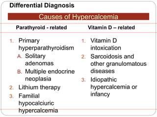
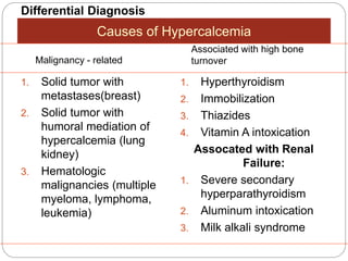
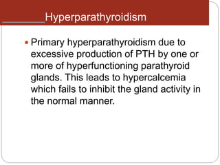
Dr syeda Hussan e Zahra
Sr surgical unit 2
DISORDERS OF THE
PARATHYROID GLANDS
 Parathyroid glands
are normally the
size of a rice grain
 Each gland: 5 to 7
mm(6X2X2 mm).
 Wt 40-50mg.
yellowish-Brown firm
gland
DISORDERS OF THE PARATHYROID GLANDS
 Maintenance of calcium, phosphate and
magnesium homeostasis is under the
influence of two polypeptide hormones;
parathyroid hormone(PTH), and
calcitonin (CT), as well as a sterol
hormone, 1,25 dihydroxy
cholecalciferol (1,25 (OH)2D3)
 These hormones regulate the flow of
minerals in and out of the extracellular
fluid compartments through their actions
on intestine, kidneys, and bones.
 The PTH acts directly on the bones and
kidneys and indirectly on the intestine
through its effect on the synthesis of
1,25 (OH)2D3. Its production is regulated
by the concentration of serum ionized
calcium.
Lowering of the serum calcium levels
will induce an increased rate of
parathyroid hormone secretion
 Calcitonin is released by the “C” cells
(parafollicular cells in the thyroid gland)
in response to small increases in plasma
ionic calcium. It acts on the kidney and
bones to restore the level of calcium to
just below a normal set point which in
turn inhibits secretion of the hormone.
PTH/Calcium Homeostasis
Low circulating
serum calcium
concentrations
stimulate the
parathyroid glands to
secrete PTH, which
mobilizes calcium
from bones by
osteoclastic
stimulation. PTH also
stimulates the
kidneys to reabsorb
calcium and to
convert 25-
hydroxyvitamin D3
(produced in the
liver) to the active
form,
1,25-
dihydroxyvitamin D3,
which stimulates GI
calcium absorption.
High serum calcium
concentrations have
a negative feedback
Differential Diagnosis
1. Primary
hyperparathyroidism
A. Solitary
adenomas
B. Multiple endocrine
neoplasia
2. Lithium therapy
3. Familial
hypocalciuric
hypercalcemia
1. Vitamin D
intoxication
2. Sarcoidosis and
other granulomatous
diseases
3. Idiopathic
hypercalcemia or
infancy
Causes of Hypercalcemia
Parathyroid - related Vitamin D – related
Differential Diagnosis
1. Solid tumor with
metastases(breast)
2. Solid tumor with
humoral mediation of
hypercalcemia (lung
kidney)
3. Hematologic
malignancies (multiple
myeloma, lymphoma,
leukemia)
1. Hyperthyroidism
2. Immobilization
3. Thiazides
4. Vitamin A intoxication
Assocated with Renal
Failure:
1. Severe secondary
hyperparathyroidism
2. Aluminum intoxication
3. Milk alkali syndrome
Causes of Hypercalcemia
Malignancy - related
Associated with high bone
turnover
**Primary hyperparathyroidism
and cancer account for 90% of
cases of hypercalcemia
Hyperparathyroidism
 Primary hyperparathyroidism due to
excessive production of PTH by one or
more of hyperfunctioning parathyroid
glands. This leads to hypercalcemia
which fails to inhibit the gland activity in
the normal manner.
 The cause of primary hyperparathyroidism is
unknown. A genetic factor may be involved.
The clonal origin of most parathyroid
adenomas suggests a defect at the level of
the gene controlling the regulation and/or
expression of parathyroid hormone.
Hyperparathyroidism
The incidence of the disease increases
dramatically after the age of 50 and it is
2-4 folds more common in women.
A single adenoma occurs in about 80%
of patients with primary
hyperparathyroidism.
Four glands hyperplasia account for
15-20% of cases.
A parathyroid carcinoma could be the
etiology in a rare incidence of less then
1%.
Clinical Features:
The two major sites of potential
complications are the bones and the
kidneys.
The kidneys may have renal stones
(nephrolithiasis) or diffuse deposition of
calcium-phosphate complexes in the
parachyma (nephrocalcinosis).
Now a days such complications are
seen less commonly and around 20% of
patients or less show such
complications.
Clinical Features
Other symptoms include;
 muscle weakness,
 easy fatigability,
 peptic ulcer disease,
 pancreatitis,
 hypertension,
 gout and pseudogout,
 Anemia,
 Depression have been associated with
primary hyperparathyroidism.
Diagnosis
The presence of established
hypercalcaemia in more than
one serum measurement
accompanied by elevated
immunoreactive PTH is
characteristic (iPTH)
Diagnosis
Serum phosphate is usually low but may
be normal. Hypercalcaemia is common
and blood alkaline phosphatase (of bone
origin) and the urinary hydroxyproline
concentrations are commonly elevated
when the bones are involved.
Nephrogenous CAMP is elevated in about
80% of patients but the test is rarely used
because of technical difficulties
 In skeleton a condition called osteitis
fibrosa cystica could occur with
subperiosteal resorption of the distal
phalanges,
 distal tappering of the clavicles,
 “salt and pepper” appearance of the
skull
 bone cysts and brown tumors of the
long bones. Such overt bone disease
even though typical of primary
hyperparathyroidism is very rarely
Radiograph
Subperiosteal resorption
salt and pepper
brown tumors
Other Diagnostic tests
 Ultrasonography
 MRI
 CT
 Tehcnichum99m scan (MIBI )
Pre-operative localization of the abnormal
parathyroid gland(s):
Scintigraphy Images
Traditional Sestamibi
Sestamibi-SPECT
Treatment
A large proportion of patients have
“biochemical” hyperparathyroidism but
with prolonged follow up they progress to
overt clinical presentation. Resection of
the parathyroid lesion is curative with
recurrences observed mainly in the
multiple glandular disease.
Medical Treatment of the
hypercalcaemia
 In acute severe forms the main stay of
therapy is adequate hydration with
saline
 Forced diuresis by diuretics to increase
the urinary excretion of calcium rapidly
along with sodium
 Prevent its reabsorption by the renal
tubules.
Surgery
 Surgical treatment should be considered
in all cases with established diagnosis of
primary hyperparathyroidism.
 During surgery the surgeon identifies all
four parathyroid glands followed by the
removal of enlarged parathyroid or 3 ½
glands in multiple glandular disease.
Surgical Candidacy
 Symptomatic primary HPT
 Serum calcium greater than 1mg/dL above
the upper limit of the reference range
 24 hour urine calcium greater than 400 mg
 Creatinine clearance reduced by more than
30% compared with age-matched subjects
 Marked reduced Bone density
 Age under 50
 -Urinary calculi
Secondary Hyperparathyroidism
An increase in PTH secretion which is
adaptive and unrelated to intrinsic disease
of the parathyroid glands is called
secondary hyperparathyroidism. This is
due to chronic stimulation of the
parathyroid glands by a chronic decrease
in the ionic calcium level in the blood
Causes of Secondary
hyperparathyroidism
 Secondary hyperparathyroidism is usually
seen in patients with renal failure.
 Rickets/osteomalacia.
 Intestinal malabsorption
Tertiary hyperparathyroidism
Chronic kidney failure or in
people who have had a
kidney transplant.
Hypoparathyroidism
Deficient secretion of PTH which
manifests itself biochemically by
hypocalcemia, hyperphospatemia
diminished or absent circulating iPTH and
clinically the symptoms of neuromuscular
hyperactivity.
Hypoparathyroidism
 Surgical hypoparathyroidism – the
commonest
After anterior neck exploration for
thyroidectomy, abnormal parathyroid gland
removal, excision of a neck lesion. It could
be due to the removal of the parathyroid
glands or due to interruption of blood
supply to the glands.
Causes:
Hypoparathyroidism
 Idiopathic hypoparathyroidism
 A form occuring at an early age (genetic origin) with
autosomal recessive mode of transmission “multiple
endocrine deficiency –APECED syndrome”
 “Juvenile familial endocrinopathy”
 Functional hypoparathyroidism
 In patients who has chronic hypomagnesaemia of
various causes.
 Magnesium is necessary for the PTH release from the
glands and also for the peripheral action of the PTH.
Causes:
Major Causes of Chronic
Hypocalcemia other than
Hypoparathyroidism
 Dietary deficiency of vitamin D or
calcium
 Decreased intestinal absorption of
vitamin D or calcium due to primary
small bowel disease, short bowel
syndrome, and post-gastrectomy
syndrome.
 Drugs that cause rickets or
osteomalacia such as phenytoin,
phenobarbital, cholestyramine, and
laxative.
Major causes of chronic hypocalcemia
other than parathyroidal
hypoparathyroidism
 States of tissue resistance to vitamin D
 Excessive intake of inorganic phosphate
compunds
 Psudohypoparathyroidism
 Severe hypomagnesemia
 Chronic renal failure
Hypoparathyroidism
A. Neuromuscular
 The rate of decrease in serum calcium is the
major determinant for the development of
neuromuscular complications.
 When nerves are exposed to low levels of
calcium they show abnormal neuronal
function which may include decrease
threshold of excitation, repetitive response
to a single stimulus and rarely continuous
activity.
Clinical Features:
Hypoparathyroidism
A. Neuromuscular
 Parasthesia
 Tetany
 Hyperventilation
 Adrenergic symptoms
 Convulsion (More common in young people)
 Signs of latent tetany
 Chvostek sign
 Trousseau sign
 Extrapyramidal signs (due to basal ganglia
calcification)
Clinical Features:
Hypoparathyroidism
B. Other clinical manifestation
1. Posterio lenticular cataract
2. Cardiac manifestation:
Prolonged QT interval in the ECG
Resistance to digitalis
Hypotension
Refractory heart failure with cardiomegally
can occur.
Clinical Features:
Hypoparathyroidism
B. Other clinical manifestation
3. Dental Manifestation
Abnormal enamel formation with delayed or absent
dental eruption and defective dental root formation.
4. Malabsorption syndrome
Presumably secondary to decreased calcium level
and may lead to steatorrhoea with long standing
untreated disease.
Clinical Features:
Hypoparathyroidism
In the absence of renal failure the
presence of hypocalcaemia with
hyperphosphataemia is virtually
diagnostic of hypoparathyroidism.
Undetectable serum iPTH confirms the
diagnosis or it can be detectable if the
assay is very sensitive.
Diagnosis:
Hypoparathyroidism
The mainstay of treatment is a
combination of oral calcium with
pharmacological doses of vitamin D or its
potent analogues. Phosphate restriction
in diet may also be useful with or without
aluminum hydroxide gel to lower serum
phosphate level.
Treatment:
Emergency Treatment for Hypocalcaemic
Calcium should be given
parenterally till adequate serum
calcium level is obtained and then
vitamin D supplementation with oral
calcium should be initiated.
Tetany:
Emergency Treatment for Hypocalcemia
In patients with hyperparathyroidism
and severe bone disease who
undergo successful
parathyroidectomy than
hypocalcaemia may be severe and
parenteral calcium infusion with later
supplementation with oral calcium
and vitamin D.
Hungry bone syndrome:
Pseudohypoparathyroidism
A rare familial disorders with target tissue
resistance to PTH. There is
hypocalcaemia, hyperphosphataemia,
with increased parathyroid gland
function. There is also a variety of
congenital defects in the growth and
development of skeleton including:
 Short statue
 Short metacarpal and metatarsal bones
Pseudohypoparathysoidism
The diagnosis is established when low
serum calcium level with
hyperphosphataemia is associated with
increased serum iPTH as well as
diminished nephrogenous CAMP and
phosphature response to PTH
administration
Pseudopseudohypoparathyroidis
m
In
pseudopseudohypoparathyroidi
sm they have the
developmental defects without
the biochemical abnormalities.
Typical biochemical features of
hypoparathyroidism
True Pseudo
Se Ca2+
Phosphate
Alkaline
phosphatase
PTH
N N

disordersoftheparathyroidglands discussuion.pptx

  • 1.
    Dr syeda Hussane Zahra Sr surgical unit 2 DISORDERS OF THE PARATHYROID GLANDS
  • 2.
     Parathyroid glands arenormally the size of a rice grain  Each gland: 5 to 7 mm(6X2X2 mm).  Wt 40-50mg. yellowish-Brown firm gland
  • 3.
    DISORDERS OF THEPARATHYROID GLANDS  Maintenance of calcium, phosphate and magnesium homeostasis is under the influence of two polypeptide hormones; parathyroid hormone(PTH), and calcitonin (CT), as well as a sterol hormone, 1,25 dihydroxy cholecalciferol (1,25 (OH)2D3)
  • 4.
     These hormonesregulate the flow of minerals in and out of the extracellular fluid compartments through their actions on intestine, kidneys, and bones.
  • 5.
     The PTHacts directly on the bones and kidneys and indirectly on the intestine through its effect on the synthesis of 1,25 (OH)2D3. Its production is regulated by the concentration of serum ionized calcium.
  • 6.
    Lowering of theserum calcium levels will induce an increased rate of parathyroid hormone secretion
  • 7.
     Calcitonin isreleased by the “C” cells (parafollicular cells in the thyroid gland) in response to small increases in plasma ionic calcium. It acts on the kidney and bones to restore the level of calcium to just below a normal set point which in turn inhibits secretion of the hormone.
  • 8.
    PTH/Calcium Homeostasis Low circulating serumcalcium concentrations stimulate the parathyroid glands to secrete PTH, which mobilizes calcium from bones by osteoclastic stimulation. PTH also stimulates the kidneys to reabsorb calcium and to convert 25- hydroxyvitamin D3 (produced in the liver) to the active form, 1,25- dihydroxyvitamin D3, which stimulates GI calcium absorption. High serum calcium concentrations have a negative feedback
  • 10.
    Differential Diagnosis 1. Primary hyperparathyroidism A.Solitary adenomas B. Multiple endocrine neoplasia 2. Lithium therapy 3. Familial hypocalciuric hypercalcemia 1. Vitamin D intoxication 2. Sarcoidosis and other granulomatous diseases 3. Idiopathic hypercalcemia or infancy Causes of Hypercalcemia Parathyroid - related Vitamin D – related
  • 11.
    Differential Diagnosis 1. Solidtumor with metastases(breast) 2. Solid tumor with humoral mediation of hypercalcemia (lung kidney) 3. Hematologic malignancies (multiple myeloma, lymphoma, leukemia) 1. Hyperthyroidism 2. Immobilization 3. Thiazides 4. Vitamin A intoxication Assocated with Renal Failure: 1. Severe secondary hyperparathyroidism 2. Aluminum intoxication 3. Milk alkali syndrome Causes of Hypercalcemia Malignancy - related Associated with high bone turnover
  • 12.
    **Primary hyperparathyroidism and canceraccount for 90% of cases of hypercalcemia
  • 13.
    Hyperparathyroidism  Primary hyperparathyroidismdue to excessive production of PTH by one or more of hyperfunctioning parathyroid glands. This leads to hypercalcemia which fails to inhibit the gland activity in the normal manner.
  • 14.
     The causeof primary hyperparathyroidism is unknown. A genetic factor may be involved. The clonal origin of most parathyroid adenomas suggests a defect at the level of the gene controlling the regulation and/or expression of parathyroid hormone.
  • 15.
    Hyperparathyroidism The incidence ofthe disease increases dramatically after the age of 50 and it is 2-4 folds more common in women. A single adenoma occurs in about 80% of patients with primary hyperparathyroidism. Four glands hyperplasia account for 15-20% of cases. A parathyroid carcinoma could be the etiology in a rare incidence of less then 1%.
  • 16.
    Clinical Features: The twomajor sites of potential complications are the bones and the kidneys. The kidneys may have renal stones (nephrolithiasis) or diffuse deposition of calcium-phosphate complexes in the parachyma (nephrocalcinosis). Now a days such complications are seen less commonly and around 20% of patients or less show such complications.
  • 17.
    Clinical Features Other symptomsinclude;  muscle weakness,  easy fatigability,  peptic ulcer disease,  pancreatitis,  hypertension,  gout and pseudogout,  Anemia,  Depression have been associated with primary hyperparathyroidism.
  • 18.
    Diagnosis The presence ofestablished hypercalcaemia in more than one serum measurement accompanied by elevated immunoreactive PTH is characteristic (iPTH)
  • 19.
    Diagnosis Serum phosphate isusually low but may be normal. Hypercalcaemia is common and blood alkaline phosphatase (of bone origin) and the urinary hydroxyproline concentrations are commonly elevated when the bones are involved. Nephrogenous CAMP is elevated in about 80% of patients but the test is rarely used because of technical difficulties
  • 20.
     In skeletona condition called osteitis fibrosa cystica could occur with subperiosteal resorption of the distal phalanges,  distal tappering of the clavicles,  “salt and pepper” appearance of the skull  bone cysts and brown tumors of the long bones. Such overt bone disease even though typical of primary hyperparathyroidism is very rarely Radiograph
  • 21.
  • 22.
  • 23.
  • 24.
    Other Diagnostic tests Ultrasonography  MRI  CT  Tehcnichum99m scan (MIBI ) Pre-operative localization of the abnormal parathyroid gland(s):
  • 25.
  • 26.
    Treatment A large proportionof patients have “biochemical” hyperparathyroidism but with prolonged follow up they progress to overt clinical presentation. Resection of the parathyroid lesion is curative with recurrences observed mainly in the multiple glandular disease.
  • 27.
    Medical Treatment ofthe hypercalcaemia  In acute severe forms the main stay of therapy is adequate hydration with saline  Forced diuresis by diuretics to increase the urinary excretion of calcium rapidly along with sodium  Prevent its reabsorption by the renal tubules.
  • 28.
    Surgery  Surgical treatmentshould be considered in all cases with established diagnosis of primary hyperparathyroidism.  During surgery the surgeon identifies all four parathyroid glands followed by the removal of enlarged parathyroid or 3 ½ glands in multiple glandular disease.
  • 29.
    Surgical Candidacy  Symptomaticprimary HPT  Serum calcium greater than 1mg/dL above the upper limit of the reference range  24 hour urine calcium greater than 400 mg  Creatinine clearance reduced by more than 30% compared with age-matched subjects  Marked reduced Bone density  Age under 50  -Urinary calculi
  • 30.
    Secondary Hyperparathyroidism An increasein PTH secretion which is adaptive and unrelated to intrinsic disease of the parathyroid glands is called secondary hyperparathyroidism. This is due to chronic stimulation of the parathyroid glands by a chronic decrease in the ionic calcium level in the blood
  • 31.
    Causes of Secondary hyperparathyroidism Secondary hyperparathyroidism is usually seen in patients with renal failure.  Rickets/osteomalacia.  Intestinal malabsorption
  • 32.
    Tertiary hyperparathyroidism Chronic kidneyfailure or in people who have had a kidney transplant.
  • 33.
    Hypoparathyroidism Deficient secretion ofPTH which manifests itself biochemically by hypocalcemia, hyperphospatemia diminished or absent circulating iPTH and clinically the symptoms of neuromuscular hyperactivity.
  • 34.
    Hypoparathyroidism  Surgical hypoparathyroidism– the commonest After anterior neck exploration for thyroidectomy, abnormal parathyroid gland removal, excision of a neck lesion. It could be due to the removal of the parathyroid glands or due to interruption of blood supply to the glands. Causes:
  • 35.
    Hypoparathyroidism  Idiopathic hypoparathyroidism A form occuring at an early age (genetic origin) with autosomal recessive mode of transmission “multiple endocrine deficiency –APECED syndrome”  “Juvenile familial endocrinopathy”  Functional hypoparathyroidism  In patients who has chronic hypomagnesaemia of various causes.  Magnesium is necessary for the PTH release from the glands and also for the peripheral action of the PTH. Causes:
  • 36.
    Major Causes ofChronic Hypocalcemia other than Hypoparathyroidism  Dietary deficiency of vitamin D or calcium  Decreased intestinal absorption of vitamin D or calcium due to primary small bowel disease, short bowel syndrome, and post-gastrectomy syndrome.  Drugs that cause rickets or osteomalacia such as phenytoin, phenobarbital, cholestyramine, and laxative.
  • 37.
    Major causes ofchronic hypocalcemia other than parathyroidal hypoparathyroidism  States of tissue resistance to vitamin D  Excessive intake of inorganic phosphate compunds  Psudohypoparathyroidism  Severe hypomagnesemia  Chronic renal failure
  • 38.
    Hypoparathyroidism A. Neuromuscular  Therate of decrease in serum calcium is the major determinant for the development of neuromuscular complications.  When nerves are exposed to low levels of calcium they show abnormal neuronal function which may include decrease threshold of excitation, repetitive response to a single stimulus and rarely continuous activity. Clinical Features:
  • 39.
    Hypoparathyroidism A. Neuromuscular  Parasthesia Tetany  Hyperventilation  Adrenergic symptoms  Convulsion (More common in young people)  Signs of latent tetany  Chvostek sign  Trousseau sign  Extrapyramidal signs (due to basal ganglia calcification) Clinical Features:
  • 40.
    Hypoparathyroidism B. Other clinicalmanifestation 1. Posterio lenticular cataract 2. Cardiac manifestation: Prolonged QT interval in the ECG Resistance to digitalis Hypotension Refractory heart failure with cardiomegally can occur. Clinical Features:
  • 41.
    Hypoparathyroidism B. Other clinicalmanifestation 3. Dental Manifestation Abnormal enamel formation with delayed or absent dental eruption and defective dental root formation. 4. Malabsorption syndrome Presumably secondary to decreased calcium level and may lead to steatorrhoea with long standing untreated disease. Clinical Features:
  • 42.
    Hypoparathyroidism In the absenceof renal failure the presence of hypocalcaemia with hyperphosphataemia is virtually diagnostic of hypoparathyroidism. Undetectable serum iPTH confirms the diagnosis or it can be detectable if the assay is very sensitive. Diagnosis:
  • 43.
    Hypoparathyroidism The mainstay oftreatment is a combination of oral calcium with pharmacological doses of vitamin D or its potent analogues. Phosphate restriction in diet may also be useful with or without aluminum hydroxide gel to lower serum phosphate level. Treatment:
  • 44.
    Emergency Treatment forHypocalcaemic Calcium should be given parenterally till adequate serum calcium level is obtained and then vitamin D supplementation with oral calcium should be initiated. Tetany:
  • 45.
    Emergency Treatment forHypocalcemia In patients with hyperparathyroidism and severe bone disease who undergo successful parathyroidectomy than hypocalcaemia may be severe and parenteral calcium infusion with later supplementation with oral calcium and vitamin D. Hungry bone syndrome:
  • 46.
    Pseudohypoparathyroidism A rare familialdisorders with target tissue resistance to PTH. There is hypocalcaemia, hyperphosphataemia, with increased parathyroid gland function. There is also a variety of congenital defects in the growth and development of skeleton including:  Short statue  Short metacarpal and metatarsal bones
  • 48.
    Pseudohypoparathysoidism The diagnosis isestablished when low serum calcium level with hyperphosphataemia is associated with increased serum iPTH as well as diminished nephrogenous CAMP and phosphature response to PTH administration
  • 49.
    Pseudopseudohypoparathyroidis m In pseudopseudohypoparathyroidi sm they havethe developmental defects without the biochemical abnormalities.
  • 50.
    Typical biochemical featuresof hypoparathyroidism True Pseudo Se Ca2+ Phosphate Alkaline phosphatase PTH N N