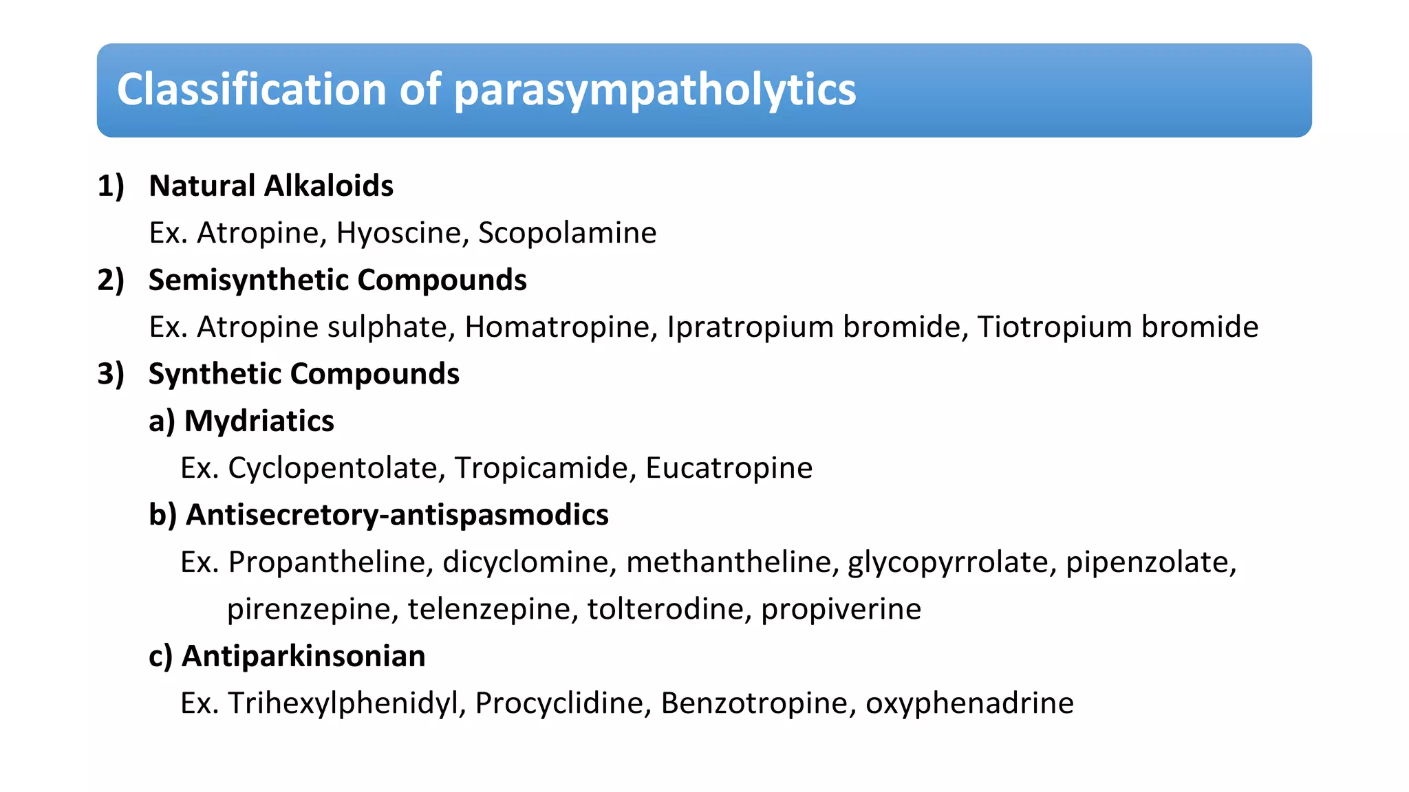
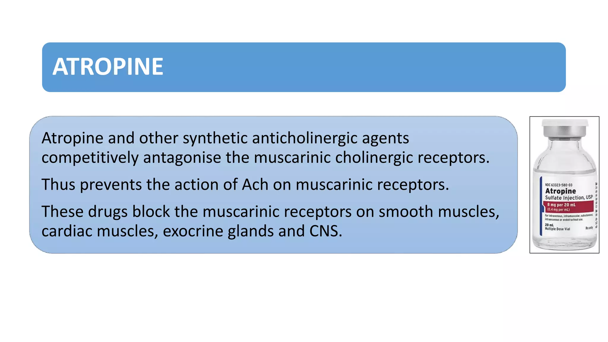
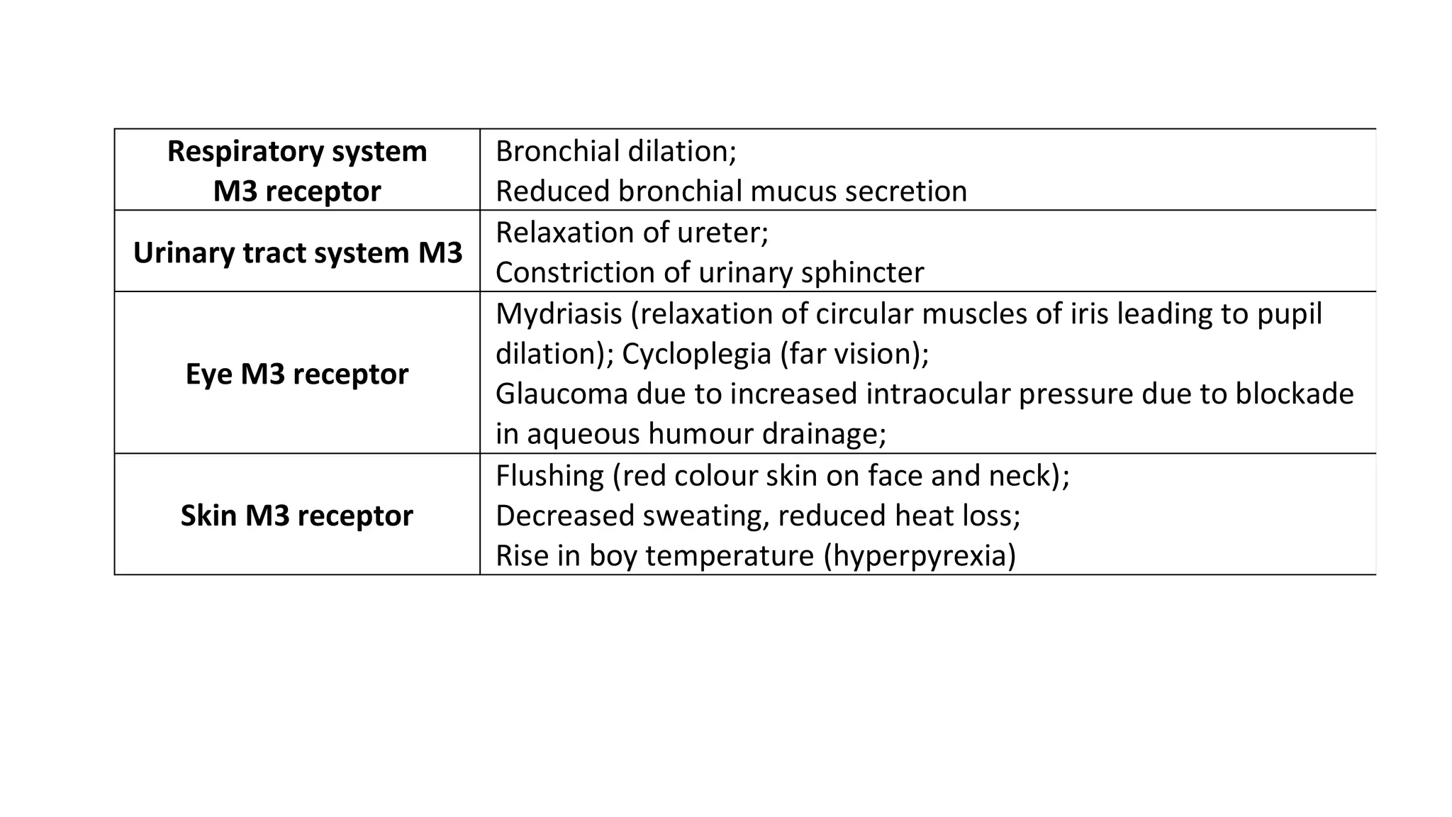
Parasympatholytics are the drugs that block or inhibit the actions of acetylcholine at postganglionic nerve endings and cholinergic receptors. They are also referred to as anticholinergics or cholinergic blocking agents or antispasmodics.
Anticholinergic drugs include atropine and related drugs- atropine is the prototype. Atropine is obtained from the plant Atropa belladonna. Atropine and scopolamine (hyoscine) are the belladonna alkaloids. They compete with acetylcholine for muscarinic receptors and block this receptors-they are muscarinic antagonists.
The parasympathetic division typically acts in opposition to the sympathetic autonomic nervous system through negative feedback control.
This action is a complementary response, causing a balance of sympathetic and parasympathetic responses.
Overall, the parasympathetic outflow results in the conservation and restoration of energy, reduction in heart rate and blood pressure, facilitation of digestion and absorption of nutrients, and excretion of waste products.
These are drugs that produce actions similar to that of Acetylcholine hence known as parasympathomimetics.
They act either by directly interacting with cholinergic receptors or by increasing the availability of Acetylcholine at these sites.
Sympatholytic drugs (Adrenergic blockers) bind to the adrenergic receptors and prevent the action of adrenergic drugs.
These are drugs which block the actions of sympathetic division or catecholamines (adrenaline and noradrenaline).
They are competitive antagonists at both α and β adrenergic receptors.
Med chem lecture on Anticholinergic drugs for B.Pharm level in Nepal
Content from Foye's Principle of medicinal chemistry, my own thoughts and some articles
Neurohumoral transmission in CNS-
The term neurohumoral transmission designates the transfer of a nerve impulse from a presynaptic to a postsynaptic neuron by means of a humoral agent e.g. a biogenic amine, an amino acid or a peptide.
Biosynthesis and catabolism of acetylcholine by Dheeraj gargDheeraj Aggarwal
Acetylcholine (ACh) is an organic chemical that functions in the brain and body of many types of animals (and humans) as a neurotransmitter—a chemical message released by nerve cells to send signals to other cells, such as neurons, muscle cells and gland cells.
miotics and mydriatics presentation m&mmparthsaraf55
Optometrists are well-acquainted with the two opposing muscles in the iris, the sphincter and the dilator, as we witness their effects daily in clinical practice. Pupil constriction (miosis) can either be stimulated by contraction of the iris sphincter or by relaxation of the iris dilator. On the other hand, pupil dilation (mydriasis) can either be stimulated by contraction of the iris dilator or by relaxation of the iris sphincter.
Miotic and mydriatic drops work by acting on these different muscles of the iris. The drops are able to control pupil size by targeting two parts of the autonomic nervous system: the sympathetic and parasympathetic systems. Let’s review their function and clinical role to better understand their present uses and why some of these agents are undergoing re-evaulation for potential new ones.
Behind the Scenes
The sympathetic pathway, mainly responsible for pupil mydriasis, involves a three-neuron pathway.1,2 The first neuron begins in the hypothalamus and descends through the midbrain to synapse onto a specific area of the spinal cord, known as the ciliospinal center of Budge. This synapse is located between the C8 and T2 vertebrae. The second neuron, which is the preganglionic neuron, exits the spinal cord, ascends through the thorax and synapses near the apex of the lung into the superior cervical ganglion. The third postganglionic neuron travels to the cavernous sinus and enters the orbit through the short and long ciliary nerves, synapsing to the iris dilator.1,2
Contrarily, the parasympathetic pathway is mainly responsible for pupil miosis.1,3 Pupil constriction starts when light enters the retina and activates the retinal ganglion cells—the beginning of the afferent arm—which then transmit their impulses into the optic nerve. This stimulus travels to the optic chiasm, through the optic tract and eventually reaches the pretectal nucleus. The impulses from the pretectal nucleus begin the efferent arm, which projects to the Edinger-Westphal nucleus. The Edinger-Westphal nucleus gives rise to preganglionic fibers, which then synapse with postganglionic neurons in the ciliary ganglion. Postganglionic neurons leave the ciliary ganglion to innervate the iris sphincter.1,3
Tropicamide has a strong mydriatic effect.
Tropicamide has a strong mydriatic effect. Click image to enlarge.
Behind the Scenes
The sympathetic pathway, mainly responsible for pupil mydriasis, involves a three-neuron pathway.1,2 The first neuron begins in the hypothalamus and descends through the midbrain to synapse onto a specific area of the spinal cord, known as the ciliospinal center of Budge. This synapse is located between the C8 and T2 vertebrae. The second neuron, which is the preganglionic neuron, exits the spinal cord, ascends through the thorax and synapses near the apex of the lung into the superior cervical ganglion. The third postganglionic neuron travels to the cavernous sinus and enters the orbit through the short and long ciliary n
drug relative to eyes with their meiotic and mydriatic effect.
In the presentation discus about spasm of accommodation and cycloplegic action on eye . pharmacological action , dosage also discussed of condition developed on eye i.e. Glaucoma
Pharmacology Experiment based Questions With Answer KeysA M O L D E O R E
MSBTE Pharmacology Practical Exam for Diploma in pharmacy students in Maharashtra.
Experimental pharmacology for D. Pharmacy Students
Pharmacology Experiment based Questions
PCI New Syllabus ER2020
Course Code: 20056
Antidepressants are a class of medication used to treat major depressive disorder, anxiety disorders, chronic pain conditions and to help manage addictions. Common side-effects of antidepressants include dry mouth, weight gain, dizziness, headaches, sexual dysfunction, and emotional blunting
Anatomy and physiology are two of the most basic terms and areas of study in the life sciences. Anatomy refers to the internal and external structures of the body and their physical relationships, whereas physiology refers to the study of the functions of those structures.
Animal cells are typical of the eukaryotic cell, enclosed by a plasma membrane and containing a membrane-bound nucleus and organelles. Unlike the eukaryotic cells of plants and fungi, animal cells do not have a cell wall. This feature was lost in the distant past by the single-celled organisms that gave rise to the kingdom Animalia. Most cells, both animal and plant, range in size between 1 and 100 micrometers and are thus visible only with the aid of a microscope.
All living organisms are made of cells and cellular products. The cell is the smallest structural, functional, and biological unit of all living organisms. It can capable of biosynthesis, replication and energy transformation. All cellular organelles carry out specific functions that are necessary for the normal functioning of the cell. Animal cells work together and function interdependently. Human cells vary in size, shape, and function. Most animal cells are so small they can only be seen with the aid of a microscope. Based on function, there are more than 200 different kinds of animal cells that help each system contribute to the homeostasis of the entire body. Despite their many differences, human cells have several similar structural features: a cell membrane, a nucleus, and cytoplasm and cell organelles.
The term “opiate” refers only to substances with morphine-like activity that are structurally related to morphine. Opioids are sometimes referred to as “narcotic analgesics” and opioid receptor antagonists as “narcotic antagonists”
Your sympathetic nervous system is best known for its role in responding to dangerous or stressful situations.
In these situations, your sympathetic nervous system activates to speed up your heart rate, deliver more blood to areas of your body that need more oxygen or other responses to help your get out of danger.
Its nerve fibers arise from the thoracic and lumbar regions of the spinal cord.
The autonomic ganglia are the synapses between preganglionic and postganglionic neurons. The postganglionic axons then go to the visceral effectors.
Acetylcholine is a neurotransmitter releases in the preganglionic nerve endings and Noradrenaline at postganglionic nerve endings.
The drugs which mimic the action sympathetic division are called sympathomimetics.
They show similar actions as that of catecholamines.
Sympathomimetic
They act by either by directly interacting with adrenergic receptors (alpha or beta) or stimulation of the adrenergic nerve endings.
The digestive system is made up of the gastrointestinal tract—also called the GI tract or digestive tract—and the liver, pancreas, and gallbladder. ... The hollow organs that make up the GI tract are the mouth, esophagus, stomach, small intestine, large intestine, and anus.
Hemostasis or haemostasis is a process to prevent and stop bleeding, meaning to keep blood within a damaged blood vessel (the opposite of hemostasis is hemorrhage). It is the first stage of wound healing. This involves coagulation, blood changing from a liquid to a gel.
Aminocaproates.
Antifibrinolytic Agents.
Estrogens, Conjugated (USP)
Hemostatics.
Tranexamic Acid.
Aprotinin.
Deamino Arginine Vasopressin
Sulfonamides (sulphonamides) are a group of man-made (synthetic) medicines that contain the sulfonamide chemical group. They may also be called sulfa drugs. Many people use the term sulfonamide imprecisely to refer only to antibiotics that have a sulfonamide functional group in their chemical structure.
The endocrine system is a messenger system comprising feedback loops of the hormones released by internal glands of an organism directly into the circulatory system, regulating distant target organs. In vertebrates, the hypothalamus is the neural control center for all endocrine systems.
Anticoagulants are used to treat and prevent blood clots that may occur in your blood vessels. Blood clots can block blood vessels (an artery or a vein). A blocked artery stops blood and oxygen from getting to a part of your body (for example, to a part of the heart, brain or lungs).
A tissue is a group of similar cells that are specialized for a particular function.
The four basic fundamental types of body tissues are
1. Epithelial tissue
2. Connective tissue
3. Muscular tissue
4. Nervous tissue
Each type of tissue is characterized by specific functions. These tissues contribute to the overall health and maintenance of the body. These tissues combine to form organs. The various organs make up the systems of the body that allow us to function and survive in our complex world. Histology is the science that deals with the study of tissues.
In biology, the tissue is a cellular organizational level between cells and a complete organ. A tissue is an ensemble of similar cells and their extracellular matrix from the same origin that together carry out a specific function. Organs are then formed by the functional grouping together of multiple tissues.
The English word "tissue" derives from the French word "tissue", meaning that something that is "woven", from the verb tisse, "to weave".
Career scope and opportunities
Pharmacy is the health profession that links the health sciences with the chemical sciences, and it is charged with ensuring the safe and effective use of medication. The scope of pharmacy practice includes compounding and dispensing medications, and it also related to more modern services like patient care, including clinical services, reviewing medications for safety and efficacy, and providing drug information.
The demand for pharma graduates is high in sectors like - healthcare, research, manufacturing, medical marketing, pharmacovigilance etc. As a pharma graduate, you can take up job roles like - drugs inspector, drugs controller, hospital pharmacist etc.
Ever hear the term "bronchial asthma" and wonder what it means? When people talk about bronchial asthma, they are really talking about asthma, a chronic inflammatory disease of the airways that causes periodic "attacks" of coughing, wheezing, shortness of breath, and chest tightness.
According to the CDC, more than 25 million Americans, including 6.8 million children under age 18, suffer with asthma today.
Allergies are strongly linked to asthma and to other respiratory diseases such as chronic sinusitis, middle ear infections, and nasal polyps. Most interestingly, a recent analysis of people with asthma showed that those who had both allergies and asthma were much more likely to have nighttime awakening due to asthma, miss work because of asthma, and require more powerful medications to control their symptoms.
Asthma is associated with mast cells, eosinophils, and T lymphocytes. Mast cells are the allergy-causing cells that release chemicals like histamine. Histamine is the substance that causes nasal stuffiness and dripping in a cold or hay fever, constriction of airways in asthma, and itchy areas in a skin allergy. Eosinophils are a type of white blood cell associated with allergic disease. T lymphocytes are also white blood cells associated with allergy and inflammation.
These cells, along with other inflammatory cells, are involved in the development of airway inflammation in asthma that contributes to the airway hyperresponsiveness, airflow limitation, respiratory symptoms, and chronic disease. In certain individuals, the inflammation results in the feelings of chest tightness and breathlessness that's felt often at night (nocturnal asthma) or in the early morning hours. Others only feel symptoms when they exercise (called exercise-induced asthma). Because of the inflammation, the airway hyperresponsiveness occurs as a result of specific triggers.
These are substances produced by a wide variety of cells in the body, having strong biological activity. Autacoids generally act locally at the site of synthesis and release. So they have also been called ‘local hormones’. They have short duration of action. They usually exert their action at the site of inflammation, lesion and injury.
The autacoids also differ from circulating hormones in that they are produced by many tissues rather than in specific endocrine glands.
The classical autacoids are— Ex.
Histamine, Serotonin
Prostaglandins, Leukotriene, Heparin, Endothelins
Bradykinin, Angiotensin, Eicosanoids
Interleukins, TNFα (tissue necrosis factor),
Platelet activating factor
The cell is the smallest structural, functional, and biological unit of all living organisms. It can capable of biosynthesis, replication and energy transformation.
ANATOMY
Anatomy is the study of the structure or morphology of the body and the physical relationship between body parts.
PHYSIOLOGY
Physiology is the study of the functions of body parts, what they do, and how they do it.
Within the body, there are different levels of structural organization and complexity.
Drug dependence
It is a physical or psychological condition resulting from repeated administration of mood-altering drugs.
It is a state characterized by a compulsion to take the drug on a continuous or periodic basis in order to experience its euphoriogenic effects.
If a mood-altering drug is unavailable, then the individual develops certain withdrawal symptoms.
Physical dependence + Psychological dependence
Drug addiction
Drug habituation
Drug abuse
Habit-forming drug
General anesthesia is a medically induced reversible loss of consciousness and loss of protective reflexes over the entire body, resulting from the administration of general anesthetic agents. The optimal combination of these agents for any given patient and procedure is typically selected by an anesthesiologist.
General anesthesia has many purposes including:
Pain relief (analgesia)
Blocking memory of the procedure (amnesia)
Producing unconsciousness
Inhibiting normal body reflexes to make surgery safe and easier to perform
Relaxing the muscles of the body
Explore natural remedies for syphilis treatment in Singapore. Discover alternative therapies, herbal remedies, and lifestyle changes that may complement conventional treatments. Learn about holistic approaches to managing syphilis symptoms and supporting overall health.
micro teaching on communication m.sc nursing.pdfAnurag Sharma
Microteaching is a unique model of practice teaching. It is a viable instrument for the. desired change in the teaching behavior or the behavior potential which, in specified types of real. classroom situations, tends to facilitate the achievement of specified types of objectives.
Tom Selleck Health: A Comprehensive Look at the Iconic Actor’s Wellness Journeygreendigital
Tom Selleck, an enduring figure in Hollywood. has captivated audiences for decades with his rugged charm, iconic moustache. and memorable roles in television and film. From his breakout role as Thomas Magnum in Magnum P.I. to his current portrayal of Frank Reagan in Blue Bloods. Selleck's career has spanned over 50 years. But beyond his professional achievements. fans have often been curious about Tom Selleck Health. especially as he has aged in the public eye.
Follow us on: Pinterest
Introduction
Many have been interested in Tom Selleck health. not only because of his enduring presence on screen but also because of the challenges. and lifestyle choices he has faced and made over the years. This article delves into the various aspects of Tom Selleck health. exploring his fitness regimen, diet, mental health. and the challenges he has encountered as he ages. We'll look at how he maintains his well-being. the health issues he has faced, and his approach to ageing .
Early Life and Career
Childhood and Athletic Beginnings
Tom Selleck was born on January 29, 1945, in Detroit, Michigan, and grew up in Sherman Oaks, California. From an early age, he was involved in sports, particularly basketball. which played a significant role in his physical development. His athletic pursuits continued into college. where he attended the University of Southern California (USC) on a basketball scholarship. This early involvement in sports laid a strong foundation for his physical health and disciplined lifestyle.
Transition to Acting
Selleck's transition from an athlete to an actor came with its physical demands. His first significant role in "Magnum P.I." required him to perform various stunts and maintain a fit appearance. This role, which he played from 1980 to 1988. necessitated a rigorous fitness routine to meet the show's demands. setting the stage for his long-term commitment to health and wellness.
Fitness Regimen
Workout Routine
Tom Selleck health and fitness regimen has evolved. adapting to his changing roles and age. During his "Magnum, P.I." days. Selleck's workouts were intense and focused on building and maintaining muscle mass. His routine included weightlifting, cardiovascular exercises. and specific training for the stunts he performed on the show.
Selleck adjusted his fitness routine as he aged to suit his body's needs. Today, his workouts focus on maintaining flexibility, strength, and cardiovascular health. He incorporates low-impact exercises such as swimming, walking, and light weightlifting. This balanced approach helps him stay fit without putting undue strain on his joints and muscles.
Importance of Flexibility and Mobility
In recent years, Selleck has emphasized the importance of flexibility and mobility in his fitness regimen. Understanding the natural decline in muscle mass and joint flexibility with age. he includes stretching and yoga in his routine. These practices help prevent injuries, improve posture, and maintain mobilit
Factory Supply Best Quality Pmk Oil CAS 28578–16–7 PMK Powder in Stockrebeccabio
Factory Supply Best Quality Pmk Oil CAS 28578–16–7 PMK Powder in Stock
Telegram: bmksupplier
signal: +85264872720
threema: TUD4A6YC
You can contact me on Telegram or Threema
Communicate promptly and reply
Free of customs clearance, Double Clearance 100% pass delivery to USA, Canada, Spain, Germany, Netherland, Poland, Italy, Sweden, UK, Czech Republic, Australia, Mexico, Russia, Ukraine, Kazakhstan.Door to door service
Hot Selling Organic intermediates
New Directions in Targeted Therapeutic Approaches for Older Adults With Mantl...i3 Health
i3 Health is pleased to make the speaker slides from this activity available for use as a non-accredited self-study or teaching resource.
This slide deck presented by Dr. Kami Maddocks, Professor-Clinical in the Division of Hematology and
Associate Division Director for Ambulatory Operations
The Ohio State University Comprehensive Cancer Center, will provide insight into new directions in targeted therapeutic approaches for older adults with mantle cell lymphoma.
STATEMENT OF NEED
Mantle cell lymphoma (MCL) is a rare, aggressive B-cell non-Hodgkin lymphoma (NHL) accounting for 5% to 7% of all lymphomas. Its prognosis ranges from indolent disease that does not require treatment for years to very aggressive disease, which is associated with poor survival (Silkenstedt et al, 2021). Typically, MCL is diagnosed at advanced stage and in older patients who cannot tolerate intensive therapy (NCCN, 2022). Although recent advances have slightly increased remission rates, recurrence and relapse remain very common, leading to a median overall survival between 3 and 6 years (LLS, 2021). Though there are several effective options, progress is still needed towards establishing an accepted frontline approach for MCL (Castellino et al, 2022). Treatment selection and management of MCL are complicated by the heterogeneity of prognosis, advanced age and comorbidities of patients, and lack of an established standard approach for treatment, making it vital that clinicians be familiar with the latest research and advances in this area. In this activity chaired by Michael Wang, MD, Professor in the Department of Lymphoma & Myeloma at MD Anderson Cancer Center, expert faculty will discuss prognostic factors informing treatment, the promising results of recent trials in new therapeutic approaches, and the implications of treatment resistance in therapeutic selection for MCL.
Target Audience
Hematology/oncology fellows, attending faculty, and other health care professionals involved in the treatment of patients with mantle cell lymphoma (MCL).
Learning Objectives
1.) Identify clinical and biological prognostic factors that can guide treatment decision making for older adults with MCL
2.) Evaluate emerging data on targeted therapeutic approaches for treatment-naive and relapsed/refractory MCL and their applicability to older adults
3.) Assess mechanisms of resistance to targeted therapies for MCL and their implications for treatment selection
Prix Galien International 2024 Forum ProgramLevi Shapiro
June 20, 2024, Prix Galien International and Jerusalem Ethics Forum in ROME. Detailed agenda including panels:
- ADVANCES IN CARDIOLOGY: A NEW PARADIGM IS COMING
- WOMEN’S HEALTH: FERTILITY PRESERVATION
- WHAT’S NEW IN THE TREATMENT OF INFECTIOUS,
ONCOLOGICAL AND INFLAMMATORY SKIN DISEASES?
- ARTIFICIAL INTELLIGENCE AND ETHICS
- GENE THERAPY
- BEYOND BORDERS: GLOBAL INITIATIVES FOR DEMOCRATIZING LIFE SCIENCE TECHNOLOGIES AND PROMOTING ACCESS TO HEALTHCARE
- ETHICAL CHALLENGES IN LIFE SCIENCES
- Prix Galien International Awards Ceremony
Title: Sense of Taste
Presenter: Dr. Faiza, Assistant Professor of Physiology
Qualifications:
MBBS (Best Graduate, AIMC Lahore)
FCPS Physiology
ICMT, CHPE, DHPE (STMU)
MPH (GC University, Faisalabad)
MBA (Virtual University of Pakistan)
Learning Objectives:
Describe the structure and function of taste buds.
Describe the relationship between the taste threshold and taste index of common substances.
Explain the chemical basis and signal transduction of taste perception for each type of primary taste sensation.
Recognize different abnormalities of taste perception and their causes.
Key Topics:
Significance of Taste Sensation:
Differentiation between pleasant and harmful food
Influence on behavior
Selection of food based on metabolic needs
Receptors of Taste:
Taste buds on the tongue
Influence of sense of smell, texture of food, and pain stimulation (e.g., by pepper)
Primary and Secondary Taste Sensations:
Primary taste sensations: Sweet, Sour, Salty, Bitter, Umami
Chemical basis and signal transduction mechanisms for each taste
Taste Threshold and Index:
Taste threshold values for Sweet (sucrose), Salty (NaCl), Sour (HCl), and Bitter (Quinine)
Taste index relationship: Inversely proportional to taste threshold
Taste Blindness:
Inability to taste certain substances, particularly thiourea compounds
Example: Phenylthiocarbamide
Structure and Function of Taste Buds:
Composition: Epithelial cells, Sustentacular/Supporting cells, Taste cells, Basal cells
Features: Taste pores, Taste hairs/microvilli, and Taste nerve fibers
Location of Taste Buds:
Found in papillae of the tongue (Fungiform, Circumvallate, Foliate)
Also present on the palate, tonsillar pillars, epiglottis, and proximal esophagus
Mechanism of Taste Stimulation:
Interaction of taste substances with receptors on microvilli
Signal transduction pathways for Umami, Sweet, Bitter, Sour, and Salty tastes
Taste Sensitivity and Adaptation:
Decrease in sensitivity with age
Rapid adaptation of taste sensation
Role of Saliva in Taste:
Dissolution of tastants to reach receptors
Washing away the stimulus
Taste Preferences and Aversions:
Mechanisms behind taste preference and aversion
Influence of receptors and neural pathways
Impact of Sensory Nerve Damage:
Degeneration of taste buds if the sensory nerve fiber is cut
Abnormalities of Taste Detection:
Conditions: Ageusia, Hypogeusia, Dysgeusia (parageusia)
Causes: Nerve damage, neurological disorders, infections, poor oral hygiene, adverse drug effects, deficiencies, aging, tobacco use, altered neurotransmitter levels
Neurotransmitters and Taste Threshold:
Effects of serotonin (5-HT) and norepinephrine (NE) on taste sensitivity
Supertasters:
25% of the population with heightened sensitivity to taste, especially bitterness
Increased number of fungiform papillae
Title: Sense of Smell
Presenter: Dr. Faiza, Assistant Professor of Physiology
Qualifications:
MBBS (Best Graduate, AIMC Lahore)
FCPS Physiology
ICMT, CHPE, DHPE (STMU)
MPH (GC University, Faisalabad)
MBA (Virtual University of Pakistan)
Learning Objectives:
Describe the primary categories of smells and the concept of odor blindness.
Explain the structure and location of the olfactory membrane and mucosa, including the types and roles of cells involved in olfaction.
Describe the pathway and mechanisms of olfactory signal transmission from the olfactory receptors to the brain.
Illustrate the biochemical cascade triggered by odorant binding to olfactory receptors, including the role of G-proteins and second messengers in generating an action potential.
Identify different types of olfactory disorders such as anosmia, hyposmia, hyperosmia, and dysosmia, including their potential causes.
Key Topics:
Olfactory Genes:
3% of the human genome accounts for olfactory genes.
400 genes for odorant receptors.
Olfactory Membrane:
Located in the superior part of the nasal cavity.
Medially: Folds downward along the superior septum.
Laterally: Folds over the superior turbinate and upper surface of the middle turbinate.
Total surface area: 5-10 square centimeters.
Olfactory Mucosa:
Olfactory Cells: Bipolar nerve cells derived from the CNS (100 million), with 4-25 olfactory cilia per cell.
Sustentacular Cells: Produce mucus and maintain ionic and molecular environment.
Basal Cells: Replace worn-out olfactory cells with an average lifespan of 1-2 months.
Bowman’s Gland: Secretes mucus.
Stimulation of Olfactory Cells:
Odorant dissolves in mucus and attaches to receptors on olfactory cilia.
Involves a cascade effect through G-proteins and second messengers, leading to depolarization and action potential generation in the olfactory nerve.
Quality of a Good Odorant:
Small (3-20 Carbon atoms), volatile, water-soluble, and lipid-soluble.
Facilitated by odorant-binding proteins in mucus.
Membrane Potential and Action Potential:
Resting membrane potential: -55mV.
Action potential frequency in the olfactory nerve increases with odorant strength.
Adaptation Towards the Sense of Smell:
Rapid adaptation within the first second, with further slow adaptation.
Psychological adaptation greater than receptor adaptation, involving feedback inhibition from the central nervous system.
Primary Sensations of Smell:
Camphoraceous, Musky, Floral, Pepperminty, Ethereal, Pungent, Putrid.
Odor Detection Threshold:
Examples: Hydrogen sulfide (0.0005 ppm), Methyl-mercaptan (0.002 ppm).
Some toxic substances are odorless at lethal concentrations.
Characteristics of Smell:
Odor blindness for single substances due to lack of appropriate receptor protein.
Behavioral and emotional influences of smell.
Transmission of Olfactory Signals:
From olfactory cells to glomeruli in the olfactory bulb, involving lateral inhibition.
Primitive, less old, and new olfactory systems with different path
Ethanol (CH3CH2OH), or beverage alcohol, is a two-carbon alcohol
that is rapidly distributed in the body and brain. Ethanol alters many
neurochemical systems and has rewarding and addictive properties. It
is the oldest recreational drug and likely contributes to more morbidity,
mortality, and public health costs than all illicit drugs combined. The
5th edition of the Diagnostic and Statistical Manual of Mental Disorders
(DSM-5) integrates alcohol abuse and alcohol dependence into a single
disorder called alcohol use disorder (AUD), with mild, moderate,
and severe subclassifications (American Psychiatric Association, 2013).
In the DSM-5, all types of substance abuse and dependence have been
combined into a single substance use disorder (SUD) on a continuum
from mild to severe. A diagnosis of AUD requires that at least two of
the 11 DSM-5 behaviors be present within a 12-month period (mild
AUD: 2–3 criteria; moderate AUD: 4–5 criteria; severe AUD: 6–11 criteria).
The four main behavioral effects of AUD are impaired control over
drinking, negative social consequences, risky use, and altered physiological
effects (tolerance, withdrawal). This chapter presents an overview
of the prevalence and harmful consequences of AUD in the U.S.,
the systemic nature of the disease, neurocircuitry and stages of AUD,
comorbidities, fetal alcohol spectrum disorders, genetic risk factors, and
pharmacotherapies for AUD.
The prostate is an exocrine gland of the male mammalian reproductive system
It is a walnut-sized gland that forms part of the male reproductive system and is located in front of the rectum and just below the urinary bladder
Function is to store and secrete a clear, slightly alkaline fluid that constitutes 10-30% of the volume of the seminal fluid that along with the spermatozoa, constitutes semen
A healthy human prostate measures (4cm-vertical, by 3cm-horizontal, 2cm ant-post ).
It surrounds the urethra just below the urinary bladder. It has anterior, median, posterior and two lateral lobes
It’s work is regulated by androgens which are responsible for male sex characteristics
Generalised disease of the prostate due to hormonal derangement which leads to non malignant enlargement of the gland (increase in the number of epithelial cells and stromal tissue)to cause compression of the urethra leading to symptoms (LUTS
2. Parasympatholytics
•Parasympatholytics are the drugs which block or
inhibit the actions of acetylcholine at postganglionic
nerve endings and cholinergic receptors.
•They are also referred as anticholinergics or
cholinergic blocking agents or antispasmodics.
3. Anticholinergic drugs include atropine
and related drugs- atropine is the
prototype.
Atropine is obtained from the plant
Atropa belladonna. Atropine and
scopolamine (hyoscine) are the
belladonna alkaloids.
They compete with acetylcholine for
muscarinic receptors and block these
receptors-they are muscarinic
antagonists.
5. ATROPINE
Atropine and other synthetic anticholinergic agents
competitively antagonise the muscarinic cholinergic receptors.
Thus prevents the action of Ach on muscarinic receptors.
These drugs block the muscarinic receptors on smooth muscles,
cardiac muscles, exocrine glands and CNS.
6. Mechanism of action of Atropine
Acetylcholine
Atropine binds
to the same receptor
sites as Acetylcholine
and prevents
Acetylcholine
from binding
smooth muscles, cardiac muscles,
exocrine glands and CNS.
Muscarinic
receptors
7. Tissue/ system Pharmacological effects
Central nervous system
M1 receptor
In small doses, stimulates respiratory centre, and
antiparkinsonism action;
In large doses: Disorientation; incoordination, sedation,
depression, stupor, coma
Heart
M2 receptor
In low doses, produces bradycardia;
In large doses, produces tachycardia;
Increased cardiac output due vagus nerve stimulation
GIT system
M3 receptor
Decreased salivation;
Reduced GIT motility;
Decreased gastric, pancreatic, intestinal and biliary secretions
8. Respiratory system
M3 receptor
Bronchial dilation;
Reduced bronchial mucus secretion
Urinary tract system M3
Relaxation of ureter;
Constriction of urinary sphincter
Eye M3 receptor
Mydriasis (relaxation of circular muscles of iris leading to pupil
dilation); Cycloplegia (far vision);
Glaucoma due to increased intraocular pressure due to blockade
in aqueous humour drainage;
Skin M3 receptor
Flushing (red colour skin on face and neck);
Decreased sweating, reduced heat loss;
Rise in boy temperature (hyperpyrexia)
14. • The smooth muscles of the iris of the eye have a dual nerve supply
(parasympathetic and sympathetic nerve supply).
• The parasympathetic supply is carried out via oculomotor nerve and
innervates circular muscles of iris.
• The stimulation of these muscles causes miosis (pupil constriction).
• The sympathetic supply is carried out via spinal nerve and innervates
radial muscles of iris.
• The stimulation of radial muscles causes mydriasis (pupil dilation).
15. When atropine is instilled in to eye (1-2% solution), a
dilation of the pupil (mydriasis) and cycloplegia occurs.
The lens becomes less convex and eye is fixed for distant
vision (far vision).
• The onset of action of atropine for mydriasis action is 10-20 min.
• The duration of action of atropine for mydriasis is 2 hours to 2 days.
• The cycloplegic effect of atropine begins within 30 min and last for 24-
48 hours.
16.
17. Therapeutic uses
• CNS disorders: they are used to reduce tremors and rigidity of
Parkinsonism’s disease.
• As antispasmodics: they relax the spasm of smooth muscles of the
intestinal (as antidiarrheal action), urinary and biliary tracts.
Ipratropium is used as bronchodilator in cases of bronchial asthma.
• As anti-secretory agent: they are used to reduce gastric secretion in
cases of peptic ulcer and to reduce sweating in tuberculosis patients
with profuse midnight sweating.
18. • As preanesthetic medication: atropine is sued to reduce salivary and
bronchial mucus secretions. Hence used as preanesthetic medication.
• Ophthalmic uses: atropine is used to produce mydriasis and cycloplegia for
measurement of refractive errors, and other diagnostic procedures. They
are also used in treatment corneal ulcers, choroiditis and iridocyclitis.
• Atropine is a cardiac stimulant hence used in heart block due to digitalis
toxicity.
• In urinary incontinence (enuresis)
• Atropine sulphate is used as antidote in Organophospharus poisoning due
to its antimuscarinic actions.
19. Dose:
• Atropine sulphate (oral, SC, eye drops) 0.5 mg to 1 mg.
• for eye drops 1-2% solution may be used.
20. Adverse drug reactions of atropine
• Dryness of mouth and throat (xerostomia): due to reduction in
salivation, leading to dysphagia (difficulty for swallowing).
• The skin is dry, hot and red, especially in the region of the face and
neck (flushing).
• The body temperature may be raised due to decreased sweating and
reduced heat loss leading to hyperpyrexia.
• Increased in intraocular pressure leading to glaucoma.
• Blurred vision due to paralysis of circular muscles and ciliary bodies of
iris (pupil dilation i.e. mydriasis). Photophobia may result due to
widely dilated pupils.
21. • Urinary retention may occur due to loss of bladder tone especially in
elder male patients with prostate enlargement. (Urinary retention is
defined as the inability to completely or partially empty the bladder.
Suffering from urinary retention means you may be unable to start
urination, or if you are able to start, you can't fully empty your
bladder)
• Palpitation and tachycardia may occur due to blockade of cardiac
vagus nerve.
• Constipation occurs due to reduced GIT motility and peristalsis.
22. Contraindications
• Atropine is contraindicated in patients with narrow angle glaucoma,
angina pectoris, congestive heart failure and prostate enlargement.
23. ACUTE BELLADONNA POISONING
It may occur accidentally by ingestion of leaves/ roots of Atropa
belladonna or seeds of Dhatura stromanium or overdose of atropine
or scopolamine or anticholinergic drugs.
24. Symptoms
M1 blockade actions: Hallucinations, mania, delirium, heavy breathing, stupor,
respiratory collapse, coma and death
M2 blockade actions: Tachycardia, palpitation
M3 blockade actions: Severe dryness of mouth and throat
Dysphagia (difficulty for swallowing) and extreme thirst
Wide pupillary dilation (mydriasis)
Blurred vision, photophobia
Respiratory collapse
Redness of skin, flushing, skin rash
Rise in body temperature (hyperpyrexia)
Urinary urgency, difficulty for micturition, urinary retention
Muscle incoordination
25. Management of belladonna poisoning
• The patient should be hospitalised immediately.
• Artificial ventilator must be given.
• The antidote of choice is Physostigmine salicylate. Intravenous injection of 1-4 mg
of physostigmine salicylate controls delirium (reduced awareness of surroundings)
and coma.
• Hyperpyrexia may be treated by an ice cap and cold water sponging on whole
body.
• Respiratory stimulants like caffeine may be given to control respiratory collapse.
• Diazepam may be given if mental symptoms are disturbance.
• Other supportive measures are to be given.
26. Atropine produces photophobia. Why?
• Atropine is anticholinergic (antimuscarinic) agent with wide
range of actions. In eye, it blocks muscarinic M3 receptors on
iris hence causes pupil dilation (mydriasis), and cycloplegia.
• As atropine causes paralysis of circular muscles of iris and
ciliary body, hence intensity of light entering in the eye is not
adjusted. Photophobia is fear from light or irritation in bright
light (blinking of eyelids).
• Thus in bright light miosis could not occur, which makes
individual to avoid bright light and prefer dim dark light. In
this way, Atropine produces photophobia.
27. Atropine substitutes are preferred over plain
atropine. Why?
• Atropine is natural belladonna alkaloid which blocks all the
muscarinic receptors (antimuscarinic agent) viz. M1, M2 and M3.
Thus atropine gives generalized effects on the organs/system. In
other words, it is non-selective and irrational.
• Atropine substitutes are semisynthetic or synthetic drugs which
chemically resemble to atropine. They produce more selective
action i.e. they are selective block either M1 or M2 or M3
receptors.
• In order to produce target specific effects, atropine substitutes are
preferred over plain atropine.
30. MIOSIS
• Decrease pupil size due to constriction of circular muscle and dilation
of radial muscles by parasympathetic stimulation (M3)
• Miotics: these drugs decrease the pupil size by pupil constriction.
• For example-parasympathomimetics: pilocarpine, physostigmine,
neostigmine
• Uses: in open angle glaucoma
• ADR: Spams of accommodation
31. MYDRIASIS
• Increase pupil size due to dilation of circular muscle and constriction of
radial muscles by sympathetic stimulation (alpha-1)
• Mydriatic: these drugs increase the pupil size by pupil dilation.
• For example-
• Sympathomimetics: Ephedrine, phenylephrine
• Parasympatholytics: atropine, homatropine, Cyclopentolate, Tropicamide.
• Uses: For testing error of refraction, ophthalmic diagnosis,
• ADR: Cycloplegia, Photophobia
32.
33. THANKING YOU
Prof. Amol B. Deore
Department of Pharmacology
MVP’s Institute of Pharmaceutical Sciences, Nashik