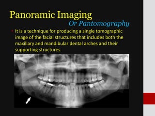
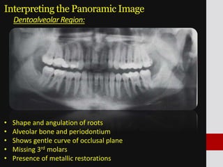
Panoramic Imaging
Panoramic Imaging
• It is a technique for producing a single tomographic
image of the facial structures that includes both the
maxillary and mandibular dental arches and their
supporting structures.
Or Pantomography
Panoramic Imaging
Or Pantomography
Panoramic Imaging
Advantages:
• Broad coverage
• Low patient radiation dose
• Convenience of the examination
• Useful in patients who are unable to open their mouths
• Short time required: 3-4 mins
• Useful visual aid
Panoramic Imaging
Indications:
• Evaluation of trauma
• Location of third molars
• Extensive dental or osseous disease
• Known or suspected large lesions
• Tooth development
• Retained teeth or root tips
• TMJ pain
• Study of sinuses
• Developmental anomalies
Panoramic Imaging
Disadvantages:
• Magnification, Geometric distortion &
overlapped images.
• Resolution of fine anatomic details of peri-apical
area and periodontal structures is less.
• The spinal cord superimpose the anterior region.
• Common to have overlapped teeth images
especially in premolars.
• Expensive
Principles of Panoramic Image Formation
• First described by Numata and independently by Paatero
in the late 1940s
• Movement of the film and objects about 2 fixed centers
of rotation.
Principles of Panoramic Image Formation
Movement of the film and objects (A, B, C, and D) about two
fixed centers of rotation. Pb, Lead collimator
Principles of Panoramic Image Formation
Movement of the film and x-ray source about one fixed
center of rotation. Pb, Lead collimator
Principles of Panoramic Image Formation
Movement of the film and x-ray source about a shifting
center of rotation. Pb, Lead collimator
Principles of Panoramic Image Formation
Movement of the x-ray source and beam.
Image Layer
• Is a 3-D curved zone, or “focal
trough” where the structures
lying within this layer are
reasonably well defined on final
panoramic image.
• The images seen on the OPG
consists largely of anatomic
structures located within the
focal trough
• Objects out of focal trough are
blurred, magnified, reduced or
distorted.
• The shape of the focal trough
varies with the brand of
equipments used.
Or Focal Trough
Image Layer
Factors that affect image layer size:
• Arc path
• Velocity of the receptor and x-ray tube head
• Alignment of the x-ray beam
• Collimator width
Image Layer
• Mandible supporting a metal ring positioned at the center of
the focal trough.
• The incisal edges of the mandibular teeth are indexed by a
bite rod-positioning device.
• The Mandible is positioned at the center of the trough.
Image Layer
• Ring 5 mm anterior to Focal Trough
Image Layer
• Ring 5 mm posterior to Focal Trough
Image Layer
• This distortions result from the reciprocal horizontal
movements of the receptor and x-ray source.
• When the mandible is displaced to the lingual side of its
optimal position in the image layer, the beam passes more
slowly through it. The images of the structures are elongated
horizontally thus, appear wider.
• When the mandible is displaced toward the buccal, the beam
passes at a rate faster than normal through the structures,
thus appear thinner.
Interpreting the Panoramic Image
The FOUR DiagnosticRegionsin a Panoramic Image:
MaxillaryRegion
MandibularRegionDentoalveolarRegion
TMJ,includingretromaxillary
andcervicalregion
Interpreting the Panoramic Image
Maxillary Region:
Interpreting the Panoramic Image
Maxillary Region:
Interpreting the Panoramic Image
MandibularRegion:
Interpreting the Panoramic Image
MandibularRegion:
Interpreting the Panoramic Image
DentoalveolarRegion:
• Shape and angulation of roots
• Alveolar bone and periodontium
• Shows gentle curve of occlusal plane
• Missing 3rd molars
• Presence of metallic restorations
Interpreting the Panoramic Image
SoftTissueImages:
Interpreting the Panoramic Image
SoftTissueImages:
Interpreting the Panoramic Image
Airspace:
Interpreting the Panoramic Image
Airspace:
Panoramic imaging

Panoramic imaging

  • 1.
  • 2.
    Panoramic Imaging • Itis a technique for producing a single tomographic image of the facial structures that includes both the maxillary and mandibular dental arches and their supporting structures. Or Pantomography
  • 3.
  • 4.
    Panoramic Imaging Advantages: • Broadcoverage • Low patient radiation dose • Convenience of the examination • Useful in patients who are unable to open their mouths • Short time required: 3-4 mins • Useful visual aid
  • 5.
    Panoramic Imaging Indications: • Evaluationof trauma • Location of third molars • Extensive dental or osseous disease • Known or suspected large lesions • Tooth development • Retained teeth or root tips • TMJ pain • Study of sinuses • Developmental anomalies
  • 6.
    Panoramic Imaging Disadvantages: • Magnification,Geometric distortion & overlapped images. • Resolution of fine anatomic details of peri-apical area and periodontal structures is less. • The spinal cord superimpose the anterior region. • Common to have overlapped teeth images especially in premolars. • Expensive
  • 7.
    Principles of PanoramicImage Formation • First described by Numata and independently by Paatero in the late 1940s • Movement of the film and objects about 2 fixed centers of rotation.
  • 8.
    Principles of PanoramicImage Formation Movement of the film and objects (A, B, C, and D) about two fixed centers of rotation. Pb, Lead collimator
  • 9.
    Principles of PanoramicImage Formation Movement of the film and x-ray source about one fixed center of rotation. Pb, Lead collimator
  • 10.
    Principles of PanoramicImage Formation Movement of the film and x-ray source about a shifting center of rotation. Pb, Lead collimator
  • 11.
    Principles of PanoramicImage Formation Movement of the x-ray source and beam.
  • 12.
    Image Layer • Isa 3-D curved zone, or “focal trough” where the structures lying within this layer are reasonably well defined on final panoramic image. • The images seen on the OPG consists largely of anatomic structures located within the focal trough • Objects out of focal trough are blurred, magnified, reduced or distorted. • The shape of the focal trough varies with the brand of equipments used. Or Focal Trough
  • 13.
    Image Layer Factors thataffect image layer size: • Arc path • Velocity of the receptor and x-ray tube head • Alignment of the x-ray beam • Collimator width
  • 14.
    Image Layer • Mandiblesupporting a metal ring positioned at the center of the focal trough. • The incisal edges of the mandibular teeth are indexed by a bite rod-positioning device. • The Mandible is positioned at the center of the trough.
  • 15.
    Image Layer • Ring5 mm anterior to Focal Trough
  • 16.
    Image Layer • Ring5 mm posterior to Focal Trough
  • 17.
    Image Layer • Thisdistortions result from the reciprocal horizontal movements of the receptor and x-ray source. • When the mandible is displaced to the lingual side of its optimal position in the image layer, the beam passes more slowly through it. The images of the structures are elongated horizontally thus, appear wider. • When the mandible is displaced toward the buccal, the beam passes at a rate faster than normal through the structures, thus appear thinner.
  • 18.
    Interpreting the PanoramicImage The FOUR DiagnosticRegionsin a Panoramic Image: MaxillaryRegion MandibularRegionDentoalveolarRegion TMJ,includingretromaxillary andcervicalregion
  • 19.
    Interpreting the PanoramicImage Maxillary Region:
  • 20.
    Interpreting the PanoramicImage Maxillary Region:
  • 21.
    Interpreting the PanoramicImage MandibularRegion:
  • 22.
    Interpreting the PanoramicImage MandibularRegion:
  • 23.
    Interpreting the PanoramicImage DentoalveolarRegion: • Shape and angulation of roots • Alveolar bone and periodontium • Shows gentle curve of occlusal plane • Missing 3rd molars • Presence of metallic restorations
  • 24.
    Interpreting the PanoramicImage SoftTissueImages:
  • 25.
    Interpreting the PanoramicImage SoftTissueImages:
  • 26.
  • 27.