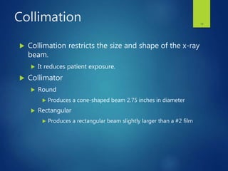
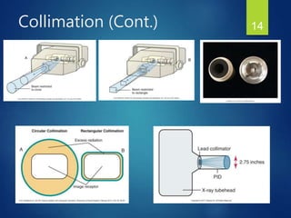
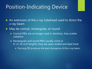
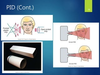
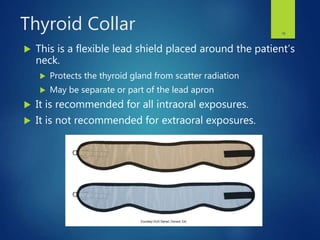
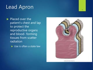
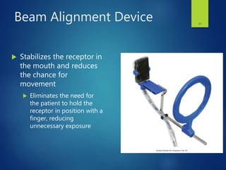
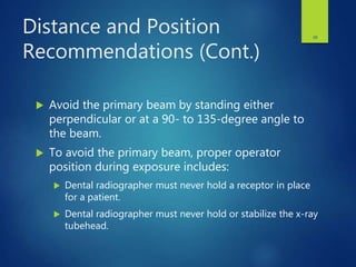
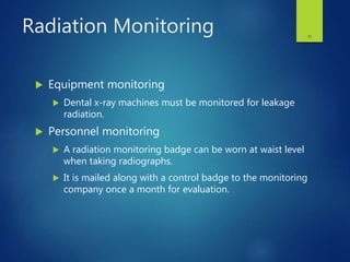
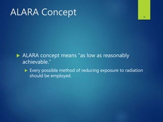
RADIATION PROTECTION
CHAPTER 5
1
Learning Objectives
Lesson 5.1: Radiation Protection
1. Define the key terms associated with radiation protection.
2. Describe in detail the basics of patient protection before x-ray
exposure.
3. Discuss the different types of filtration, and state the recommended
total filtration for dental x-ray machines operating above and
below 70 kV.
4. Describe the collimator used in dental x-ray machines and state the
recommended diameter of the useful beam at the patient’s skin.
2
Learning Objectives
Lesson 5.1: Radiation Protection
(Cont.)
5. List six ways to protect the patient from excessive radiation during
x-ray exposure.
6. Describe the importance of receptor handling and processing after
patient exposure to x-radiation.
7. Discuss operator protection in terms of adequate distance,
shielding, and avoidance of the useful beam.
8. Describe personnel and equipment monitoring devices used to
detect radiation.
3
Learning Objectives
Lesson 5.1: Radiation Protection
(Cont.)
9. Discuss radiation exposure guidelines, including radiation safety
legislation, maximum permissible dose (MPD), and the ALARA
concept.
10. Discuss with the dental patient radiation protection steps used
before, during, and after exposure to x-radiation.
4
Introduction
 Purpose
 To discuss patient protection before, during,
and after exposure to x-rays
 To detail operator protection methods
 To present radiation exposure and safety
guidelines
5
Patient Protection
 X-radiation causes biologic changes in living
cells; it adversely affects all living tissue.
 Our goal is to minimize the amount of radiation
received by the patient and maximize the benefits.
 Techniques may be used before, during, and
after the procedure to protect the patient.
6
Before Exposure
 Prescribing dental radiographs
 Proper equipment
7
Prescribing Dental Images
 Professional judgment is used to determine the
number, type, and frequency of dental
radiographs.
 The ADA Council on Scientific Affairs, in conjunction
with the U.S. Department of Health and Human Services,
Public Health Service, Food and Drug Administration
(FDA) has adopted guidelines for prescribing the
number, type, and frequency of dental images.
 See Table 5-1 on page 44
8
Proper Equipment
 Use of equipment that complies with state and
federal radiation guidelines will minimize the
radiation a patient receives.
 Filtration
 Inherent filtration
 Added filtration
 Total filtration
 Collimation
 Position-indicating device
9
Inherent Filtration
 Inherent filtration takes place when the primary
beam passes through the glass window of the x-
ray tube, the insulating oil, and the tubehead seal.
 Is equivalent to approximately 0.5 to 1.0 mm of aluminum
 Does not meet the standards regulated by state and
federal law, so additional filtration is required
10
Added Filtration
 An aluminum disk is placed between the collimator and
the tubehead seal to filter out longer wavelength, lower
energy x-rays from the x-ray beam.
 Filtration results in a higher-energy and more penetrating useful
beam.
11
Total Filtration
 The sum of inherent and added filtration
 Regulated by state and federal laws
 Machines operating at or below 70 kVp
 Require a minimum total of 1.5 mm aluminum filtration
 Machines operating above 70 kVp
 Require a minimum total of 2.5 mm aluminum filtration
12
Collimation
 Collimation restricts the size and shape of the x-ray
beam.
 It reduces patient exposure.
 Collimator
 Round
 Produces a cone-shaped beam 2.75 inches in diameter
 Rectangular
 Produces a rectangular beam slightly larger than a #2 film
13
Collimation (Cont.) 14
Position-Indicating Device
 An extension of the x-ray tubehead used to direct the
x-ray beam
 May be conical, rectangular, or round
 Conical PIDs are no longer used in dentistry; they scatter
radiation.
 Rectangular and round PIDs usually come in
8- or 16-inch lengths; they are open ended and lead lined.
 The long PID produces the least divergence of the x-ray beam.
15
PID (Cont.) 16
During Exposure
 Thyroid collar
 Lead apron
 Image receptors
 Beam alignment devices
 Exposure factor selection
 Proper technique
17
Thyroid Collar
 This is a flexible lead shield placed around the patient’s
neck.
 Protects the thyroid gland from scatter radiation
 May be separate or part of the lead apron
 It is recommended for all intraoral exposures.
 It is not recommended for extraoral exposures.
18
Lead Apron
 Placed over the
patient’s chest and lap
to protect the
reproductive organs
and blood- forming
tissues from scatter
radiation
 Use is often a state law
19
Image Receptors
 Digital image receptors require less radiation exposure
of the patient.
 Use of a digital receptor is the most effective method
of reducing a patient’s exposure to radiation.
 Fast film
 D-speed and F-speed film are now the two speeds on the
market.
 F-speed (Kodak Insight) is the faster of the two.
20
Beam Alignment Device
 Stabilizes the receptor in
the mouth and reduces
the chance for
movement
 Eliminates the need for
the patient to hold the
receptor in position with a
finger, reducing
unnecessary exposure
21
Exposure Factor Selection
 Adjustment of kVp, milliamperage, and time
settings on the control panel to limit the amount of
x-radiation exposure received by the patient
 On most units, the kilovolt peak and milliamperage are
preset by the manufacturer and cannot be adjusted.
22
Proper Technique
 Nondiagnostic images must be retaken, resulting
in additional radiation exposure for the patient.
 Re-exposure of an image must be avoided at all
times.
23
After Exposure
 Proper receptor handling
 Artifacts caused by improper film handling result in
nondiagnostic films.
 Proper film processing and image retrieval
 Improper film processing may necessitate retakes,
needlessly exposing the patient to excess
x-radiation.
24
Operator Protection
 Protection guidelines
 Radiation monitoring
25
Protection Guidelines
 The dental radiographer must avoid the primary
beam.
 Protection guidelines include:
 Distance recommendations
 Position recommendations
 Shielding recommendations
26
Distance and Position
Recommendations
 Maintain an adequate distance during exposure.
 The dental radiographer must maintain a distance of at
least 6 feet from the tubehead during an exposure.
27
Distance and Position
Recommendations (Cont.)
 Avoid the primary beam by standing either
perpendicular or at a 90- to 135-degree angle to
the beam.
 To avoid the primary beam, proper operator
position during exposure includes:
 Dental radiographer must never hold a receptor in place
for a patient.
 Dental radiographer must never hold or stabilize the x-ray
tubehead.
28
29
Shielding Recommendations
 Dental office design may include walls to
protect the operator from primary and scatter
radiation.
 Protective barriers may be incorporated into the
office design.
30
Radiation Monitoring
 Equipment monitoring
 Dental x-ray machines must be monitored for leakage
radiation.
 Personnel monitoring
 A radiation monitoring badge can be worn at waist level
when taking radiographs.
 It is mailed along with a control badge to the monitoring
company once a month for evaluation.
31
Radiation Exposure
Guidelines
 Radiation safety legislation
 Maximum permissible dose
 Cumulative occupational dose
 ALARA concept
32
Radiation Safety Legislation
 Radiation Control for Health and Safety Act, 1968
 Enacted to standardize the operation of x-ray
equipment
 Consumer-Patient Radiation Health and Safety
Act, 1981
 Enacted to address the issues of educating and
certifying operators of radiographic equipment
33
Maximum Permissible Dose
(MPD)
 MPD is the maximum dose equivalent that a body
is permitted to receive in a specific period.
 MPD for occupationally exposed persons is 50 mSv/year
(0.05 Sv/year or 5.0 rem/year).
 For nonoccupationally exposed persons, it is 1 mSv/year
(0.1 rem/year).
 For occupationally exposed pregnant women, MPD is 0.5
mSv per month during the pregnancy months.
34
Cumulative Occupational
Dose
 Cumulative occupational dose is the dose
accumulated over a lifetime.
 An individual’s cumulative occupational effective dose
should not exceed the worker’s age multiplied by 10
mSv.
35
ALARA Concept
 ALARA concept means “as low as reasonably
achievable.”
 Every possible method of reducing exposure to radiation
should be employed.
36
Radiation Protection
and Patient Education
 What you say to the patient before, during and
after the procedure
 Patient education may take the form of an
informal conversation or printed literature
 Dental radiographer must be prepared to explain
exactly how patients are protected before, during,
and after x-ray exposure
37
Questions?
38

Chapter 5: Radiation Protection

  • 1.
  • 2.
    Learning Objectives Lesson 5.1:Radiation Protection 1. Define the key terms associated with radiation protection. 2. Describe in detail the basics of patient protection before x-ray exposure. 3. Discuss the different types of filtration, and state the recommended total filtration for dental x-ray machines operating above and below 70 kV. 4. Describe the collimator used in dental x-ray machines and state the recommended diameter of the useful beam at the patient’s skin. 2
  • 3.
    Learning Objectives Lesson 5.1:Radiation Protection (Cont.) 5. List six ways to protect the patient from excessive radiation during x-ray exposure. 6. Describe the importance of receptor handling and processing after patient exposure to x-radiation. 7. Discuss operator protection in terms of adequate distance, shielding, and avoidance of the useful beam. 8. Describe personnel and equipment monitoring devices used to detect radiation. 3
  • 4.
    Learning Objectives Lesson 5.1:Radiation Protection (Cont.) 9. Discuss radiation exposure guidelines, including radiation safety legislation, maximum permissible dose (MPD), and the ALARA concept. 10. Discuss with the dental patient radiation protection steps used before, during, and after exposure to x-radiation. 4
  • 5.
    Introduction  Purpose  Todiscuss patient protection before, during, and after exposure to x-rays  To detail operator protection methods  To present radiation exposure and safety guidelines 5
  • 6.
    Patient Protection  X-radiationcauses biologic changes in living cells; it adversely affects all living tissue.  Our goal is to minimize the amount of radiation received by the patient and maximize the benefits.  Techniques may be used before, during, and after the procedure to protect the patient. 6
  • 7.
    Before Exposure  Prescribingdental radiographs  Proper equipment 7
  • 8.
    Prescribing Dental Images Professional judgment is used to determine the number, type, and frequency of dental radiographs.  The ADA Council on Scientific Affairs, in conjunction with the U.S. Department of Health and Human Services, Public Health Service, Food and Drug Administration (FDA) has adopted guidelines for prescribing the number, type, and frequency of dental images.  See Table 5-1 on page 44 8
  • 9.
    Proper Equipment  Useof equipment that complies with state and federal radiation guidelines will minimize the radiation a patient receives.  Filtration  Inherent filtration  Added filtration  Total filtration  Collimation  Position-indicating device 9
  • 10.
    Inherent Filtration  Inherentfiltration takes place when the primary beam passes through the glass window of the x- ray tube, the insulating oil, and the tubehead seal.  Is equivalent to approximately 0.5 to 1.0 mm of aluminum  Does not meet the standards regulated by state and federal law, so additional filtration is required 10
  • 11.
    Added Filtration  Analuminum disk is placed between the collimator and the tubehead seal to filter out longer wavelength, lower energy x-rays from the x-ray beam.  Filtration results in a higher-energy and more penetrating useful beam. 11
  • 12.
    Total Filtration  Thesum of inherent and added filtration  Regulated by state and federal laws  Machines operating at or below 70 kVp  Require a minimum total of 1.5 mm aluminum filtration  Machines operating above 70 kVp  Require a minimum total of 2.5 mm aluminum filtration 12
  • 13.
    Collimation  Collimation restrictsthe size and shape of the x-ray beam.  It reduces patient exposure.  Collimator  Round  Produces a cone-shaped beam 2.75 inches in diameter  Rectangular  Produces a rectangular beam slightly larger than a #2 film 13
  • 14.
  • 15.
    Position-Indicating Device  Anextension of the x-ray tubehead used to direct the x-ray beam  May be conical, rectangular, or round  Conical PIDs are no longer used in dentistry; they scatter radiation.  Rectangular and round PIDs usually come in 8- or 16-inch lengths; they are open ended and lead lined.  The long PID produces the least divergence of the x-ray beam. 15
  • 16.
  • 17.
    During Exposure  Thyroidcollar  Lead apron  Image receptors  Beam alignment devices  Exposure factor selection  Proper technique 17
  • 18.
    Thyroid Collar  Thisis a flexible lead shield placed around the patient’s neck.  Protects the thyroid gland from scatter radiation  May be separate or part of the lead apron  It is recommended for all intraoral exposures.  It is not recommended for extraoral exposures. 18
  • 19.
    Lead Apron  Placedover the patient’s chest and lap to protect the reproductive organs and blood- forming tissues from scatter radiation  Use is often a state law 19
  • 20.
    Image Receptors  Digitalimage receptors require less radiation exposure of the patient.  Use of a digital receptor is the most effective method of reducing a patient’s exposure to radiation.  Fast film  D-speed and F-speed film are now the two speeds on the market.  F-speed (Kodak Insight) is the faster of the two. 20
  • 21.
    Beam Alignment Device Stabilizes the receptor in the mouth and reduces the chance for movement  Eliminates the need for the patient to hold the receptor in position with a finger, reducing unnecessary exposure 21
  • 22.
    Exposure Factor Selection Adjustment of kVp, milliamperage, and time settings on the control panel to limit the amount of x-radiation exposure received by the patient  On most units, the kilovolt peak and milliamperage are preset by the manufacturer and cannot be adjusted. 22
  • 23.
    Proper Technique  Nondiagnosticimages must be retaken, resulting in additional radiation exposure for the patient.  Re-exposure of an image must be avoided at all times. 23
  • 24.
    After Exposure  Properreceptor handling  Artifacts caused by improper film handling result in nondiagnostic films.  Proper film processing and image retrieval  Improper film processing may necessitate retakes, needlessly exposing the patient to excess x-radiation. 24
  • 25.
    Operator Protection  Protectionguidelines  Radiation monitoring 25
  • 26.
    Protection Guidelines  Thedental radiographer must avoid the primary beam.  Protection guidelines include:  Distance recommendations  Position recommendations  Shielding recommendations 26
  • 27.
    Distance and Position Recommendations Maintain an adequate distance during exposure.  The dental radiographer must maintain a distance of at least 6 feet from the tubehead during an exposure. 27
  • 28.
    Distance and Position Recommendations(Cont.)  Avoid the primary beam by standing either perpendicular or at a 90- to 135-degree angle to the beam.  To avoid the primary beam, proper operator position during exposure includes:  Dental radiographer must never hold a receptor in place for a patient.  Dental radiographer must never hold or stabilize the x-ray tubehead. 28
  • 29.
  • 30.
    Shielding Recommendations  Dentaloffice design may include walls to protect the operator from primary and scatter radiation.  Protective barriers may be incorporated into the office design. 30
  • 31.
    Radiation Monitoring  Equipmentmonitoring  Dental x-ray machines must be monitored for leakage radiation.  Personnel monitoring  A radiation monitoring badge can be worn at waist level when taking radiographs.  It is mailed along with a control badge to the monitoring company once a month for evaluation. 31
  • 32.
    Radiation Exposure Guidelines  Radiationsafety legislation  Maximum permissible dose  Cumulative occupational dose  ALARA concept 32
  • 33.
    Radiation Safety Legislation Radiation Control for Health and Safety Act, 1968  Enacted to standardize the operation of x-ray equipment  Consumer-Patient Radiation Health and Safety Act, 1981  Enacted to address the issues of educating and certifying operators of radiographic equipment 33
  • 34.
    Maximum Permissible Dose (MPD) MPD is the maximum dose equivalent that a body is permitted to receive in a specific period.  MPD for occupationally exposed persons is 50 mSv/year (0.05 Sv/year or 5.0 rem/year).  For nonoccupationally exposed persons, it is 1 mSv/year (0.1 rem/year).  For occupationally exposed pregnant women, MPD is 0.5 mSv per month during the pregnancy months. 34
  • 35.
    Cumulative Occupational Dose  Cumulativeoccupational dose is the dose accumulated over a lifetime.  An individual’s cumulative occupational effective dose should not exceed the worker’s age multiplied by 10 mSv. 35
  • 36.
    ALARA Concept  ALARAconcept means “as low as reasonably achievable.”  Every possible method of reducing exposure to radiation should be employed. 36
  • 37.
    Radiation Protection and PatientEducation  What you say to the patient before, during and after the procedure  Patient education may take the form of an informal conversation or printed literature  Dental radiographer must be prepared to explain exactly how patients are protected before, during, and after x-ray exposure 37
  • 38.

Editor's Notes

  • #6 In addition, this chapter includes a discussion of patient education about radiation protection.
  • #7 With the use of proper patient protection techniques, the amount of x-radiation received by a dental patient can be minimized.
  • #8 Patient protection measures can be used before any x-radiation exposure.
  • #9 Every patient should be evaluated on an individual basis to determine what x-ray images are necessary. Proper prescribing of dental x-ray images can minimize the amount of radiation exposure. Refer students to Table 5-1.
  • #10 The x-ray tubehead should be properly equipped with aluminum filters, lead collimator, and PID.
  • #11 Inherent filtration is composed of part of the x-ray tube and tube housing. Filtration is necessary to absorb the low-energy x-ray photons emitted by the tube before they hit their target.
  • #12 For added filtration, thin sheets of metal are placed in the path of the x-ray beam. Refer students to Figures 5-1 and 5-2.
  • #13 Total filtration is regulated by state and federal laws.
  • #14 Where is the collimator located? (Between the x-ray tube and the position-indicating device) Refer students to Figures 5-3 through 5-6.
  • #16 For infection control purposes, the PID, along with the tubehead, should be covered with plastic barriers and changed for each patient. The end of the PID should be positioned close to the extraoral portion of the film holder and lined up correctly in all three planes to ensure accurate exposure and image production on the film. Refer students to Figures 5-7 through 5-9.
  • #18 As a dental radiographer, you want to limit the amount of radiation received by the patient. Proper selection of exposure factors and good technique further protect the patient from excess radiation exposure.
  • #19 The thyroid collar and lead apron should be used on all patients regardless of their age, sex, or number of films being taken. Refer students to Figure 5-10.
  • #20 Many states mandate the use of the lead apron. Dental radiographers should ensure that the lead apron and thyroid collar are stored properly and not folded, since creases may crack the lead. Care should be taken not to touch the apron with the same gloves that were used to place the film in the patient’s mouth, as this will cause cross-contamination, compromising infection control. The radiographer should ensure that the patient removes any oral piercings, eyeglasses, earrings, and removable dental prostheses that may distort or interfere with the desired image. Refer students to Figure 5-11.
  • #21 The size of the silver bromide crystals is the main factor in determining the film speed. The larger the crystals, the faster the film. When digital sensors are not used, fast film is the most effective method of reducing a patient’s exposure to x-radiation. Name a slight disadvantage to using F-speed film? (Larger silver bromide crystals result in a slightly decreased contrast.)
  • #22 Film-holding devices also assist the operator in properly positioning the film and PID, thus decreasing the chance for retakes. What may be placed in areas of missing teeth to help stabilize the film-holding instrument? (Cotton rolls) Refer students to Figure 5-12.
  • #23 A setting of 60 to 80 kVp keeps patient exposure to a minimum.
  • #24 Retakes are a major cause of unnecessary radiation to patients and must be avoided. Having to retake images wastes time and is not appreciated by most patients. If a retake is ordered, the dental radiographer should know how to correct the error that resulted in the need for a retake.
  • #25 From the time the films are exposed until they are processed, careful handling is crucial.
  • #26 Dental radiographers must accept responsibility for their own radiation safety.
  • #27 To keep radiation exposure to zero, the dental radiographer must carefully follow the safety guidelines and use radiation monitoring devices.
  • #28 Ideally, the radiographer should leave the room or take a position behind a suitable barrier or wall during exposures. When maintaining this distance is not possible, a protective barrier must be used.
  • #29 Refer students to Figure 5-13. Dental radiographers working in an unfamiliar office or treatment room should familiarize themselves with the proper position to stand in while taking radiographs.
  • #31 There may be a lead wall to stand behind, or there may be a need to walk around the corner with a portable exposure button. There may be an exposure button positioned on a wall outside the room used for radiographs.
  • #32 These monitoring devices are small and light enough to be placed on the breast pocket of a lab jacket without interfering with normal duties. Monitoring devices are usually sent to the agency monthly and a new one is returned to the operators along with a report to the dentist showing radiation exposure results for the individual. Refer students to Figure 5-14.
  • #33 Radiation exposure guidelines have been established to protect the patient and operator from excess radiation. Strict adherence to radiation exposure guidelines is mandatory for all dental radiographers.
  • #34 Legislation has been established at both the state and federal levels to protect the patient, operator, and general public from radiation hazards. Radiation legislation varies greatly from state to state, and the dental radiographer must be familiar with the laws that apply to his or her workplace.
  • #35 The amount of radiation to the whole body carries very little chance of injury. Dental personnel should strive for an occupational dose of 0 by adhering to strict radiation protection practices. National Council on Radiation Protection and Measurements (NCRP) publishes these standards; the International Commission on Radiological Protection (ICRP) also publishes recommendations, but the NCRP and ICRP do not always agree on recommended dose limits.
  • #36 The age of 18 is the minimum required age of a person who works with radiation. For a 60-year-old worker, the NCRP would recommend a cumulative occupational dose of no more than 60 × 10 mSv = 600 mSv (0.6 Sv).
  • #37 What are some precautions taken to abide by the ALARA concept? (Radiographs should be ordered only for diagnostic purposes; use the lowest possible kVp, mA, and exposure time; use F-speed film or digital radiography; use a longer PID with a rectangular shape; use a tubehead with an aluminum filter and a lead collimator; use a lead apron and thyroid collar; use film-holding devices; avoid retakes; and test equipment for efficiency and proper functioning.)
  • #38 It may be useful to have educational brochures on hand that explain the importance of diagnostic dental radiographs and how their benefits far outweigh the minimal risks. Some dental facilities also have informative videos that can help educate patients about dental radiographs.