3. Tomography
imaging by sectioning, through a body, by moving an x-ray source and the
film in opposite directions during the exposure.
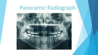
Panorama
An unobstructed and wide view of an extensive area in all directions.
Pantamography
aka Panoramic/rotational radiography. A technique for producing a single
tomogramphic image of the facial structures, including maxilla mandible and
supporting structures.
4. Working Principle
Employs sconagraphy (slit beam)&tomography
Panoramic imaging is a technique for producing a single tomographic
image of the facial structures that includes both maxillary and mandibular
arch and their supporting structure.
It is a curvilinear variant of conventional tomography and is based on the
principal of the reciprocal movement of an x-ray source and an image
receptor around a central point or plane called the image layer in which the
object of interest is located.
5.
6.
7. Indications
As a substitute for full mouth intraoral periapical radiographs
Evaluation of trauma
Evaluation of tooth development in mixed dentition for children
Orthodontic treatment
Developmental anomalies
Third molars
Large lesions like cyst, tumors.
Detection of fractures
Generalized disease
Inability to tolerate intraoral films
Assessment for surgical procedure
8. Contraindications
Panoramic film are not as Defined or sharp as the images seen
on intraoral films.
*Can’t use where require
-Fine anatomical details
-Small carious lesions
Fine structures of the marginal periodontium
Periapical diseases
For equal magnification
9. Advantages
Well-tolerated by patients
Minimal time to expose when compared to intraoral
radiographs
Broad anatomical coverage
Relatively low patient dose
For object localization in conjunction with occlusal
radiography.
10. Disadvantages
Resolution is not as good as intraoral films.
This results in loss of detail
Superimposition of real/double images
Only objects in focal trough are seen clearly
Distortion of image
– Overlapped teeth
– Magnification
– Objects of interest outside of focal trough are distorted and blurred.
12. Hard tissue land mark of the Maxilla and
surrounding structures
Mastoid process
Styloid process
External auditory meatus
Mandibular fossa
Articular eminence
Lateral pterygoid plate
Pterygomaxillary fissure
Maxillary tuberosity
Infraorbital foramen
Orbit
Incisive canal and foramen
Anterior nasal spine
Nasal cavity
Nasal septum
Hard palate
Maxillary sinus and its floor
Zygomatic process of maxilla
Zygoma
Hamulus
Dentition
13. Hard tissue land mark of the Mandible and
surrounding structures
Mandibular condyle
Condylar notch
Coronoid process
Ramus
Mandibular foramen
Lingula
Mandibular canal
Mental foramen
Mental ridge
Mental fossa
Lingual foramen
Genial tubercle
Inferior border of mandible
Mylohyoid ridge
Internal oblique ridge
External oblique ridge
Angle of the mandible
Dentition
14.
15.
16.
17. Air spaces
Palatoglossal air space
Nasopharyngeal air
space
Glossopharyngeal air
space
Soft tissue images
• Tongue
• Ear lobes
• Nasal cartridge
• Soft palate
• Nasolabial
• Soft palate
• Nasolabial folds
• Soft palate and uvula
• Lip line
18. Ghost/artifactual shadows
Cervical vertebrae
Body, condyle and ramus of the contralateral side of the mandible
Palate
Chin rest
(R)or(L) markers of the machine
Neck chains
Napkin chains
Earrings, tongue rings
Shoulder straps of protective apron
27. INTERPRETATION
Assess the periphery and corners of the image
Examine the outer cortices of the mandible
Examine the cortices of the maxilla
Examine the zygomatic bones and arches
Assess the internal density of the maxillary sinuses
Assess the structures of the nasal cavity and the palates
Examine bone the pattern of the maxilla and mandible
Alveolar processes and teeth