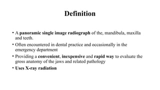
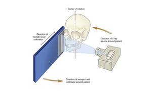
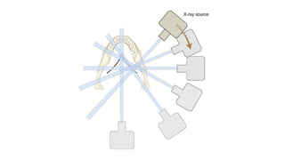
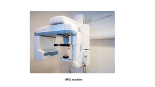
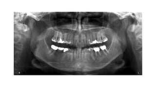
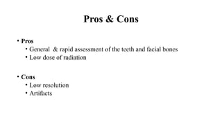
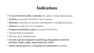
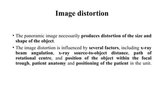
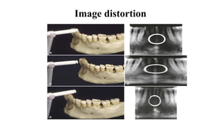
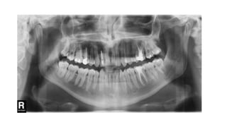
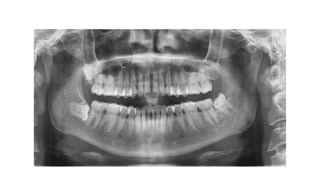
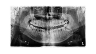
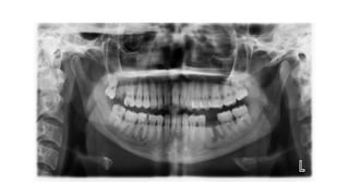
Orthopantomography
(OPG)
Dr Fazel Rahman FAIZI
Definition
• A panoramic single image radiograph of the, mandibula, maxilla
and teeth.
• Often encountered in dental practice and occasionally in the
emergency department
• Providing a convenient, inexpensive and rapid way to evaluate the
gross anatomy of the jaws and related pathology
• Uses X-ray radiation
OPG machine
Pros & Cons
• Pros
• General & rapid assessment of the teeth and facial bones
• Low dose of radiation
• Cons
• Low resolution
• Artifacts
Indications
• General dental health evaluation for caries or pulp origin disease
• Trauma assessment for tooth or jaw fractures
• Infection evaluation of sinusitis, periodontitis or periapical disease
• Tumour or radicular cyst evaluation
• Temporomandibular joint assessment for disease.
• Foreign body localization
• Salivary stone identification
• Growth and development monitoring of paediatric teeth for
location, shape, angle, supernumerary tooth,
• Initial and progressive evaluation of orthodontic treatment
Focal trough
• A three-dimensional curved zone, or “image layer,” where the
structures lying within this zone are reasonably well defined on the final
panoramic image.
• Structures seen on a panoramic image are primarily those located
within the focal trough.
• Images are most clear in the middle and become less clear further from
the central line.
• Objects outside the focal trough are blurred, magnified, or reduced
in size and are sometimes distorted to the extent of not being
recognizable.
Focal trough
Image distortion
• The panoramic image necessarily produces distortion of the size and
shape of the object.
• The image distortion is influenced by several factors, including x-ray
beam angulation, x-ray source-to-object distance, path of
rotational centre, and position of the object within the focal
trough, patient anatomy and positioning of the patient in the unit.
Image distortion
Interpretation of panaromic image
• Under negatoscope or in compute
1.Dentition
2.Mid facial region or upper jaw
3.Lower jaw
4.Soft tissues
5.Check peripheries
Dentition
• Count the teeth; note the missing
• Supernumerary and impacted teeth
• Caries and abscess
• Fractures
• Lucent or sclerotic lesions
Mid facial region/upper jaw
• Facial bones, sinuses and soft tissue
• Facial bones; temporal, zygomatic, mandibles, maxilla, frontal,
ethmoid and maxillary sinuses, nasal septum, concha
• Look for fracture
• Cystic or sclerotic lesion
• Maxillary sinuses
• Nasal cavity
Lower jaw
• Mandibular condyles and TMJ
• Coronoid process
• Ramus and body of mandible
• Alveolar processes
Periphery/soft tissues
• Hyoid bone
• Spine
• Pharynx
• Zygomatic arch
Thank you

OPG. PRESENTATION FOR NEW DENTAL STUDENTS

  • 1.
  • 2.
    Definition • A panoramicsingle image radiograph of the, mandibula, maxilla and teeth. • Often encountered in dental practice and occasionally in the emergency department • Providing a convenient, inexpensive and rapid way to evaluate the gross anatomy of the jaws and related pathology • Uses X-ray radiation
  • 5.
  • 7.
    Pros & Cons •Pros • General & rapid assessment of the teeth and facial bones • Low dose of radiation • Cons • Low resolution • Artifacts
  • 8.
    Indications • General dentalhealth evaluation for caries or pulp origin disease • Trauma assessment for tooth or jaw fractures • Infection evaluation of sinusitis, periodontitis or periapical disease • Tumour or radicular cyst evaluation • Temporomandibular joint assessment for disease. • Foreign body localization • Salivary stone identification • Growth and development monitoring of paediatric teeth for location, shape, angle, supernumerary tooth, • Initial and progressive evaluation of orthodontic treatment
  • 9.
    Focal trough • Athree-dimensional curved zone, or “image layer,” where the structures lying within this zone are reasonably well defined on the final panoramic image. • Structures seen on a panoramic image are primarily those located within the focal trough. • Images are most clear in the middle and become less clear further from the central line. • Objects outside the focal trough are blurred, magnified, or reduced in size and are sometimes distorted to the extent of not being recognizable.
  • 10.
  • 11.
    Image distortion • Thepanoramic image necessarily produces distortion of the size and shape of the object. • The image distortion is influenced by several factors, including x-ray beam angulation, x-ray source-to-object distance, path of rotational centre, and position of the object within the focal trough, patient anatomy and positioning of the patient in the unit.
  • 12.
  • 13.
    Interpretation of panaromicimage • Under negatoscope or in compute 1.Dentition 2.Mid facial region or upper jaw 3.Lower jaw 4.Soft tissues 5.Check peripheries
  • 14.
    Dentition • Count theteeth; note the missing • Supernumerary and impacted teeth • Caries and abscess • Fractures • Lucent or sclerotic lesions
  • 15.
    Mid facial region/upperjaw • Facial bones, sinuses and soft tissue • Facial bones; temporal, zygomatic, mandibles, maxilla, frontal, ethmoid and maxillary sinuses, nasal septum, concha • Look for fracture • Cystic or sclerotic lesion • Maxillary sinuses • Nasal cavity
  • 16.
    Lower jaw • Mandibularcondyles and TMJ • Coronoid process • Ramus and body of mandible • Alveolar processes
  • 17.
    Periphery/soft tissues • Hyoidbone • Spine • Pharynx • Zygomatic arch
  • 23.