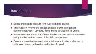
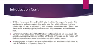
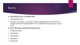
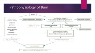
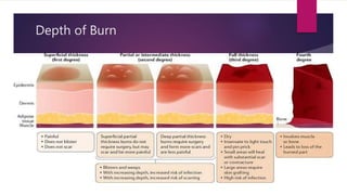
Burn & It’s Management in
Children
TOPIC PRESENTED BY:
DR. ANICK SAHA SHUVO (MS PHASE-A, PAED. SURGERY)
Introduction
 Burns and scalds account for 6% of pediatric injuries.
 The majority involve pre-school children, burns being most
common between 1-2 years, flame burns between 5-18 years.
 House fires are the cause of most fatal burns with smoke inhalation
being the immediate cause of death in many cases.
 Scalds are most associated with hot drinks in toddlers, also occur
with over heated bath water and hot cooking oil.
Introduction Cont.
 Children have nearly 3 times BSA:BM ratio of adults. Consequently, greater fluid
requirements and more evaporative water loss than adults. children <2yr have
thinner layers of skin and insulating sub cutaneous tissue than older children and
adults. Burn that may appear partial thickness may instead be a full thickness
burn.
 Generally, burns less than 15% of the body surface area are not associated with
an extensive capillary leak and children with burns of this size can be treated with
fluid administration and close observation of their hydration status.
 Recommended end points are also higher in children, with urine output closer to
1 mL/kg/h being a more appropriate goal.
Burns
 Severity of burn is related with:
1. Temperature and
2. Duration of contact. e.g. At 44˚c tissue damage occurs with 6hrs of
contact with heat source while At 70˚c epidermal injury occurs in just
1sec.
 Burn Wounds caused by exposure to:
1. Excessive heat
2. Chemicals
3. Fire/steam
4. Radiation
5. Electricity
Pathophysiology of Burn
Depth of Burn
Depth as per Cause
Assessment of Depth of Burn
Major Burn defined in Different ways
Measurement Charts
 Rule of Nines: Quick estimate of percent of burn
 Lund and Browder: More accurate assessment tool
Useful chart for children – considers the head size
proportion.
 Rule of Palms: Good for estimating small patches of
burn wound
Modified Wallace Rule of Nines
Lund & Browder Chart
Key Action on Presentation
 Weigh pt. or perform accurate estimate.
 Assess BSA
 Establish time and mode of burn injury and note time presentation.
 Resuscitate according to ATLS guidelines i.e., ABCs
 Establish access rapidly.
 Give analgesia.
 Contact burn & plastic surgeon.
 Consider need to protect airway and intubate.
 If housefire or possibility of inhalational give 100% o2 and measure COHb levels
Urgent Management
 As the primary survey is starting, give high flow O2 from face mask with a reservoir bag.
 A cervical collar should be applied if potential injury spine from a fall or escape.
 Cooling the burn wound –cold running water for 15-20 min, avoid making pt. hypothermic.
 Prevent hypothermia-there is disruption to thermoregulation with a significant burn.
 Insert min 2 peripheral cannula in unburnt skin if possible.
Urgent Management Cont.
 Fluid resuscitate according to protocol
 Insert urinary catheter in all pts>15% BSA.
 Fast the pt. and insert NG tube for all pts with>15% BSA, all intubated pts, head and
neck burns, younger children >10% BSA.
 Adequate analgesia-IV opioids.
 Emergency wound management e.g. cling film or clean non-adhesive dressing.
 Escharotomy if indicated e.g., circumferential burns around limbs or trunk.
Vitals to Monitor
1. Pulse
2. Urine Output (1ml/kg/hour)
3. Mental Clarity
4. ABG
5. Body Temperature
6. Distal Extremity – Color, warmth
7. Capillary Refill
8. Breath Sound
(F/U: 1st 24 hours 6 hourly then 12 hourly)
Investigation
 FBC
 Clotting studies (PT, APTT)
 Electrolytes, renal and liver function.
 RBS (4 hourly for 1st 24 hours)
 CK if suspicion of significant tissue damage.
 Blood grouping, Cross – match if early surgery anticipated
 Wound Swab for C/S
 CXR P/A View
Indication of Intubation
 Evidence of possible airway compromise:
1. Burn to head and neck with swelling. stridor, hoarse voice, swollen lips,
singed facial, nasal or head hairs, carbonaceous mat in or around mouth
or nose or sputum
2. Unconscious
3. If complex/severe burns which require significant interventions.
Indication of Intubation Cont.
 RSI (Rapid Sequence Intubation)
 Cuffed ETT used, as chest wall
compliance may be reduced resulting in
significant leak.
 Intubation should be performed by
experienced individual cause failed
attempts can create edema and further
obstruct the airway
Fluid Resuscitation - Formulas
Type of Fluid Used
 Ringers Lactate
 Hartman Solution
 Human Albumin (0.5 × percentage body surface area burnt × weight = one portion)
 FFP
(In children with burn over 10% TBSA needs I/V fluid, If oral fluid are to be
used salt must be added. Daily requirement fluid will be continued after
bolus fluid)
Subsequent Management
1. Surgery and dressings
2. Airway/ventilation
3. Nutrition
4. Antibiotics
5. Miscellaneous
Surgery & Dressing
 Escharotomy may be needed for circumferential burns to limbs, neck or trunk.
 Early surgical debridement of necrotic tissue is preferred as early grafting is
associated with improved outcome.
 Scrubbing of affected skin is also frequently undertaken.
 Blood loss during operative sessions can be massive
Escharotomy Indications
 Indications are:
1. Circulation to distal limb is in danger due to swelling.
2. Progressive loss of sensation / motion in hand / foot.
3. Progressive loss of pulses in the distal extremity by
palpation or doppler.
4. In circumferential chest burn, patient might not be
able to expand his chest enough to ventilate, and
might need escharotomy of the skin of the chest
Nutrition
 NPO for 6 hours >30%
 Early enteral nutrition (Southerland Formula Feeding)
 Aim for a high calorie, high protein intake.
Total Calorie = (60🞩Kg) + (35Kcal 🞩%TBSA)
 Supplement with parenteral if enteral feeding is not well tolerated.
 Only trace element supplements can be given.
 If severe burns to perineum, consider creation of an abdominal stoma to
prevent fecal contamination.
Antibiotics
 Prophylactic antibiotics should be avoided, rather perioperative or
according to C/S should be used.
 Increased temp is universal after a severe burn and doesn’t always
mean infection.
 Monitor WBC count, check frequent cultures.
 There is no evidence to support routine line changes unless there is
clear suspicion of line - related sepsis.
Burn Wound Management
 Regular dressing of burn wounds is needed to remove the exudates or debris, to control
infection and prevention of wound desiccation. Small superficial burns managed in outpatient
setting. Deep burns are needed to be hospitalized.
 Following topical medications are generally used in the dressing of burn wounds-
1. Silver sulfadiazine - Broad antibacterial spectrum; painless application
2. Aqueous 0.5% silver nitrate - Broad- spectrum coverage, including fungi
3. Petrolatum - Bland and nontoxic
4. Mafenide acetate - Broad antibacterial spectrum; penetrates eschar best
5. Various debriding enzymes - Useful in selected partial-thickness wounds
6. Various antibiotic ointments - Useful in many superficial partial-thickness wounds
Burn Wound Management Cont.
 Recently various membranes are used also:
1. Various hydrocolloid dressings - Provide vapor and bacteria barrier
while absorbing wound exudate
2. Various impregnated gauzes - Provide vapor and bacteria barrier
while allowing drainage
3. Various semipermeable membranes - Provide vapor and bacteria
barrier
Anesthetic Consideration
 Minor changes of dressings are often performed on ward with sedation
and analgesia.
 ICU pt’s are transferred to theater with sedative and analgesic infusions
continuing.
 If I/V access is present, I/V induction is appropriate otherwise inhalational
induction. Suxamethonium is best avoided from 5-15 days post burn
because of risk of severe hyperkalemia.
 Application of ECG dots, oximeter probes, NIBP cuffs may be difficult.
 Dressing around the trunk may worsen ventilation.
Grafting
1. Patients with larger injuries generally needs grafting to close the wound and to
prevent post burn contracture.
2. If wounds cover more than 40% TBSA, this may require staged procedures. If the
wounds involve more than 50% of the body surface, achieving immediate
autograft closure is often impossible.
3. When autograft material is exhausted, temporary biologic closure is achieved with
human allograft or other temporary wound closure material. Wounds are later
resurfaced with autograft when donor sites have healed.
4. Temporary skin substitutes provide protection from mechanical trauma. But they
are expensive and fragile.
Complication of Burn
Immediate Delayed Late
Shock Wound Infection Hypertrophied Scar
Renal Failure Septicemia Keloid
Resp. Failure Protein Losing Enteropathy Contracture
Pneumonia Cerebral Damage Marjoling’s Ulcer
Laryngeal Oedema
Acute GI Ulcer
Hypothermia
MOSF
Inhalational Injury
 Inhalational injury is caused by the minute
particles within thick smoke, which, because of
their small size, are not filtered by the upper
airway, causing an intense reaction in the alveoli.
 This chemical pneumonitis causes oedema within
the alveolar sacs and decreasing gaseous
exchange.
 It is very significant effect on the mortality of any
burn patient.
Curling Ulcer
 Acute ulcerative gastro duodenal disease
 Occur within 24 hours after burn
 Due to reduced GI blood flow and mucosal damage
 Treat pts with H2 blockers, mucoprotectants and early
enteral nutrition
 Watch for sudden drop in hemoglobin
Criteria of Transferring to Burn Center
 Burn >15% TBSA adult, >10% TBSA child, >5% if FT in child.
 Burn to face, hands, feet, genitalia, perinium or major joints.
 Electrical and chemical burns.
 Inhalational injury.
 Circumferential burn to the limbs or chest.
 Patients at the extremes of age.
 Pt’s with poor medical condition, which may complicate treatment.
Rehabilitation
1. The final phase of burn care is rehabilitation and reconstruction.
2. Regular movement of the affected joints - First done passively then
actively. When patient is beginning to recover, he/she is allowed to play.
3. Minimize hypertrophic scars- by scar massage, compression garments,
topical silicone, steroid injections.
4. Management of pruritus
5. Reconstruction and management of post burn contractures
6. Mental support
Paediatric burn management

Paediatric burn management

  • 1.
    Burn & It’sManagement in Children TOPIC PRESENTED BY: DR. ANICK SAHA SHUVO (MS PHASE-A, PAED. SURGERY)
  • 2.
    Introduction  Burns andscalds account for 6% of pediatric injuries.  The majority involve pre-school children, burns being most common between 1-2 years, flame burns between 5-18 years.  House fires are the cause of most fatal burns with smoke inhalation being the immediate cause of death in many cases.  Scalds are most associated with hot drinks in toddlers, also occur with over heated bath water and hot cooking oil.
  • 3.
    Introduction Cont.  Childrenhave nearly 3 times BSA:BM ratio of adults. Consequently, greater fluid requirements and more evaporative water loss than adults. children <2yr have thinner layers of skin and insulating sub cutaneous tissue than older children and adults. Burn that may appear partial thickness may instead be a full thickness burn.  Generally, burns less than 15% of the body surface area are not associated with an extensive capillary leak and children with burns of this size can be treated with fluid administration and close observation of their hydration status.  Recommended end points are also higher in children, with urine output closer to 1 mL/kg/h being a more appropriate goal.
  • 4.
    Burns  Severity ofburn is related with: 1. Temperature and 2. Duration of contact. e.g. At 44˚c tissue damage occurs with 6hrs of contact with heat source while At 70˚c epidermal injury occurs in just 1sec.  Burn Wounds caused by exposure to: 1. Excessive heat 2. Chemicals 3. Fire/steam 4. Radiation 5. Electricity
  • 5.
  • 6.
  • 7.
  • 8.
  • 9.
    Major Burn definedin Different ways
  • 10.
    Measurement Charts  Ruleof Nines: Quick estimate of percent of burn  Lund and Browder: More accurate assessment tool Useful chart for children – considers the head size proportion.  Rule of Palms: Good for estimating small patches of burn wound
  • 11.
  • 12.
  • 13.
    Key Action onPresentation  Weigh pt. or perform accurate estimate.  Assess BSA  Establish time and mode of burn injury and note time presentation.  Resuscitate according to ATLS guidelines i.e., ABCs  Establish access rapidly.  Give analgesia.  Contact burn & plastic surgeon.  Consider need to protect airway and intubate.  If housefire or possibility of inhalational give 100% o2 and measure COHb levels
  • 14.
    Urgent Management  Asthe primary survey is starting, give high flow O2 from face mask with a reservoir bag.  A cervical collar should be applied if potential injury spine from a fall or escape.  Cooling the burn wound –cold running water for 15-20 min, avoid making pt. hypothermic.  Prevent hypothermia-there is disruption to thermoregulation with a significant burn.  Insert min 2 peripheral cannula in unburnt skin if possible.
  • 15.
    Urgent Management Cont. Fluid resuscitate according to protocol  Insert urinary catheter in all pts>15% BSA.  Fast the pt. and insert NG tube for all pts with>15% BSA, all intubated pts, head and neck burns, younger children >10% BSA.  Adequate analgesia-IV opioids.  Emergency wound management e.g. cling film or clean non-adhesive dressing.  Escharotomy if indicated e.g., circumferential burns around limbs or trunk.
  • 16.
    Vitals to Monitor 1.Pulse 2. Urine Output (1ml/kg/hour) 3. Mental Clarity 4. ABG 5. Body Temperature 6. Distal Extremity – Color, warmth 7. Capillary Refill 8. Breath Sound (F/U: 1st 24 hours 6 hourly then 12 hourly)
  • 17.
    Investigation  FBC  Clottingstudies (PT, APTT)  Electrolytes, renal and liver function.  RBS (4 hourly for 1st 24 hours)  CK if suspicion of significant tissue damage.  Blood grouping, Cross – match if early surgery anticipated  Wound Swab for C/S  CXR P/A View
  • 18.
    Indication of Intubation Evidence of possible airway compromise: 1. Burn to head and neck with swelling. stridor, hoarse voice, swollen lips, singed facial, nasal or head hairs, carbonaceous mat in or around mouth or nose or sputum 2. Unconscious 3. If complex/severe burns which require significant interventions.
  • 19.
    Indication of IntubationCont.  RSI (Rapid Sequence Intubation)  Cuffed ETT used, as chest wall compliance may be reduced resulting in significant leak.  Intubation should be performed by experienced individual cause failed attempts can create edema and further obstruct the airway
  • 20.
  • 21.
    Type of FluidUsed  Ringers Lactate  Hartman Solution  Human Albumin (0.5 × percentage body surface area burnt × weight = one portion)  FFP (In children with burn over 10% TBSA needs I/V fluid, If oral fluid are to be used salt must be added. Daily requirement fluid will be continued after bolus fluid)
  • 22.
    Subsequent Management 1. Surgeryand dressings 2. Airway/ventilation 3. Nutrition 4. Antibiotics 5. Miscellaneous
  • 23.
    Surgery & Dressing Escharotomy may be needed for circumferential burns to limbs, neck or trunk.  Early surgical debridement of necrotic tissue is preferred as early grafting is associated with improved outcome.  Scrubbing of affected skin is also frequently undertaken.  Blood loss during operative sessions can be massive
  • 24.
    Escharotomy Indications  Indicationsare: 1. Circulation to distal limb is in danger due to swelling. 2. Progressive loss of sensation / motion in hand / foot. 3. Progressive loss of pulses in the distal extremity by palpation or doppler. 4. In circumferential chest burn, patient might not be able to expand his chest enough to ventilate, and might need escharotomy of the skin of the chest
  • 25.
    Nutrition  NPO for6 hours >30%  Early enteral nutrition (Southerland Formula Feeding)  Aim for a high calorie, high protein intake. Total Calorie = (60🞩Kg) + (35Kcal 🞩%TBSA)  Supplement with parenteral if enteral feeding is not well tolerated.  Only trace element supplements can be given.  If severe burns to perineum, consider creation of an abdominal stoma to prevent fecal contamination.
  • 26.
    Antibiotics  Prophylactic antibioticsshould be avoided, rather perioperative or according to C/S should be used.  Increased temp is universal after a severe burn and doesn’t always mean infection.  Monitor WBC count, check frequent cultures.  There is no evidence to support routine line changes unless there is clear suspicion of line - related sepsis.
  • 27.
    Burn Wound Management Regular dressing of burn wounds is needed to remove the exudates or debris, to control infection and prevention of wound desiccation. Small superficial burns managed in outpatient setting. Deep burns are needed to be hospitalized.  Following topical medications are generally used in the dressing of burn wounds- 1. Silver sulfadiazine - Broad antibacterial spectrum; painless application 2. Aqueous 0.5% silver nitrate - Broad- spectrum coverage, including fungi 3. Petrolatum - Bland and nontoxic 4. Mafenide acetate - Broad antibacterial spectrum; penetrates eschar best 5. Various debriding enzymes - Useful in selected partial-thickness wounds 6. Various antibiotic ointments - Useful in many superficial partial-thickness wounds
  • 28.
    Burn Wound ManagementCont.  Recently various membranes are used also: 1. Various hydrocolloid dressings - Provide vapor and bacteria barrier while absorbing wound exudate 2. Various impregnated gauzes - Provide vapor and bacteria barrier while allowing drainage 3. Various semipermeable membranes - Provide vapor and bacteria barrier
  • 29.
    Anesthetic Consideration  Minorchanges of dressings are often performed on ward with sedation and analgesia.  ICU pt’s are transferred to theater with sedative and analgesic infusions continuing.  If I/V access is present, I/V induction is appropriate otherwise inhalational induction. Suxamethonium is best avoided from 5-15 days post burn because of risk of severe hyperkalemia.  Application of ECG dots, oximeter probes, NIBP cuffs may be difficult.  Dressing around the trunk may worsen ventilation.
  • 30.
    Grafting 1. Patients withlarger injuries generally needs grafting to close the wound and to prevent post burn contracture. 2. If wounds cover more than 40% TBSA, this may require staged procedures. If the wounds involve more than 50% of the body surface, achieving immediate autograft closure is often impossible. 3. When autograft material is exhausted, temporary biologic closure is achieved with human allograft or other temporary wound closure material. Wounds are later resurfaced with autograft when donor sites have healed. 4. Temporary skin substitutes provide protection from mechanical trauma. But they are expensive and fragile.
  • 31.
    Complication of Burn ImmediateDelayed Late Shock Wound Infection Hypertrophied Scar Renal Failure Septicemia Keloid Resp. Failure Protein Losing Enteropathy Contracture Pneumonia Cerebral Damage Marjoling’s Ulcer Laryngeal Oedema Acute GI Ulcer Hypothermia MOSF
  • 32.
    Inhalational Injury  Inhalationalinjury is caused by the minute particles within thick smoke, which, because of their small size, are not filtered by the upper airway, causing an intense reaction in the alveoli.  This chemical pneumonitis causes oedema within the alveolar sacs and decreasing gaseous exchange.  It is very significant effect on the mortality of any burn patient.
  • 33.
    Curling Ulcer  Acuteulcerative gastro duodenal disease  Occur within 24 hours after burn  Due to reduced GI blood flow and mucosal damage  Treat pts with H2 blockers, mucoprotectants and early enteral nutrition  Watch for sudden drop in hemoglobin
  • 34.
    Criteria of Transferringto Burn Center  Burn >15% TBSA adult, >10% TBSA child, >5% if FT in child.  Burn to face, hands, feet, genitalia, perinium or major joints.  Electrical and chemical burns.  Inhalational injury.  Circumferential burn to the limbs or chest.  Patients at the extremes of age.  Pt’s with poor medical condition, which may complicate treatment.
  • 35.
    Rehabilitation 1. The finalphase of burn care is rehabilitation and reconstruction. 2. Regular movement of the affected joints - First done passively then actively. When patient is beginning to recover, he/she is allowed to play. 3. Minimize hypertrophic scars- by scar massage, compression garments, topical silicone, steroid injections. 4. Management of pruritus 5. Reconstruction and management of post burn contractures 6. Mental support