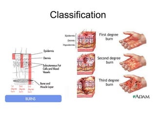
- The document discusses the anatomy, classification, pathophysiology, assessment, and management of pediatric burns. It describes the layers of the skin and how burns are classified based on depth. Management of minor burns involves debriding dead tissue, evaluating the wound, and applying semiocclusive dressings. Major burns require IV fluids and special attention to risks like hypothermia and fluid imbalance due to immature kidneys in young children.
This Presentation is about burn in children it's defination ,causes , classification , methods of estimation of TBSA of burn , diagnose , medical , surgical and nursing management and complications.
BURN - Presented By Mohammed Haroon Rashid Haroon Rashid
Subject is Medical Surgical Nursing - II & Topic is Burn, Presented by Mohammed Haroon Rashid Basci B.Sc Nursing 3rd Year in Florence College of Nursing
neonatal hypothermia is a very emergency condition. if we identify this in early stage we can save the life of neonate. all should know about the maintaining the temperature if the neonate is in our home.
Defines Exchange Transfusion, the Aims, and indications of Exchange Transfusion. Articles required, choice of donor, the procedure of exchange transfusion. Post transfusion care and the complications that can occur due to exchange transfusion. The Ppt also describes the special considerations during the procedure.
medical surgical nursing , nursing management of burn patients, it includes definition, classification of burn injury, clinical manifestaion, assessment of burn injury , management of patient with burn, care given to the patient.
Brief description about what are burns, structure of skin, how we can classify burns based upon mechanism and differential diagnosis ,pathophysiology of burn, rule of 9, general and systemic response to burns, complications, fluid resuscitation, parkland formula, monitoring of resuscitation
this slide is selection important part of thermal burn topic in
Tintinalli's Emergency Medicine
with this presentation you can have a great present in your collage.
Molluscum contagiosum Made Extremely SimpleDrYusraShabbir
A brief description of a very common viral infection affecting children and adults. Molluscum Contagious is an infectious contagious disease. Useful information regarding the symptoms and treatment of the rash are available for medical students, doctors, dermatologists, ophthalmologists, gynaecologist, pediatricians and nurses. Helpful for studying for exams. Reference: Rooks, Textbook of Dermatology
Objective: At the end of this unit, the students will be able to:
Describe internationally accepted rights of child
Discuss national policies, legislation and agencies related to child welfare
Explain National Health Programs related to child health
Enumerate changing trends in child health
Outline child morbidity and mortality
Describe the ethics in Pediatric Nursing
At the end of unit 2, the students will be able to:
Appreciate the differences between children and adult
Describe the hospital environment for a sick child
Explain the impact of hospitalization on child
Discuss the grief and bereavement
Outline the role of a child health nurse
Explain the principles of pre- and post-operative care for children
Perform pain assessment in children
Here is the updated list of Top Best Ayurvedic medicine for Gas and Indigestion and those are Gas-O-Go Syp for Dyspepsia | Lavizyme Syrup for Acidity | Yumzyme Hepatoprotective Capsules etc
- Video recording of this lecture in English language: https://youtu.be/kqbnxVAZs-0
- Video recording of this lecture in Arabic language: https://youtu.be/SINlygW1Mpc
- Link to download the book free: https://nephrotube.blogspot.com/p/nephrotube-nephrology-books.html
- Link to NephroTube website: www.NephroTube.com
- Link to NephroTube social media accounts: https://nephrotube.blogspot.com/p/join-nephrotube-on-social-media.html
These simplified slides by Dr. Sidra Arshad present an overview of the non-respiratory functions of the respiratory tract.
Learning objectives:
1. Enlist the non-respiratory functions of the respiratory tract
2. Briefly explain how these functions are carried out
3. Discuss the significance of dead space
4. Differentiate between minute ventilation and alveolar ventilation
5. Describe the cough and sneeze reflexes
Study Resources:
1. Chapter 39, Guyton and Hall Textbook of Medical Physiology, 14th edition
2. Chapter 34, Ganong’s Review of Medical Physiology, 26th edition
3. Chapter 17, Human Physiology by Lauralee Sherwood, 9th edition
4. Non-respiratory functions of the lungs https://academic.oup.com/bjaed/article/13/3/98/278874
NVBDCP.pptx Nation vector borne disease control programSapna Thakur
NVBDCP was launched in 2003-2004 . Vector-Borne Disease: Disease that results from an infection transmitted to humans and other animals by blood-feeding arthropods, such as mosquitoes, ticks, and fleas. Examples of vector-borne diseases include Dengue fever, West Nile Virus, Lyme disease, and malaria.
Muktapishti is a traditional Ayurvedic preparation made from Shoditha Mukta (Purified Pearl), is believed to help regulate thyroid function and reduce symptoms of hyperthyroidism due to its cooling and balancing properties. Clinical evidence on its efficacy remains limited, necessitating further research to validate its therapeutic benefits.
Integrating Ayurveda into Parkinson’s Management: A Holistic ApproachAyurveda ForAll
Explore the benefits of combining Ayurveda with conventional Parkinson's treatments. Learn how a holistic approach can manage symptoms, enhance well-being, and balance body energies. Discover the steps to safely integrate Ayurvedic practices into your Parkinson’s care plan, including expert guidance on diet, herbal remedies, and lifestyle modifications.
Local Advanced Lung Cancer: Artificial Intelligence, Synergetics, Complex Sys...Oleg Kshivets
Overall life span (LS) was 1671.7±1721.6 days and cumulative 5YS reached 62.4%, 10 years – 50.4%, 20 years – 44.6%. 94 LCP lived more than 5 years without cancer (LS=2958.6±1723.6 days), 22 – more than 10 years (LS=5571±1841.8 days). 67 LCP died because of LC (LS=471.9±344 days). AT significantly improved 5YS (68% vs. 53.7%) (P=0.028 by log-rank test). Cox modeling displayed that 5YS of LCP significantly depended on: N0-N12, T3-4, blood cell circuit, cell ratio factors (ratio between cancer cells-CC and blood cells subpopulations), LC cell dynamics, recalcification time, heparin tolerance, prothrombin index, protein, AT, procedure type (P=0.000-0.031). Neural networks, genetic algorithm selection and bootstrap simulation revealed relationships between 5YS and N0-12 (rank=1), thrombocytes/CC (rank=2), segmented neutrophils/CC (3), eosinophils/CC (4), erythrocytes/CC (5), healthy cells/CC (6), lymphocytes/CC (7), stick neutrophils/CC (8), leucocytes/CC (9), monocytes/CC (10). Correct prediction of 5YS was 100% by neural networks computing (error=0.000; area under ROC curve=1.0).
Adv. biopharm. APPLICATION OF PHARMACOKINETICS : TARGETED DRUG DELIVERY SYSTEMSAkankshaAshtankar
MIP 201T & MPH 202T
ADVANCED BIOPHARMACEUTICS & PHARMACOKINETICS : UNIT 5
APPLICATION OF PHARMACOKINETICS : TARGETED DRUG DELIVERY SYSTEMS By - AKANKSHA ASHTANKAR
Title: Sense of Smell
Presenter: Dr. Faiza, Assistant Professor of Physiology
Qualifications:
MBBS (Best Graduate, AIMC Lahore)
FCPS Physiology
ICMT, CHPE, DHPE (STMU)
MPH (GC University, Faisalabad)
MBA (Virtual University of Pakistan)
Learning Objectives:
Describe the primary categories of smells and the concept of odor blindness.
Explain the structure and location of the olfactory membrane and mucosa, including the types and roles of cells involved in olfaction.
Describe the pathway and mechanisms of olfactory signal transmission from the olfactory receptors to the brain.
Illustrate the biochemical cascade triggered by odorant binding to olfactory receptors, including the role of G-proteins and second messengers in generating an action potential.
Identify different types of olfactory disorders such as anosmia, hyposmia, hyperosmia, and dysosmia, including their potential causes.
Key Topics:
Olfactory Genes:
3% of the human genome accounts for olfactory genes.
400 genes for odorant receptors.
Olfactory Membrane:
Located in the superior part of the nasal cavity.
Medially: Folds downward along the superior septum.
Laterally: Folds over the superior turbinate and upper surface of the middle turbinate.
Total surface area: 5-10 square centimeters.
Olfactory Mucosa:
Olfactory Cells: Bipolar nerve cells derived from the CNS (100 million), with 4-25 olfactory cilia per cell.
Sustentacular Cells: Produce mucus and maintain ionic and molecular environment.
Basal Cells: Replace worn-out olfactory cells with an average lifespan of 1-2 months.
Bowman’s Gland: Secretes mucus.
Stimulation of Olfactory Cells:
Odorant dissolves in mucus and attaches to receptors on olfactory cilia.
Involves a cascade effect through G-proteins and second messengers, leading to depolarization and action potential generation in the olfactory nerve.
Quality of a Good Odorant:
Small (3-20 Carbon atoms), volatile, water-soluble, and lipid-soluble.
Facilitated by odorant-binding proteins in mucus.
Membrane Potential and Action Potential:
Resting membrane potential: -55mV.
Action potential frequency in the olfactory nerve increases with odorant strength.
Adaptation Towards the Sense of Smell:
Rapid adaptation within the first second, with further slow adaptation.
Psychological adaptation greater than receptor adaptation, involving feedback inhibition from the central nervous system.
Primary Sensations of Smell:
Camphoraceous, Musky, Floral, Pepperminty, Ethereal, Pungent, Putrid.
Odor Detection Threshold:
Examples: Hydrogen sulfide (0.0005 ppm), Methyl-mercaptan (0.002 ppm).
Some toxic substances are odorless at lethal concentrations.
Characteristics of Smell:
Odor blindness for single substances due to lack of appropriate receptor protein.
Behavioral and emotional influences of smell.
Transmission of Olfactory Signals:
From olfactory cells to glomeruli in the olfactory bulb, involving lateral inhibition.
Primitive, less old, and new olfactory systems with different path
3. Related anatomy
• The skin is the largest organ in the body, ranging in area from 0.25-
1.8 m2. It is composed of 3 main layers, as follows:
• Epidermis
Outermost layer : Composed of 2 layers,
– stratum corneum- The outer layer of anucleated cornified cells, acts as
a barrier to the entrance of microorganisms and the loss of water and
electrolytes.
– Malpighian layers- The inner layer of the epidermis is composed of
viable cells that mature and differentiate into cornified cells of the
stratum corneum.
• Dermis Beneath the epidermis ; is composed of a dense
fibroelastic connective tissue stroma containing collagen, elastic
fibers, and an extracellular gel termed the ground substance. It
contains an extensive vascular and nerve network and special
glands and appendages that communicate with the overlying
epidermis.
• Subcutaneous tissue- The innermost layer of the skin is the,
composed primarily of areolar and fatty connective tissue. This layer
contains skin appendages, glands, and hair follicles.
4. Classification
Burns are divided into 4 categories, depending on the
depth of the injury, as follows:
• First-degree burns : limited to the epidermis. A typical
sunburn is a first-degree burn. It is characterized by
erythema, pain, and minor microscopic changes. Pain
usually lasts 48–72 hours, and the damaged epithelium
peels off in 5-10 days. First-degree burns do not lead to
scarring and require only local wound care.
• Second -degree burns: the point of injury extends into
the dermis, with some residual dermis remaining viable.
The healing is directly related to the amount of
undamaged dermis. Deep dermal burns lead to severe
hypertrophic scarring and may take 25–35 days to heal.
5. • Third-degree, or full-thickness, burns involve
destruction of the entire dermis, leaving only
subcutaneous tissue exposed. It is characterized by lack
of sensation in the burned skin, a leathery texture, and
no capillary refill.
• Fourth-degree burn is a term that is rarely seen in
literature and periodicals. This type of burn is usually
associated with lethal injury. Fourth-degree burns extend
beyond the subcutaneous tissue, involving the muscle,
fascia, and bone. Occasionally termed transmural burns,
these injuries often are associated with complete
transection of an extremity.
7. • The zone of coagulation occurs at the point of
maximum damage. There is irreversible tissue loss
because of the coagulation of constituent proteins.
• The zone of stasis surrounds the zone of coagulation,
and is characterized by decreased tissue perfusion. The
tissue can be salvaged, and the aim is to increase tissue
perfusion here, and prevent irreversible damage.
Prolonged hypotension, infection or oedema can result in
complete tissue loss.
• The zone of hyperaemia is the outermost area. There is
increased tissue perfusion, and in the absence of severe
sepsis or prolonged hypoperfusion, tissue will recover.
8. Pathophysiology
• Following a major burn injury, heart rate and
peripheral vascular resistance increase. This is
due to the release of catecholamines from
injured tissues, and the relative hypovolemia
that occurs from fluid volume shifts.
• Initially cardiac output decreases. At
approximately 24 hours after burn injuries (for
patients receiving fluid resuscitation) cardiac
output returns to normal, then increases to meet
the hypermetabolic needs of the body.
9. • Acute upper gastrointestinal tract erosions and
ulcers may occur in patients with severe burn
injuries.
• The lesions are termed stress or Curling ulcers.
• Painless gastrointestinal tract bleeding is the
most common clinical finding.
• Blood loss usually is minimal and can be
prevented with prophylactic administration of H2
blockers.
10. Estimation of the TBSA
• Accurate estimation of the TBSA of a burn
is essential to guide management:
The best-known method, the Wallace’s
“rule of nines,” is appropriate for use in
all adults and when a quick assessment is
needed for a child
• Palm method
• The Lund and Browder method
11. Rule of nines
o Essentially, the rule of nines divides the body into 11
areas of 9% each.
o Each arm is 9% (2), the anterior and posterior
portions of each leg are each 9% (4),
o the anterior upper and lower portions of the thorax are
9% each (2),
o the posterior upper and lower portions of the thorax
are 9% each (2), and the area including the neck and
head is 9% (1).
o The total is 9% times 11, or 99%.
o The perineum comprises the remaining 1%.
12.
13. Palm method
o A quicker, but less accurate, method for
determining burn size is to use the palm of
the patient's hand, which is roughly equivalent
to 1% of the patient's BSA.
o The palm-of-the-hand approach is most
convenient for splash-type no confluent burns
and should never be used for significant burns
(>10% BSA).
14.
15.
16. Laboratory Studies
• Screen all patients with closed-space smoke exposure
(indoor) for carbon monoxide poisoning.
• In patients with minor burns and in patients in whom
arterial puncture is not otherwise indicated, venous
carboxyhemoglobin levels are as accurate as arterial
levels.
• Evaluate CBC, typing and crossmatching blood,
carboxyhemoglobin levels, coagulation studies, basic
chemistry panel, arterial blood gas, and chest
radiograph.
• Other studies include measurement of the urine
myoglobin, serum protein, and ethanol levels; urinalysis;
and toxicology screen.
• Patients with heart disease or with cardiac risk factors
need a baseline ECG.
17. Imaging Studies
• An initial chest radiograph is essential to
demonstrate life-threatening injuries, such
as rib fractures, hemothorax, or
pneumothorax
18. Treatment
• For treatment purposes, burns can be divided
into 2 categories, eg, minor and major burns.
Minor burns
– Infants with burns over < 10% BSA and
– children with burns over < 15% BSA without
significant inhalation injury or preexisting medical
conditions
– can be treated initially without IV access.
Major burns
– Infants with burns > 10% BSA,
– children with burns over >15% BSA, or individuals
with significant inhalation injury
– should be treated with IV access and fluid
resuscitation.
20. Manage hypothermia
Compared to adults, children have a larger BSA relative
to their weight.
BSA is a major determinant of evaporative water loss in
burn patients.
As a result, children with burns have more evaporative
water loss compared by weight than do adults.
Therefore, children usually have somewhat greater fluid
needs during resuscitation.
The greater amount of evaporative water loss also leads
to greater heat loss.
The infant or child with burns is especially prone to
hypothermia; therefore, the ambient temperature should
be kept high to minimize radiant and evaporative heat
losses.
21. • Infants younger than 6 months present a special
challenge.
– Temperature is largely regulated by nonshivering
thermogenesis in this age group. This metabolic
process involves catabolism of fat stores under the
influence of norepinephrine, which requires large
amounts of oxygen.
– Prolonged periods of thermogenesis in the
hypothermic infant can lead to excessive amounts
of lactate production and metabolic acidosis.
Infants and children older than 6 months have the
ability to shiver and are not as likely to develop lactic
acidosis.
22. Fluid imbalance : Another problem unique to resuscitation of an
infant with burns is immaturity of the kidneys and inefficiency in
handling fluid overload.
– The GFR in infants does not reach adult levels until age 9-12
months due to an imbalance in maturation in tubular and
glomerular functions. During the first year of life, the infant has
approximately 50% the osmolar concentrating capacity of an
adult, and water load is handled inefficiently.
– The younger the infant, the more exaggerated is the imbalance.
During the first few weeks of life, infants are likely to retain a
larger portion of the water load administered as part of burn
resuscitation.
– The hyposmolarity of lactated Ringer solution, when used in
accordance with the Parkland formula, already accounts for the
free water needs of infants during the first 24 hours of
resuscitation. Additional hydration of young infants may result in
fluid overload.
Infection : Infants are at increased risk for developing infection
because their immune system is not completely developed.
23. Treatment of minor burns
– Debride devitalized tissue.
– Evaluate the wound for surface area and depth of
injury.
– Develop a wound care plan.
– Leave intact blisters alone. Blister fluid contains
vasoactive mediators such as thromboxane, which
can cause vasoconstriction, promote ischemia, cause
progression of the ischemic zone, and inhibit healing.
– The intact blister also serves as a physiologic
dressing against infection, and unless the blister is
overlying a large joint, the blister should be left intact.
– Blisters larger than several inches in diameter are
most likely to rupture and should be removed.
24. Treatment of minor burns
• Superficial or partial-thickness burns that are limited in size can be
treated with a semiocclusive dressing, such as Xeroform gauze. The
wound should be pink and moist and exhibit good blanching.
– Place gauze dressings on a clean wound devoid of devitalized
tissue. Apply multiple layers of gauze dressing because the
impregnated gauze offers little barrier to moisture evaporation
and wound dehydration.
– Covering wounds reduces pain considerably.
– When an extremity is involved, instruct the patient and family
regarding immobility and as much elevation as possible.
Edema, which is exacerbated when the extremity is held in the
natural position, can worsen the ischemic zone and tissue loss.
– Patients with circumferential burns are at risk for development of
compartment syndrome, which can compromise circulation to
the entire extremity.
– Incision of the burned skin (escharotomy) is the treatment for
compartment syndrome.
25. Major burns
• Patients with major burns usually benefit from
placement of a nasogastric (NG) tube and a
Foley catheter.
– Patients with burns often develop an adynamic ileus
and bowel obstruction. Decompression of the
stomach and removal of its contents via the NG tube
decreases the likelihood of significant aspiration,
which could contribute to an already tenuous
respiratory status.
– A Foley catheter is important to closely follow urinary
output, which is the best means to monitor an
adequate hydration status.
26. Major burns
• Treat patients with major burns who require fluid
resuscitation according to the Parkland formula. The
Parkland formula is a starting point for fluid
resuscitation.
– Maintain urine output (as measured with a Foley catheter) at 1.0
mL/kg/h. In younger children ( <30 kg), maintain a urine output of
1.0-1.5 mL/kg/h.
– The Parkland formula is used during the first 24 hours of fluid
resuscitation and is as follows:
– Amount of IV fluid in first 24 hours = weight in kg X 4 mL X
% BSA burned
• Administer one half of the calculated fluid during the first
8 hours
• And one half of the calculated fluid in the subsequent 16
hours.
27. PARKLAND FORMULA
Second 24hrs:
0.5ml colloid * weight in Kgs * TBSA + 2,000ml
5% dextrose in water run concurrently over the
24 hrs.
E.g. 0.5ml * 70kgs * 80% = 2,800ml colloid +
2,000ml 5% D/W yields 117ml colloid/hr.
Boluses of crystalloids or colloid may be
necessary to keep a urinary output of 0.5 to 1
ml/kg/hr.
28. • The starting time is considered to be the time at
which the burn occurred and not the time at
which medical care is initiated.
• Children aged 6 months to 5 years should
receive the fluid recommended by calculations
using the Parkland formula plus maintenance
during the first 24 hours.
• Urine output is the criterion standard for gauging
appropriate intravascular volume and hydration
status.
29. • All burn patients should receive
supplementary oxygen at the highest
concentration allowed by patient comfort.
• Maintain a high flow of oxygen until a
negative carboxyhemoglobin level result
has been returned from the laboratory.
30. Surgical Care
• Clean burned areas using isotonic sodium chloride
solution or mild soap and water, taking care to maintain
thermal homeostasis.
• Remove ruptured blisters and devitalized epidermis with
forceps and scissors or, more efficiently, by rubbing with
gauze soaked in isotonic sodium chloride solution after
an adequate level of analgesia is obtained.
• Leave intact blisters alone.
• Apply a topical antimicrobial (silver sulfadiazine,
bacitracin) followed by a sterile dressing after the burn
has been cleaned and debrided.
• Dressings should be changed and wounds cleaned daily
in outpatients.
• Incision of burned skin (escharotomy) is the treatment for
compartment syndrome.
31. Medication
• Apply silver sulfadiazine to the wound for the first 48 hours after
initial treatment.
• Dressing changes cause further debridement and aid
epithelialization. Silver sulfadiazine is most effective against gram-
positive organisms and is relatively ineffective against
pseudomonads. If the wound develops a greenish drainage or a
sweet smell, consider Pseudomonas aeruginosa colonization or
infection.
• Bacitracin ointment for small- and moderate-sized burns. Bacitracin
is as effective as silver sulfadiazine and is somewhat easier to
apply. Bacitracin should always be used for burns located above the
clavicles because silver sulfadiazine occasionally causes cosmetic
complications (eg, skin pigmentation changes).
• Mafenide acetate cream provides more complete coverage against
Pseudomonas and other gram-negative species. Unlike silver
sulfadiazine, mafenide acetate cream is painful on application
because it is hyperosmolar. Extensive wounds covered with
mafenide acetate may develop metabolic acidosis.
32. Analgesic agents
• Morphine sulfate
• Patients who have sustained anything more than localized burns ( <5% BSA) should
receive systemic analgesics. Morphine is a good choice for analgesia. All opioids
work well, but morphine has a relatively long half-life. Patients with significant burns
require an NG tube, limiting the use of PO medications. IM injections are painful and
repeated injections can increase muscle damage in patients who are already
susceptible to rhabdomyolysis due to muscle breakdown. Blood flow may be
decreased to tissues, another reason to avoid IM/SC injections.
• Dose
• 0.1 mg/kg IV q2-4h prn
• Fentanyl (Sublimaze)
• The shortest acting opioid, fentanyl has a minimal effect on histamine release. Of all
opioids, fentanyl releases histamine the least and is the safest in patients with
hyperactive airway disease. Consider continuous infusion because of the short half-
life of fentanyl.
• Dose
• 1-2 mcg/kg/dose IV q30-60min
33. • Verify tetanus immunization and
administer tetanus toxoid (0.5 mL) if
indicated.
• Patients with more than 50% of the body
surface burned should receive tetanus
immune globulin.
34. Vaccines
• Active immunization increases resistance
to infection. Vaccines consist of
microorganisms or cellular components,
which act as antigens.
• Administration of the vaccine stimulates
the production of antibodies
35. • Tetanus immune globulin (Hyper-Tet) and tetanus toxoid
• Tetanus immune globulin (TIG) is used for passive immunization of any person with a
wound that may be contaminated with tetanus spores.
• Tetanus toxoid is used to induce active immunity against tetanus in selected patients.
• Burns are extremely tetanus-prone wounds. All burn patients with an incomplete
immunization history should receive a dosage of tetanus toxoid (Td for adults or
children > 7 y, DTP or TD for children <7 y). Children with up-to-date primary
immunization series are considered to be up-to-date for tetanus immunization status.
If the patient has a history of complete immunization and the last immunization with
absorbed tetanus toxoid is within the last 5 years, further immunization is not
required. If the history of tetanus immunization is unknown, both Td and TIG should
be administered.
• Pediatric
• Tetanus toxoid:
<7 years: 0.5 mL IM of TD or DTP
>7 years: 0.5 mL IM of Td or Tdap for adolescents
TIG: 250 U IM
Note: When Td and TIG are administered simultaneously, administer with different
syringes in different locations
36. Complications
• Acute Tubular Necrosis of the kidneys can be
caused by myoglobin and hemoglobin released
from damaged muscles and red blood cells. This
is common in electrical burns or crush injuries
where adequate fluid resuscitation has not been
achieved.
• Fever occurs frequently in children and adults
with burns. The burned skin is a potent source of
mediators (eg, interleukin 1) that produce fevers.
Every fever spike in a patient with burns does
not require an extensive evaluation with blood
cultures and other diagnostic modalities.
37. • Wound infections are classified as cellulitis, invasive
infections, and impetigo.
Cellulitis usually begins on the second or third day after the burn
injury occurs. If the cellulitis does not resolve and continues to
expand, it is treated topically with the appropriate antibiotic,
warm soaks, and mafenide acetate cream until the infection is
controlled.
• Invasive infections can be caused by bacterial, fungal, or
viral pathogens.
• Daily examination of the entire wound is necessary to
check for signs of infection. In difficult cases, perform
biopsy on suspected lesions and submit specimens for
culture and microscopic evaluation.
• Treatment with topical antimicrobial drugs or early
excision is important for infection control.
38. Rehabilitation
• Rehabilitation involves maximizing positioning,
range of motion of all joints, limitation of
pressure necrosis, exercise, ambulation, and
assistance in daily activity.
– Pressure therapy is important and should be
implemented as part of the rehabilitation program.
Pressure therapy can reduce hypertrophic scar
formation. Garments used in pressure therapy can be
custom made and are available for different body
parts and in varying sizes.
– Pressure therapy is used to deliver consistent
pressure on scarred areas, thus shortening the time
of scar maturation and thickness and the total
affected area.
39. Prognosis
• Hypertrophic scarring refers to the development
of thickened raised skin. Scarring may be
characterized by contractures of joints.
• In general, children scar more than adults.
Patients with pigmented skin tend to scar more
than patients with lighter skin.
• Continuous wearing of pressure garments for up
to 1 year after healing has been shown to help
limit the progression of hypertrophic scarring.
The garments must be worn nearly 24 hours per
day to be effective.
40. Summary
• A burn is damage to your body's tissues caused by heat,
chemicals, electricity, sunlight or radiation. Scalds from
hot liquids and steam, building fires and flammable
liquids and gases are the most common causes of
burns.
• The three types of burns are:
– First-degree burns damage only the outer layer of skin
– Second-degree burns damage the outer layer and the layer
underneath
– Third-degree burns damage or destroy the deepest layer of skin
and tissues underneath
41. • Burns can cause swelling, blistering, scarring and, in
serious cases, shock and even death.
• They also can lead to infections because they damage
skin's protective barrier.
• Antibiotic creams can prevent or treat infections.
• After a third-degree burn, you need skin or synthetic
grafts to cover exposed tissue and encourage new skin
to grow.
• First- and second-degree burns usually heal without
grafts.