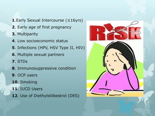
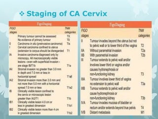
Welcome
Seminar on Cervical Cancer
 Presented By:
Dr. Anick Saha Shuvo
MBBS (RpMC)
MS Resident (P-A), Pediatric Surgery, DMCH
 Why This Awareness???
Cervical cancer has become a burning issue in today’s world. It
is the 2nd most common cancer in women worldwide & the 4th
most common cause of death in women.
Every year about 5,30,000 women acquired the disease,
among which 2,75,000 women die from the disease. That means
in every two minutes a woman die from cervical cancer all over
the world.
The highest burden of disease(upto 70%) occurs in less
developed countries where incidence & mortality rate are so high
due to lack of awareness and screening programme.
In Asia every year more than 3,14,000 new cases have been
diagnosed and more than 1,61,000 death occurs.
 Impact of CA Cervix in
Bangladesh
In Bangladesh cervical cancer is the most common
gynaecological cancer. About 70% of the hospital cases
of gynaecological cancer are cervical cancer. It is the 2nd
most frequent cancer among women age between 15-44
years. Women at risk of CA Cervix annually(>15yrs)
54.38 million.
Every year about 17,686 newly recorded case found,
among them about 10,364 women die.That means
everyday 33 women are diagnosed with cervical cancer
and 18 women dies among them.
 Statistical Diagram
 What is Cervical Cancer???
Cancer begins when cells in a part of the body start to
grow out of control.
Cervical cancer starts in the cells lining the cervix,the
lower part of the uterus (womb). This is sometimes called
the uterine cervix. The cervix connects the body of the
uterus to the vagina (birth canal).
Cervix can be divided into two main parts:
A. Endocervix: Closest part to the body of the uterus
mainly lined by glandular cells.
B. Ectocervix (Exocervix): Part next to the vagina
mainly lined by squamous cells.
 Diagram Showing Parts of Female
Genital Organ
 Continuation
Ectocervix & Endocervix meet at a place
called the transformation zone. The
exact location of the transformation zone
changes as you age and if you give
birth. Most cervical cancers begin from
this transformation zone.
 Pre-Cancerous Conditions
Cells of the cervix do not suddenly change
into cancer. Instead, the normal cells of the
cervix first gradually develop pre-cancerous
changes that turn into cancer.
Common pre-cancerous conditions are:
CIN I,II,III(Cervical Intraepithelial
Neoplasia)
A.CIN I:- LSIL (Low Grade
Squamous Intraepithelial Lesion
B.CIN II & III:- HSIL (High Grade
Squamous Intraepithelial Lesion)
 Types of Cervical Cancer
 CA Cervix can be classified from two aspects:
A. Morphological Type:-
(i) Ulcerative
(ii) Exophytic
(iii) Infiltrative
B. Histopathologcal Types:-
(i) Squamous Cell Carcinoma (80-90%)
-Keratinising
-Non-Keratinising
-Verrucous
(ii) Adenocarcinoma (5-10%)
(iii) Mixed (Adenosquamous Carcinoma)
 Continuation of Types
(iv) Others:-
-Carcinoma in situ
-Clear cell Carcinoma
-Small Cell Carcinoma
-Adenoma Malignum
-Adenocanthoma
-Mucoepidermoid
-Glassy Cell Carcinoma
 Which Factors Can lead a woman
to the risk zone???
1.Early Sexual Intercourse (≤16yrs)
2. Early age of first pregnancy
3. Multiparity
4. Low socioeconomic status
5. Infections (HPV, HSV Type II, HIV)
6. Multiple sexual partners
7. STDs
8. Immunosuppressive condition
9. OCP users
10. Smoking
11. IUCD Users
12. Use of Diethylstilbestrol (DES)
 Detail about HPV infection
The most important risk factor for CA Cervix is infection by
Human Papilloma Virus (HPV). It is found in 99.7% case of
invasive carcinoma. HPV is a relatively small double
stranded DNA virus within a spherical capsid. The capsid is
composed of two proteins the stuctural proteins are L1 & L2.
It consist of group of more than 100 related viruses. Among
these at least 30 different types of HPV target the genital
mucosa in which 15-20 are oncogenic.
Fig. Human Papilloma Virus (HPV)
 Detail about HPV infection
Cont.
HPV Type 16,18,45,31,33 are the most common types
associated with CA Cervix. Approximately 70% cervical
cancer worldwide are associated with HPV type 16 &
18.
HPV 16,18,31,45 & 33 account for >80% of cervical
cancer worldwide.
*High Risk Strains: Type 16, 18, 45, 31, 33 (Type 16
& 18 causes 70% and Type 31, 45 causes 10% cervical
carcinoma)
*Low Risk Strains: Type 6, 11
In Bangladesh prevalence of HPV is 7.9% of women
in the general population. 82.8% invasive cervical
cancers are attributed to HPV type 16 & 18.
 Transmission of HPV Virus
 Sexual Transmission:-
 Majority of anogenital HPV is sexually transmitted
 If one partner has HPV the other partner’s chance of being
infected with the same HPV type increases by >50 times.
 Sexual intercourse and/or genital skin to skin contact are the
primary routes of anogenital HPV transmission. Even using
condom does not provide complete protection.
 Self-inoculation is possible
 Non-Sexual Transmission:-
 HPV also can be transferred through non-sexual routes (un-
common) by-
1) Transmission from mother to newborn (Vertical Transmission)
2) Via finger genital contact
3) Transmission via fomites and environmental surfaces
 Pathophysiology of CA
Cervix caused by HPV
HPV is epitheliotrophic in nature. After infecting the cervical
cells HPV can have three phases:-
A. Latent phase: Usually asymptomatic.
B. Acute Infection Phase: Here HPV undergoes
vegetative replication but no integration occurs into
genome. eg. leading to chondyloma of CIN
C. Neoplastic Transformation Phase: Following
integration of oncogenic HPV DNA into the human
genome. Here HPV causes the production of 2 proteins
known as E6 and E7 which turn off some tumor
suppressor genes of normal cell. This may allow the
cervical lining cells to grow too much and to develop
changes in additional genes, which will lead to cancer.
 Mode of Spreading(Metastasis)
 Direct Extention: Growth spreads to the body of
the uterus, vaginal wall, the bladder, parametrial
vessels and the cellular tissues of the broad and
uterosacral ligament.
 Lymphatic Permeation And Embolism: The
notes are commonly involve being the bifurcation of
common iliac, external and internal iliac, obturator,
sacral and ultimately the para-aortic nodes.
 Blood Stream: Ovaries, Brain, Bones, Lungs, Liver.
 Patient’s Complaints (Symptoms)
 No symptoms at early stage
 Post coital bleeding
 Bleeding on straining
 Intermenstrual bleeding
 Foul smelling P/V discharge (Creamy/Whitish→ Dirty Brown)
 Pelvic pain of varying degree
 Leg Oedema
 Renal symptoms: Frequency of Micturation, dysuria,
Haematuria, True Urinary Incontinence.
 In case of Rectal involvement: Diarrhoea, Rectal Pain,
Bleeding per Rectum, Rectovaginal Fistula
 In case of Lung involvement: Haemoptysis, Persistent
Racking Cough
 In case of Liver involvement: Right Upper quadrant Pain &
Fullness
 Findings of Physician (Signs)
A. On general examination:
1. Anemia
2. Weight loss
B. On pelvic examination:
 Per-Vaginal Examination-
 Inspection:
1. Bleeding or foul smelling discharge or both may be see from vulva
vagina.
 Per-speculum Examination:
1. Cervix may be enlarged, ulcerated, excavated or completely
destroyed or replaced by hypertrophic mass.
 Bi-manual Examination:
1. Cervix is fixed, Hard in consistency, Friable and bleeds on touch.
These are the cardinal signs of CA Cervix.
 DRE: Rectum may be involved and found nodular induration.
Anterior wall of the rectum may found adhered with posterior fornix
 Investigation for Diagnostic
Purpose
 Cervical Biopsy
 X-ray Chest
 CT Scan
 MRI
 PET Scan
 Cystoscopy
 Proctoscopy
 Laperoscopy
 Investigation for Preventive
Purpose
1) Screening Procedure
2) Colposcopy and Colpomicroscopy
3) HPV Typing
 Screening Test
Screening with cervical cytology reduces the incidence and
mortality from cervical cancer.
 VIA Test: Visual inspection of cervix with acetic acid
(3.5%). Here acetowhite area in cervix considered as a
VIA positive.
 Continuation of Screening
 Schiller Test: In this test the cervix is stained with a
solution of Lugol’s Iodine(Iodine and Potassium Iodide)
Normal squamous epithelium of cervix contains glycogen
and stains a deep brown. Abnormal one do not contain
glycogen so don’t take stain.
 Continuation of Screening
 Pap-Smear Test: Performed by opening the vaginal
canal with a speculum then collecting cells at the outer
opening of the cervix at the transformation zone. The
collected cell are examined under microscope to look for
abnormalities. The test aims to detect potentially
precancerous change or cervical dysplasia.
 Continuation of Screening
 LBC (Liquid Based Cytology): It is a method of
preparing and processing cervical smears. In this
process the sample is collected normally by a small
brush then it is deposited into a small bottle of
preservative liquid. At lab the liquid is treated to
removed other elements before a layer of cells is placed
on a slide. This process is more informative than others.
 Staging of CA Cervix
 Figure of Staging
 Staging Procedure
Examination Findings
Physical examination i) Palpable lymph nodes
ii) Examine vagina
iii) Bimanual rectovaginal examination (Under
anaesthesia recommended)
Radiological i) Intravenous pyelogram
ii) Barium enema
iii) Chest X-ray
iv) Skeletal X-ray
Procedure
( Allowed by FIGO)
i) Biopsy
ii) Conisation
iii) Hysteroscopy
iv) Colposcopy
v) Endocervical curettage
vi) Cystoscopy
vii) Proctoscopy
Optional studies
(Information that is not allowed by
FIGO to change the clinical stage)
i) CT (axial)-97%% specific
ii) Lymphangiography
iii) USG-99% specific
iv) MRI- more sensitive
v) PET scan
vi) Radionucleotide scanning
vii) Laparoscopy
 Treatment Modalities
 Radiotherapy
 Surgery
 Combined Radiotherapy & Surgery
 Chemotherapy
 Neoadjuvent Chemotherapy
 Ultraradical Surgery & Palliation
 Treatment at a Glance
 General Treatment:
1.Nutritional Support
2.Correction of anaemia by blood transfusion
3.Control of infection by antibiotics
4.Relieve of pain by analgesics
 Primary Surgery:
1.Wertheim’s Hysterectomy: Stage I & early stage IIA
2.Pelvic Exenteration:
» Ant. Exenteration
» Post. Exenteration
» Complete Exenteration
» Laparoscopic Radical Hysterectomy
 Radiotherapy
 Chemotherapy
 Combination Therapy
 Management of Invasive Cancer of Cervix
Stage Extent Treatment
Stage Ia1 (<3mm
depth)
≤3mm with no LVSI Conisation or type I
Hysterectomy (No
lymphadenectomy)
≤3mm with LVSI Radical trachelectomy or type
II radical Hysterectomy with
pelvic node dissection
Stage Ia2(3-5mm
depth & 1mm long)
>3-5mm invasion Radical trachelectomy or type
II radical Hysterectomy with
pelvic lymphadenectomy
Stage Ib1(<4cm) >5mm invasion,
<2cm
Radical trachelectomy or type
II radical Hysterectomy with
pelvic lymphadenectomy
>5mm invasion,
>2cm
Type III radical Hysterectomy
with pelvic lymphadenectomy
Stage Extent Treatment
Stage Ib2
--
Type III Radical
Hysterectomy with pelvic
& para-aortic
lymphadenectomy or
primary chemoradiation
Stage IIa
--
Type III Radical
Hysterectomy with pelvic
& para-aortic
lymphadenectomy or
primary chemoradiation
Stage IIb, IIIa, IIIb
--
Primary chemoradiation
Stage IVa
--
Primary chemoradiation or
primary exenteration
Stage IVb
--
Primary Chemotherapy
± Radiation
 Pre-Requisite of Radiotherapy
1)From examination, anaemia & PID are to be
excluded.
2)Investigation should confirm-
-Haemoglobin Level >10gm%
-Total count of WBC >3000/cu mm
-Platelet count >1,00,000 /cu mm
-Serum Urea
-Serum Creatinine
 Radiotherapy Procedures
 External Irradiation: External Beam Radiotherapy (EBRT)
using cobalt 60 is given in short daily treatment over a period
of 3-6 weeks (Monday to Friday basis). It covers the area
extends from just above the urethral orifice to the lower
border of the fourth lumber vertebra and laterally to cover the
whole pelvis with the femoral heads protected.
 Radiotherapy Procedures
Cont.
 Intracavitary Radiotherapy (ICRT):
A. Stockholm Technique: The process involves placing
radium in applicators in the uterus and vaginal fornices for 27-30
hours & then remove it. After that give a similar course of
treatment either on 3 occasion at interval of 3 weeks.
B. Manchester Technique: Placing radium in intrauterine
tube therapy given for 72 hours on 2 occasion repeating the first
course after an interval of 1 week.
C. Low dose Rate (LDR) Therapy: Selectron was
introduced as LDR system. But now a days an HDR system is also
available.
D. High Dose Rate (HDR) Therapy: Cathetron is the
oldest Cobalt 60 based HDR but recently Californium-252 is used.
It is used now a days due to being more convenient procedure. It
can be done in outpatient basis, has short course duration, it has
less morbidity and more cost effective but equipment is more
expensive.
Procedure Amount & Type
of Radiation
No. of
Application
Duration
Stockholm
Intrauterine Tube
50mg one vaginal
ovoid 50-60mg
3
48 hours each
with gap of one
week between 1st
& 2nd and gap of
two weeks
between 2nd & 3rd
Manchester
Intrauterine Tube
50mg vaginal
colpostat 30-
50mg
2
72 fours at
interval of 1 week
Paris
Intrauterine Tube
33.3mg and two
vaginal ovoids
13.3mg
1
5 days (each day
radium removed,
cleaned &
replaced)
 Radiotherapy Procedures
Cont.
 Interstitial Brachytherapy: In patient with advanced
parametrial disease, distorted anatomy or post operative
recurrences. The interstitial perineal implant can deliver
supplementary dose following EBRT more optimally.
Parametrial implants using Radium needles were used
earlier. Currently afterloading transperineal perforated
templates with Iridium 192 and Iodine 125 are used.
 Complication of Radiotherapy
1. During Treatment:
 Diarrhoea
 Abdominal Cramps
 Nausea
 Bleeding from Bowel
 Haematuria in case of urinary infection.
2. Early Complications:
 Uterine Perforation: Occurs at the time of insertion of
the uterine tandem specially in pt. with previous
conisation or advanced disease.
 Pelvic Inflammatory Disease (PID)
 Diverticulitis
 Complication Cont.
3. Late Complications:
 Continuous formation of Arteritis & Fibrosis
 Blood loss from Pelvic Colon & Rectum
 Subacute or Acute Obstruction of Bowel
 Vesicovaginal Fistula (VVF)
 Rectovaginal Fistula (RVF)
 Narrowing & Shortening of Vagina
 Loss of Ovarian Function
 Combined Radiotherapy & Surgery
 Surgery can be operated upto Stage IIa then followed by
chemoradiation.
 Chemoradiation can be given in those stage which has
no LSVI.
 Stage IB2 disease has been shown have a higher risk of
metastatic disease and post operative radiotherapy is
recommended in these patients.
 Sometimes pre operative radiation can make surgery
possible when it would not have been otherwise.
 Chemotherapy
 Single agents used are:
 Cisplatinum (Best Response)
 Paclitaxel
 Bleomycin
 Ifosfamide
 Methotrixate
 Neoadjuvant Chemotherapy: Used in cases with
bulky tumors preoperatively & in combination with
radiotherapy.
 Ultraredical Surgery & Palliation
 General Treatment:
1) General Care
2) Relief of Pain:
» NSAIDs
» Cordoromy (Division of spinothalamic tract)
» Colostomy for rectal pain
» Drainage for pyometra
» Presacral or lumber sympathectomy
 Haemorrhage:
1) Hot vaginal douches
2) Diathermy in the bleeding site
3)Packing with application of the cytotoxic drugs
 Swelling of the leg: Controlled by elastic bandage or posture.
 Incontinence of the urine: Urinary diversion.
 Incontinence of faeces: Colostomy
 3 months interval for 1 year.
 6 months interval for next 1 year.
 Then yearly afterwards.
 Avoid polygamous relationship
 Avoid sexual promiscuity
 Maintain social and religious values
 Avoid early marriage
 Use of condoms
 Avoid smoking, betel & nuts
 Maintain proper nutrition
 Maintain hygiene
 Screening must be done after 30s
 Complications of CA Cervix
1.Haemorrhage
2.Frequent attacks of abdominal pain due to:
-Pyelitis
-Pylonephritis
-Hydronephrosis
3. Pyometra
4. VVF (Vesicovaginal Fistula)
5. RVF (Rectovaginal Fistula)
6. Uremia
7. Renal Failure
8. Cachecxia
9. Sepsis
10. Metastasis to other organ
-Lung (36%)
-Lymph Node (30%)
-Bone (16%)
-Abdominal Cavity (7%)
 Prevention of CA Cervix
 Primary Prevention:
1) Identifying high risk female
2) Identifying high risk male
3) Prophylactic HPV vaccine
4) Use of condoms
5) Removal of cervix during Hysterectomy
6) Health Education
 Secondary Prevention:
1) Screening Procedure
 Age of Administration:
◊According to WHO
1.Target Group: (9-13yrs)
2.Catch-up Group: (16-25 yrs)
3.Considering Group: 45 yrs
onwards
 Dosage Schedule:
Bivalent 0,2,6 months
Quadrivalent 0,1,6 months
*2 dose can be given sexually non exposed women
●Two type of vaccine against HPV is available:
1.Bivalent Vaccine (Cervarix)
2.Quadrivalent Vaccine (Gardasil)
● Protection Against:
Bivalent protects against HPV type:
16 and18
Quadrivalent protects against HPV type :
16,18,6 and 11
 Contraindication
 Vaccines can’t be given in some conditions like:
1) Pregnancy
2) Lactating mother
 What’s New
 Sentinel Lymph node Biopsy: During surgery for cervical cancer,
lymph nodes in the pelvis may be removed to check for cancer
spread. Instead of removing many lymph nodes, a technique called
sentinel lymph node biopsy can be used to target just the few lymph
nodes most likely to contain cancer.
 HPV Vaccines: Vaccines have been developed to prevent infection
with some of the HPV types associated with cervical cancer.
Currently available vaccines are intended to produce immunity to
HPV types 16 and 18, so that women who are exposed to these
viruses will not develop infections. Vaccines are also being developed
to prevent infection with some of the other HPV types that also
cause cancer. Studies are being done to see how well these vaccines
will reduce the risk of cervical cancer.
 Ref: Journal about CA Cervix American Cancer Society’ rev-15
 What’s New Cont.
 Targeted Therapy: As researchers have learned more about
the gene changes in cells that cause cancer, they have been able
to develop newer drugs that specifically target these changes.
These targeted drugs work differently from standard
chemotherapy drugs. They often have different (and less severe)
side effects. These drugs may be used alone or with more
traditional chemotherapy.
 Hyperthermia: Some research indicates that adding
hyperthermia to radiation may help keep the cancer from coming
back and help patients live longer. Hyperthermia is a treatment
that raises the temperature in the area where the tumor is, most
often by using radiofrequency antennae placed around the
patient.
• Ref: Journal about CA Cervix American Cancer Society’ rev-15
CA Cervix

CA Cervix

  • 1.
  • 2.
    Seminar on CervicalCancer  Presented By: Dr. Anick Saha Shuvo MBBS (RpMC) MS Resident (P-A), Pediatric Surgery, DMCH
  • 3.
     Why ThisAwareness??? Cervical cancer has become a burning issue in today’s world. It is the 2nd most common cancer in women worldwide & the 4th most common cause of death in women. Every year about 5,30,000 women acquired the disease, among which 2,75,000 women die from the disease. That means in every two minutes a woman die from cervical cancer all over the world. The highest burden of disease(upto 70%) occurs in less developed countries where incidence & mortality rate are so high due to lack of awareness and screening programme. In Asia every year more than 3,14,000 new cases have been diagnosed and more than 1,61,000 death occurs.
  • 4.
     Impact ofCA Cervix in Bangladesh In Bangladesh cervical cancer is the most common gynaecological cancer. About 70% of the hospital cases of gynaecological cancer are cervical cancer. It is the 2nd most frequent cancer among women age between 15-44 years. Women at risk of CA Cervix annually(>15yrs) 54.38 million. Every year about 17,686 newly recorded case found, among them about 10,364 women die.That means everyday 33 women are diagnosed with cervical cancer and 18 women dies among them.
  • 5.
  • 6.
     What isCervical Cancer??? Cancer begins when cells in a part of the body start to grow out of control. Cervical cancer starts in the cells lining the cervix,the lower part of the uterus (womb). This is sometimes called the uterine cervix. The cervix connects the body of the uterus to the vagina (birth canal). Cervix can be divided into two main parts: A. Endocervix: Closest part to the body of the uterus mainly lined by glandular cells. B. Ectocervix (Exocervix): Part next to the vagina mainly lined by squamous cells.
  • 7.
     Diagram ShowingParts of Female Genital Organ
  • 8.
     Continuation Ectocervix &Endocervix meet at a place called the transformation zone. The exact location of the transformation zone changes as you age and if you give birth. Most cervical cancers begin from this transformation zone.
  • 9.
     Pre-Cancerous Conditions Cellsof the cervix do not suddenly change into cancer. Instead, the normal cells of the cervix first gradually develop pre-cancerous changes that turn into cancer. Common pre-cancerous conditions are: CIN I,II,III(Cervical Intraepithelial Neoplasia) A.CIN I:- LSIL (Low Grade Squamous Intraepithelial Lesion B.CIN II & III:- HSIL (High Grade Squamous Intraepithelial Lesion)
  • 10.
     Types ofCervical Cancer  CA Cervix can be classified from two aspects: A. Morphological Type:- (i) Ulcerative (ii) Exophytic (iii) Infiltrative B. Histopathologcal Types:- (i) Squamous Cell Carcinoma (80-90%) -Keratinising -Non-Keratinising -Verrucous (ii) Adenocarcinoma (5-10%) (iii) Mixed (Adenosquamous Carcinoma)
  • 11.
     Continuation ofTypes (iv) Others:- -Carcinoma in situ -Clear cell Carcinoma -Small Cell Carcinoma -Adenoma Malignum -Adenocanthoma -Mucoepidermoid -Glassy Cell Carcinoma
  • 12.
     Which FactorsCan lead a woman to the risk zone???
  • 13.
    1.Early Sexual Intercourse(≤16yrs) 2. Early age of first pregnancy 3. Multiparity 4. Low socioeconomic status 5. Infections (HPV, HSV Type II, HIV) 6. Multiple sexual partners 7. STDs 8. Immunosuppressive condition 9. OCP users 10. Smoking 11. IUCD Users 12. Use of Diethylstilbestrol (DES)
  • 14.
     Detail aboutHPV infection The most important risk factor for CA Cervix is infection by Human Papilloma Virus (HPV). It is found in 99.7% case of invasive carcinoma. HPV is a relatively small double stranded DNA virus within a spherical capsid. The capsid is composed of two proteins the stuctural proteins are L1 & L2. It consist of group of more than 100 related viruses. Among these at least 30 different types of HPV target the genital mucosa in which 15-20 are oncogenic. Fig. Human Papilloma Virus (HPV)
  • 15.
     Detail aboutHPV infection Cont. HPV Type 16,18,45,31,33 are the most common types associated with CA Cervix. Approximately 70% cervical cancer worldwide are associated with HPV type 16 & 18. HPV 16,18,31,45 & 33 account for >80% of cervical cancer worldwide. *High Risk Strains: Type 16, 18, 45, 31, 33 (Type 16 & 18 causes 70% and Type 31, 45 causes 10% cervical carcinoma) *Low Risk Strains: Type 6, 11 In Bangladesh prevalence of HPV is 7.9% of women in the general population. 82.8% invasive cervical cancers are attributed to HPV type 16 & 18.
  • 17.
     Transmission ofHPV Virus  Sexual Transmission:-  Majority of anogenital HPV is sexually transmitted  If one partner has HPV the other partner’s chance of being infected with the same HPV type increases by >50 times.  Sexual intercourse and/or genital skin to skin contact are the primary routes of anogenital HPV transmission. Even using condom does not provide complete protection.  Self-inoculation is possible  Non-Sexual Transmission:-  HPV also can be transferred through non-sexual routes (un- common) by- 1) Transmission from mother to newborn (Vertical Transmission) 2) Via finger genital contact 3) Transmission via fomites and environmental surfaces
  • 18.
     Pathophysiology ofCA Cervix caused by HPV HPV is epitheliotrophic in nature. After infecting the cervical cells HPV can have three phases:- A. Latent phase: Usually asymptomatic. B. Acute Infection Phase: Here HPV undergoes vegetative replication but no integration occurs into genome. eg. leading to chondyloma of CIN C. Neoplastic Transformation Phase: Following integration of oncogenic HPV DNA into the human genome. Here HPV causes the production of 2 proteins known as E6 and E7 which turn off some tumor suppressor genes of normal cell. This may allow the cervical lining cells to grow too much and to develop changes in additional genes, which will lead to cancer.
  • 20.
     Mode ofSpreading(Metastasis)  Direct Extention: Growth spreads to the body of the uterus, vaginal wall, the bladder, parametrial vessels and the cellular tissues of the broad and uterosacral ligament.  Lymphatic Permeation And Embolism: The notes are commonly involve being the bifurcation of common iliac, external and internal iliac, obturator, sacral and ultimately the para-aortic nodes.  Blood Stream: Ovaries, Brain, Bones, Lungs, Liver.
  • 21.
     Patient’s Complaints(Symptoms)  No symptoms at early stage  Post coital bleeding  Bleeding on straining  Intermenstrual bleeding  Foul smelling P/V discharge (Creamy/Whitish→ Dirty Brown)  Pelvic pain of varying degree  Leg Oedema  Renal symptoms: Frequency of Micturation, dysuria, Haematuria, True Urinary Incontinence.  In case of Rectal involvement: Diarrhoea, Rectal Pain, Bleeding per Rectum, Rectovaginal Fistula  In case of Lung involvement: Haemoptysis, Persistent Racking Cough  In case of Liver involvement: Right Upper quadrant Pain & Fullness
  • 22.
     Findings ofPhysician (Signs) A. On general examination: 1. Anemia 2. Weight loss B. On pelvic examination:  Per-Vaginal Examination-  Inspection: 1. Bleeding or foul smelling discharge or both may be see from vulva vagina.  Per-speculum Examination: 1. Cervix may be enlarged, ulcerated, excavated or completely destroyed or replaced by hypertrophic mass.  Bi-manual Examination: 1. Cervix is fixed, Hard in consistency, Friable and bleeds on touch. These are the cardinal signs of CA Cervix.  DRE: Rectum may be involved and found nodular induration. Anterior wall of the rectum may found adhered with posterior fornix
  • 23.
     Investigation forDiagnostic Purpose  Cervical Biopsy  X-ray Chest  CT Scan  MRI  PET Scan  Cystoscopy  Proctoscopy  Laperoscopy
  • 24.
     Investigation forPreventive Purpose 1) Screening Procedure 2) Colposcopy and Colpomicroscopy 3) HPV Typing
  • 25.
     Screening Test Screeningwith cervical cytology reduces the incidence and mortality from cervical cancer.  VIA Test: Visual inspection of cervix with acetic acid (3.5%). Here acetowhite area in cervix considered as a VIA positive.
  • 26.
     Continuation ofScreening  Schiller Test: In this test the cervix is stained with a solution of Lugol’s Iodine(Iodine and Potassium Iodide) Normal squamous epithelium of cervix contains glycogen and stains a deep brown. Abnormal one do not contain glycogen so don’t take stain.
  • 27.
     Continuation ofScreening  Pap-Smear Test: Performed by opening the vaginal canal with a speculum then collecting cells at the outer opening of the cervix at the transformation zone. The collected cell are examined under microscope to look for abnormalities. The test aims to detect potentially precancerous change or cervical dysplasia.
  • 28.
     Continuation ofScreening  LBC (Liquid Based Cytology): It is a method of preparing and processing cervical smears. In this process the sample is collected normally by a small brush then it is deposited into a small bottle of preservative liquid. At lab the liquid is treated to removed other elements before a layer of cells is placed on a slide. This process is more informative than others.
  • 29.
     Staging ofCA Cervix
  • 30.
  • 31.
     Staging Procedure ExaminationFindings Physical examination i) Palpable lymph nodes ii) Examine vagina iii) Bimanual rectovaginal examination (Under anaesthesia recommended) Radiological i) Intravenous pyelogram ii) Barium enema iii) Chest X-ray iv) Skeletal X-ray Procedure ( Allowed by FIGO) i) Biopsy ii) Conisation iii) Hysteroscopy iv) Colposcopy v) Endocervical curettage vi) Cystoscopy vii) Proctoscopy Optional studies (Information that is not allowed by FIGO to change the clinical stage) i) CT (axial)-97%% specific ii) Lymphangiography iii) USG-99% specific iv) MRI- more sensitive v) PET scan vi) Radionucleotide scanning vii) Laparoscopy
  • 33.
     Treatment Modalities Radiotherapy  Surgery  Combined Radiotherapy & Surgery  Chemotherapy  Neoadjuvent Chemotherapy  Ultraradical Surgery & Palliation
  • 34.
     Treatment ata Glance  General Treatment: 1.Nutritional Support 2.Correction of anaemia by blood transfusion 3.Control of infection by antibiotics 4.Relieve of pain by analgesics  Primary Surgery: 1.Wertheim’s Hysterectomy: Stage I & early stage IIA 2.Pelvic Exenteration: » Ant. Exenteration » Post. Exenteration » Complete Exenteration » Laparoscopic Radical Hysterectomy  Radiotherapy  Chemotherapy  Combination Therapy
  • 35.
     Management ofInvasive Cancer of Cervix Stage Extent Treatment Stage Ia1 (<3mm depth) ≤3mm with no LVSI Conisation or type I Hysterectomy (No lymphadenectomy) ≤3mm with LVSI Radical trachelectomy or type II radical Hysterectomy with pelvic node dissection Stage Ia2(3-5mm depth & 1mm long) >3-5mm invasion Radical trachelectomy or type II radical Hysterectomy with pelvic lymphadenectomy Stage Ib1(<4cm) >5mm invasion, <2cm Radical trachelectomy or type II radical Hysterectomy with pelvic lymphadenectomy >5mm invasion, >2cm Type III radical Hysterectomy with pelvic lymphadenectomy
  • 36.
    Stage Extent Treatment StageIb2 -- Type III Radical Hysterectomy with pelvic & para-aortic lymphadenectomy or primary chemoradiation Stage IIa -- Type III Radical Hysterectomy with pelvic & para-aortic lymphadenectomy or primary chemoradiation Stage IIb, IIIa, IIIb -- Primary chemoradiation Stage IVa -- Primary chemoradiation or primary exenteration Stage IVb -- Primary Chemotherapy ± Radiation
  • 38.
     Pre-Requisite ofRadiotherapy 1)From examination, anaemia & PID are to be excluded. 2)Investigation should confirm- -Haemoglobin Level >10gm% -Total count of WBC >3000/cu mm -Platelet count >1,00,000 /cu mm -Serum Urea -Serum Creatinine
  • 39.
     Radiotherapy Procedures External Irradiation: External Beam Radiotherapy (EBRT) using cobalt 60 is given in short daily treatment over a period of 3-6 weeks (Monday to Friday basis). It covers the area extends from just above the urethral orifice to the lower border of the fourth lumber vertebra and laterally to cover the whole pelvis with the femoral heads protected.
  • 40.
     Radiotherapy Procedures Cont. Intracavitary Radiotherapy (ICRT): A. Stockholm Technique: The process involves placing radium in applicators in the uterus and vaginal fornices for 27-30 hours & then remove it. After that give a similar course of treatment either on 3 occasion at interval of 3 weeks. B. Manchester Technique: Placing radium in intrauterine tube therapy given for 72 hours on 2 occasion repeating the first course after an interval of 1 week. C. Low dose Rate (LDR) Therapy: Selectron was introduced as LDR system. But now a days an HDR system is also available. D. High Dose Rate (HDR) Therapy: Cathetron is the oldest Cobalt 60 based HDR but recently Californium-252 is used. It is used now a days due to being more convenient procedure. It can be done in outpatient basis, has short course duration, it has less morbidity and more cost effective but equipment is more expensive.
  • 41.
    Procedure Amount &Type of Radiation No. of Application Duration Stockholm Intrauterine Tube 50mg one vaginal ovoid 50-60mg 3 48 hours each with gap of one week between 1st & 2nd and gap of two weeks between 2nd & 3rd Manchester Intrauterine Tube 50mg vaginal colpostat 30- 50mg 2 72 fours at interval of 1 week Paris Intrauterine Tube 33.3mg and two vaginal ovoids 13.3mg 1 5 days (each day radium removed, cleaned & replaced)
  • 42.
     Radiotherapy Procedures Cont. Interstitial Brachytherapy: In patient with advanced parametrial disease, distorted anatomy or post operative recurrences. The interstitial perineal implant can deliver supplementary dose following EBRT more optimally. Parametrial implants using Radium needles were used earlier. Currently afterloading transperineal perforated templates with Iridium 192 and Iodine 125 are used.
  • 43.
     Complication ofRadiotherapy 1. During Treatment:  Diarrhoea  Abdominal Cramps  Nausea  Bleeding from Bowel  Haematuria in case of urinary infection. 2. Early Complications:  Uterine Perforation: Occurs at the time of insertion of the uterine tandem specially in pt. with previous conisation or advanced disease.  Pelvic Inflammatory Disease (PID)  Diverticulitis
  • 44.
     Complication Cont. 3.Late Complications:  Continuous formation of Arteritis & Fibrosis  Blood loss from Pelvic Colon & Rectum  Subacute or Acute Obstruction of Bowel  Vesicovaginal Fistula (VVF)  Rectovaginal Fistula (RVF)  Narrowing & Shortening of Vagina  Loss of Ovarian Function
  • 45.
     Combined Radiotherapy& Surgery  Surgery can be operated upto Stage IIa then followed by chemoradiation.  Chemoradiation can be given in those stage which has no LSVI.  Stage IB2 disease has been shown have a higher risk of metastatic disease and post operative radiotherapy is recommended in these patients.  Sometimes pre operative radiation can make surgery possible when it would not have been otherwise.
  • 46.
     Chemotherapy  Singleagents used are:  Cisplatinum (Best Response)  Paclitaxel  Bleomycin  Ifosfamide  Methotrixate  Neoadjuvant Chemotherapy: Used in cases with bulky tumors preoperatively & in combination with radiotherapy.
  • 47.
     Ultraredical Surgery& Palliation  General Treatment: 1) General Care 2) Relief of Pain: » NSAIDs » Cordoromy (Division of spinothalamic tract) » Colostomy for rectal pain » Drainage for pyometra » Presacral or lumber sympathectomy  Haemorrhage: 1) Hot vaginal douches 2) Diathermy in the bleeding site 3)Packing with application of the cytotoxic drugs  Swelling of the leg: Controlled by elastic bandage or posture.  Incontinence of the urine: Urinary diversion.  Incontinence of faeces: Colostomy
  • 48.
     3 monthsinterval for 1 year.  6 months interval for next 1 year.  Then yearly afterwards.
  • 49.
     Avoid polygamousrelationship  Avoid sexual promiscuity  Maintain social and religious values  Avoid early marriage  Use of condoms  Avoid smoking, betel & nuts  Maintain proper nutrition  Maintain hygiene  Screening must be done after 30s
  • 50.
     Complications ofCA Cervix 1.Haemorrhage 2.Frequent attacks of abdominal pain due to: -Pyelitis -Pylonephritis -Hydronephrosis 3. Pyometra 4. VVF (Vesicovaginal Fistula) 5. RVF (Rectovaginal Fistula) 6. Uremia 7. Renal Failure 8. Cachecxia 9. Sepsis 10. Metastasis to other organ -Lung (36%) -Lymph Node (30%) -Bone (16%) -Abdominal Cavity (7%)
  • 51.
     Prevention ofCA Cervix  Primary Prevention: 1) Identifying high risk female 2) Identifying high risk male 3) Prophylactic HPV vaccine 4) Use of condoms 5) Removal of cervix during Hysterectomy 6) Health Education  Secondary Prevention: 1) Screening Procedure
  • 52.
     Age ofAdministration: ◊According to WHO 1.Target Group: (9-13yrs) 2.Catch-up Group: (16-25 yrs) 3.Considering Group: 45 yrs onwards  Dosage Schedule: Bivalent 0,2,6 months Quadrivalent 0,1,6 months *2 dose can be given sexually non exposed women ●Two type of vaccine against HPV is available: 1.Bivalent Vaccine (Cervarix) 2.Quadrivalent Vaccine (Gardasil) ● Protection Against: Bivalent protects against HPV type: 16 and18 Quadrivalent protects against HPV type : 16,18,6 and 11
  • 53.
     Contraindication  Vaccinescan’t be given in some conditions like: 1) Pregnancy 2) Lactating mother
  • 54.
     What’s New Sentinel Lymph node Biopsy: During surgery for cervical cancer, lymph nodes in the pelvis may be removed to check for cancer spread. Instead of removing many lymph nodes, a technique called sentinel lymph node biopsy can be used to target just the few lymph nodes most likely to contain cancer.  HPV Vaccines: Vaccines have been developed to prevent infection with some of the HPV types associated with cervical cancer. Currently available vaccines are intended to produce immunity to HPV types 16 and 18, so that women who are exposed to these viruses will not develop infections. Vaccines are also being developed to prevent infection with some of the other HPV types that also cause cancer. Studies are being done to see how well these vaccines will reduce the risk of cervical cancer.  Ref: Journal about CA Cervix American Cancer Society’ rev-15
  • 55.
     What’s NewCont.  Targeted Therapy: As researchers have learned more about the gene changes in cells that cause cancer, they have been able to develop newer drugs that specifically target these changes. These targeted drugs work differently from standard chemotherapy drugs. They often have different (and less severe) side effects. These drugs may be used alone or with more traditional chemotherapy.  Hyperthermia: Some research indicates that adding hyperthermia to radiation may help keep the cancer from coming back and help patients live longer. Hyperthermia is a treatment that raises the temperature in the area where the tumor is, most often by using radiofrequency antennae placed around the patient. • Ref: Journal about CA Cervix American Cancer Society’ rev-15