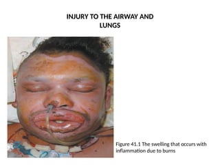
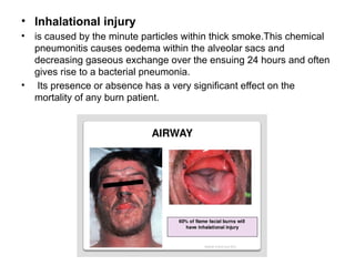
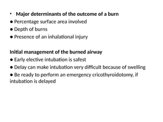
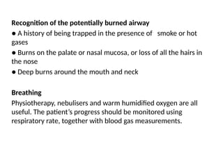
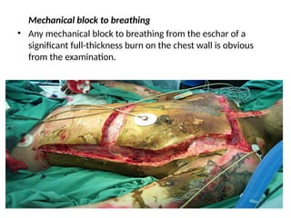
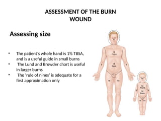
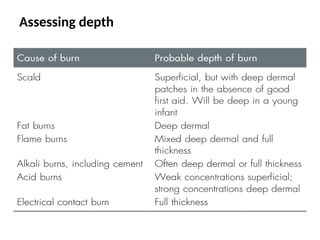
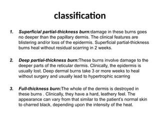
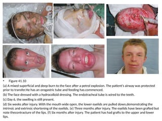
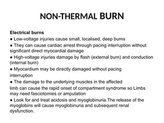
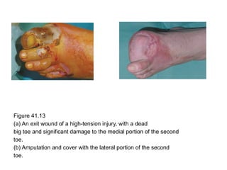
Burn
Presented by:
Yassmin Abdulameer
Yusra Mustafa
Supervised by: dr. Yaseen Alrawi
• the most common organ affected is the skin.
• However, burns can also damage the airway and lungs,
with life-threatening consequences.
• Respiratory system injuries usually occur if a person is
trapped in a burning vehicle, house, car or aeroplane
and is forced to inhale the hot and poisonous gases.
• Warning signs of burns to the respiratory system
● Burns around the face and neck
● A history of being trapped in a burning room
● Change in voice
● Stridor
INJURY TO THE AIRWAY AND
LUNGS
Figure 41.1 The swelling that occurs with
inflammation due to burns
Dangers of smoke, hot gas or steam inhalation
● Inhaled hot gases can cause supraglottic airway burns and laryngeal
oedema
● Inhaled steam can cause subglottic burns and loss of respiratory
epithelium
● Inhaled smoke particles can cause chemical alveolitis and respiratory
failure
● Inhaled poisons, such as carbon monoxide, can cause metabolic
poisoning
● Full-thickness burns to the chest can cause mechanical blockage to
rib movement
Metabolic poisoning
• There are many poisonous gases that can be given off in a fire, the
most common being carbon monoxide
• the usual cause of a person being found with altered consciousness
at the scene of a fire
• Concentrations above 10% are dangerous and need treatment with
pure oxygen for more than 24 hours.
• Death occurs with concentrations around 60%.
• Another metabolic toxin is hydrogen cyanide, which causes a
metabolic acidosis
• Inhalational injury
• is caused by the minute particles within thick smoke.This chemical
pneumonitis causes oedema within the alveolar sacs and
decreasing gaseous exchange over the ensuing 24 hours and often
gives rise to a bacterial pneumonia.
• Its presence or absence has a very significant effect on the
mortality of any burn patient.
The shock reaction after burns
● Burns produce an inflammatory reaction
● This leads to vastly increased vascular permeability
● Water, solutes and proteins move from the intra- to the extravascular
space
● The volume of fluid lost is directly proportional to the area of the burn
● Above 15% of surface area, the loss of fluid produces shock
Other complications of burns
● Infection from the burn site, lungs, gut, lines and catheters
● Malabsorption from the gut
● Circumferential burns may compromise circulation to a limb
IMMEDIATE CARE OF THE BURN
Prehospital care
The principles of prehospital care are:
● Ensure rescuer safety.
● Stop the burning process.
● Check for other injuries. A standard ABC (airway,
breathing, circulation) check followed by a rapid secondary
survey will ensure that no other significant injuries
are missed.
● Cool the burn wound..
● Give oxygen
● Elevate.
The criteria for acute admission to a burns unit:
• Suspected airway or inhalational injury
• Any burn likely to require fluid resuscitation
• Any burn likely to require surgery
• Patients with burns of any significance to the hands, face, feet or
perineum
• Patients whose psychiatric or social background makes it
inadvisable to send them home
• Any suspicion of non-accidental injury
• Any burn in a patient at the extremes of age
• Any burn with associated potentially serious sequelae, including
high-tension electrical burns and concentrated hydrofluoric acid
burns
• Hospital care
• The principles of managing an acute burn injury
are the same as in any acute trauma case:
● A, Airway control.
● B, Breathing and ventilation.
● C, Circulation.
● D, Disability – neurological status.
● E, Exposure with environmental control.
● F, Fluid resuscitation.
• Major determinants of the outcome of a burn
● Percentage surface area involved
● Depth of burns
● Presence of an inhalational injury
Initial management of the burned airway
● Early elective intubation is safest
● Delay can make intubation very difficult because of swelling
● Be ready to perform an emergency cricothyroidotomy, if
intubation is delayed
Recognition of the potentially burned airway
● A history of being trapped in the presence of smoke or hot
gases
● Burns on the palate or nasal mucosa, or loss of all the hairs in
the nose
● Deep burns around the mouth and neck
Breathing
Physiotherapy, nebulisers and warm humidified oxygen are all
useful. The patient’s progress should be monitored using
respiratory rate, together with blood gas measurements.
Mechanical block to breathing
• Any mechanical block to breathing from the eschar of a
significant full-thickness burn on the chest wall is obvious
from the examination.
ASSESSMENT OF THE BURN
WOUND
Assessing size
• The patient’s whole hand is 1% TBSA,
and is a useful guide in small burns
• The Lund and Browder chart is useful
in larger burns
• The ‘rule of nines’ is adequate for a
first approximation only
Assessing depth
classification
1. Superficial partial-thickness burn:damage in these burns goes
no deeper than the papillary dermis. The clinical features are
blistering and/or loss of the epidermis. Superficial partial-thickness
burns heal without residual scarring in 2 weeks.
2. Deep partial-thickness burn:These burns involve damage to the
deeper parts of the reticular dermis. Clinically, the epidermis is
usually lost. Deep dermal burns take 3 or more weeks to heal
without surgery and usually lead to hypertrophic scarring
3. Full-thickness burn:The whole of the dermis is destroyed in
these burns . Clinically, they have a hard, leathery feel. The
appearance can vary from that similar to the patient’s normal skin
to charred black, depending upon the intensity of the heat.
Figure 41.4
(a) A superficial partial-thickness scald 24 hours after injury. The
dermis is pink and blanches to pressure.
(b) At 2 weeks, the wound is healed but lacks pigment.
(c) At 3 months, the pigment is returning.
FLUID RESUSCITATION
• In children with burns over 10% TBSA and adults with burns over 15%
TBSA, consider the need for intravenous fluid Resuscitation
• If oral fluids are to be used, salt must be added
• Fluids needed can be calculated from a standard formula.
total percentage body surface area × weight (kg) × 4 = volume (mL).
• The key is to monitor urine output
Crystalloid resuscitation
Ringer’s lactate is the most commonly used crystalloid. Crystalloids are said to
be as effective as colloids for maintaining intravascular volume. They are also
significantly less expensive.
• Colloid resuscitation
Human albumin solution (HAS) is a commonly used colloid.Proteins should be
given after the first 12 hours of burn because, before this time, the massive fluid
shifts cause proteins to leak out of the cells.
• Hypertonic saline
It produces hyperosmolarity and hypernatraemia. This reduces the shift of
intracellular water to the extracellular space.
TREATING THE BURN WOUND
• Escharotomy
Circumferential full-thickness burns to the limbs
require emergency surgery. The tourniquet
effect of this injury is easily treated by incising
the whole length of full-thickness burns.
Figure 41.8
A full-thickness burn to the upper
limb with a mid-axial
escharotomy. The soot and debris
have been washed off.
• Thereafter, the management of the burn wound remains
the same, irrespective of the size of the injury. The burn
needs to be cleaned, and the size and depth need to be
assessed.
Principles of dressings for burns
●● Full-thickness and deep dermal burns need
antibacterial dressings to delay colonisation prior to
surgery
●● Superficial burns will heal and need simple
dressings
●● An optimal healing environment can make a
difference to
outcome in borderline depth burns
• Full-thickness burns and obvious deep dermal wounds
● Silver sulphadiazine cream (1%). This gives broad
spectrum prophylaxis against bacterial colonisation and is
particularly effective against Pseudomonas aeruginosa and
also methicillin-resistant Staphylococcus aureus.
● Silver nitrate solution (0.5%).
this is highly effective as a prophylaxis against Pseudomonas
colonisation,but it is not as active as silver sulphadiazine cream
against some of the gram-negative aerobes.
• Mafenide acetate cream.
● Silver sulphadiazine and cerium nitrate. This is also
a very useful burn dressing, especially for full-thickness
burns.
Superficial partial-thickness wounds
and mixed-depth wounds
• The simplest method of treating a superficial wound is by exposure.
The initial exudate needs to be managed by frequent changes of
clean linen around the patient but, after a few days, a dry eschar
forms, which then separates as the wound epithelialises.
• If the wound is heavily contaminated as a result of the accident,
then it is prudent to clean the wound formally under a general
anaesthetic
• With more chronic contamination,silver sulphadiazine cream
dressing for 2 or 3 days is very effective and can be changed to a
dressing that is more efficient at promoting healing after this period.
Figure 41.9
(a) A scald to the chest from boiling water, mainly
superficial but in some areas close to being deep dermal. This was
treated with a hydrocolloid dressing.
(b) There are two tiny areas of
hypertrophy indicating how close the burn was to being deep dermal.
The good first aid this patient received probably made a difference to
the outcome.
Analgesia
Acute
Analgesia is a vital part of burns management.
• Small burns,especially superficial burns, respond well to simple oral
analgesia.
• Large burns require intravenous opiates.
Subacute
• In patients with large burns, continuous analgesia is required.
Nutrition in burns patients
● Burns patients need extra feeding
● A nasogastric tube should be used in all patients with burns over 15% of
TBSA
● Removing the burn and achieving healing stops the catabolic drive
• Infection control in burns patients
● Burns patients are immunocompromised
● They are susceptible to infection from many
routes
● Sterile precautions must be rigorous
● Swabs should be taken regularly
● A rise in white blood cell count, thrombocytosis
and increasedcatabolism are warnings of infection
• Nursing care
• Physiotherapy
• Psychological
SURGERY FOR THE ACUTE BURN WOUND
• Any deep partial-thickness and full-thickness burns, except those
that are less than about 4 cm2, need surgery.
• For most burn excisions, subcutaneous injection of a dilute solution
of adrenaline 1:1 000 000 or 1:500 000 and tourniquet control are
important for controlling blood loss.
• The anaesthetist needs to be ready for significant blood loss
• in most cases, the burn excision is down to viable fat.
• Wherever possible, a skin graft should be applied immediately. With
very large burns, the use of synthetic dermis or homografts provides
temporary stable coverage and will allow complete excision of the
wound and thus reduce the burn load on the patient.
Postoperative management
• Obviously requires careful evaluation of fluid balance
and levels of haemoglobin.
• The outer dressings will quickly be soaked through
with serum and will need to be changed on a regular
basis to reduce the bacterial load within the dressing.
• Physiotherapy and splints are important in maintaining
range of movement and reducing joint contracture.
• Supervised movement by the physiotherapists, usually
under direct vision of any affected joints, should begin
after about 5 days.
Delayed reconstruction of burns
● Eyelids must be treated before exposure keratitis
arises
● Transposition flaps and Z-plasties with or without
tissue expansion are useful
● Full-thickness grafts and free flaps may be needed
for large or difficult areas
● Hypertrophy is treated with pressure garments
● Pharmacological treatment of itch is important
Figure 41.11 A transposition flap bringing normal
skin across a scarred elbow.
Figure 41.12
(a) A healed full-thickness leg burn prior to resurfacing
with Integra. (b) The burn scar has been excised and Integra applied
prior to split-thickness skin grafting. (c) Six months after Integra
resurfacing. The skin is smoother and more supple and the scar has
faded.
• Figure 41.10
(a) A mixed superficial and deep burn to the face after a petrol explosion. The patient’s airway was protected
prior to transfer.He has an orogastric tube and feeding has commenced.
(b) The face dressed with a hydrocolloid dressing. The endotracheal tube is wired to the teeth.
(c) Day 6, the swelling is still present.
(d) Six weeks after injury. With the mouth wide open, the lower eyelids are pulled down,demonstrating the
intrinsic and extrinsic shortening of the eyelids. (e) Three months after injury. The eyelids have been grafted but
note thecontracture of the lips. (f) Six months after injury. The patient has had grafts to the upper and lower
lips.
MINOR BURNS
• Blisters
Leaving a ruptured blister is not advised.
• Initial cleaning of the burn wound
Washing the burn wound with chlorhexidine solution is ideal for this purpose.
• Topical agents
For initial management of minor burns that are superficial or partial thickness,
dressings with a non-adherent material,such as Vaseline-impregnated gauze
or Mepitel are often sufficient.These dressings are left in place for 5 days.
Silver sulphadiazine (1%) or Flamazine® is the most commonly used topical
agent.
Synthetic burn wound dressings are popular as they:
● decrease pain associated with dressings;
● improve healing times;
● decrease outpatient appointments;
● lower overall costs.
Healing of burn wounds
Burns that are being managed conservatively should be
healed within 3 weeks. If there are no signs of re-
epithelialisation in this time, the wound requires
debridement and grafting.
• Dressing
Sequales
• Infection
in the minor burn should be tackled very aggressively as it is known to
convert a superficial burn to a partial- thickness burn and a partial- to a deep
partial-thickness.
It should be managed using a combination of topical and systemic
agents. Debridement and skin grafting should also be considered.
• Itching
Antihistamines, analgesics, moisturising creams, aloe vera and antibiotics
have all been tried with varying degrees of success.
• Traumatic blisters
The healed burn wound is prone to getting traumatic blisters because the
new epithelium is very fragile. Non-adherent dressings usually suffice;
regular moisturisation is also useful in this condition.
NON-THERMAL BURN
Electrical burns
● Low-voltage injuries cause small, localised, deep burns
● They can cause cardiac arrest through pacing interruption without
significant direct myocardial damage
● High-voltage injuries damage by flash (external burn) and conduction
(internal burn)
● Myocardium may be directly damaged without pacing
interruption
● The damage to the underlying muscles in the affected
limb can cause the rapid onset of compartment syndrome so Limbs
may need fasciotomies or amputation
● Look for and treat acidosis and myoglobinuria.The release of the
myoglobins will cause myoglobinuria and subsequent renal
dysfunction.
Figure 41.13
(a) An exit wound of a high-tension injury, with a dead
big toe and significant damage to the medial portion of the second
toe.
(b) Amputation and cover with the lateral portion of the second
toe.
Chemical injuries
• Ultimately,there are two aspects to a chemical injury. The first is the
physical destruction of the skin and the second is any poisoning
caused by systemic absorption.
• The initial management of any chemical injury is copious lavage
with water. There are only a handful of chemicals for which water is
not helpful, for example phosphorus
• The more common injuries are caused by either acids or alkalis.
Alkalis are usually the more destructive and are especially
dangerous if they have come into contact with the eyes.
• One acid that is a common cause of acid burns is hydrofluoric acid.
• The initial management is with calcium gluconate gel. It is best not
to split-skin graft these hydrofluoric acid wounds initially, but to do
this at a delayed stage.
Ionising radiation injury
• These injuries can be divided into groups depending on
whether radiation exposure was to the whole body or
localised.
• The management of localised radiation damage is usually
conservative until the true extent of the tissue injury is
apparent.Should this damage have caused an ulcer, then
excision and coverage with vascularised tissue is required.
• Non-lethal radiation has a number of systemic effects related
to the gut mucosa and immune system dysfunction. Other
than giving iodine tablets, the management of these injuries is
supportive.
Cold injuries
principally divided into two types:
acute cold injuries from industrial accidents and frostbite.
Exposure to liquid nitrogen and other such liquids will cause
epidermal and dermal destruction. The assessment of depth of injury is
more difficult, so it is rare to make the decision for surgery early.
Frostbite injuries affect the peripheries in cold climates.
The initial treatment is with rapid rewarming in a bath at 42°C. The cold
injury produces delayed microvascular damage
similar to that of cardiac reperfusion injury. The level of damage is
difficult to assess, and surgery usually does not play a role in its
management, which is conservative, until there is absolute
demarcation of the level of injury.

burn - Copy (2).pMMMMMMMMMMMMMMMMMMMMMMMptx

  • 1.
    Burn Presented by: Yassmin Abdulameer YusraMustafa Supervised by: dr. Yaseen Alrawi
  • 2.
    • the mostcommon organ affected is the skin. • However, burns can also damage the airway and lungs, with life-threatening consequences. • Respiratory system injuries usually occur if a person is trapped in a burning vehicle, house, car or aeroplane and is forced to inhale the hot and poisonous gases. • Warning signs of burns to the respiratory system ● Burns around the face and neck ● A history of being trapped in a burning room ● Change in voice ● Stridor
  • 3.
    INJURY TO THEAIRWAY AND LUNGS Figure 41.1 The swelling that occurs with inflammation due to burns
  • 4.
    Dangers of smoke,hot gas or steam inhalation ● Inhaled hot gases can cause supraglottic airway burns and laryngeal oedema ● Inhaled steam can cause subglottic burns and loss of respiratory epithelium ● Inhaled smoke particles can cause chemical alveolitis and respiratory failure ● Inhaled poisons, such as carbon monoxide, can cause metabolic poisoning ● Full-thickness burns to the chest can cause mechanical blockage to rib movement
  • 5.
    Metabolic poisoning • Thereare many poisonous gases that can be given off in a fire, the most common being carbon monoxide • the usual cause of a person being found with altered consciousness at the scene of a fire • Concentrations above 10% are dangerous and need treatment with pure oxygen for more than 24 hours. • Death occurs with concentrations around 60%. • Another metabolic toxin is hydrogen cyanide, which causes a metabolic acidosis
  • 6.
    • Inhalational injury •is caused by the minute particles within thick smoke.This chemical pneumonitis causes oedema within the alveolar sacs and decreasing gaseous exchange over the ensuing 24 hours and often gives rise to a bacterial pneumonia. • Its presence or absence has a very significant effect on the mortality of any burn patient.
  • 7.
    The shock reactionafter burns ● Burns produce an inflammatory reaction ● This leads to vastly increased vascular permeability ● Water, solutes and proteins move from the intra- to the extravascular space ● The volume of fluid lost is directly proportional to the area of the burn ● Above 15% of surface area, the loss of fluid produces shock Other complications of burns ● Infection from the burn site, lungs, gut, lines and catheters ● Malabsorption from the gut ● Circumferential burns may compromise circulation to a limb
  • 8.
    IMMEDIATE CARE OFTHE BURN Prehospital care The principles of prehospital care are: ● Ensure rescuer safety. ● Stop the burning process. ● Check for other injuries. A standard ABC (airway, breathing, circulation) check followed by a rapid secondary survey will ensure that no other significant injuries are missed. ● Cool the burn wound.. ● Give oxygen ● Elevate.
  • 9.
    The criteria foracute admission to a burns unit: • Suspected airway or inhalational injury • Any burn likely to require fluid resuscitation • Any burn likely to require surgery • Patients with burns of any significance to the hands, face, feet or perineum • Patients whose psychiatric or social background makes it inadvisable to send them home • Any suspicion of non-accidental injury • Any burn in a patient at the extremes of age • Any burn with associated potentially serious sequelae, including high-tension electrical burns and concentrated hydrofluoric acid burns
  • 10.
    • Hospital care •The principles of managing an acute burn injury are the same as in any acute trauma case: ● A, Airway control. ● B, Breathing and ventilation. ● C, Circulation. ● D, Disability – neurological status. ● E, Exposure with environmental control. ● F, Fluid resuscitation.
  • 11.
    • Major determinantsof the outcome of a burn ● Percentage surface area involved ● Depth of burns ● Presence of an inhalational injury Initial management of the burned airway ● Early elective intubation is safest ● Delay can make intubation very difficult because of swelling ● Be ready to perform an emergency cricothyroidotomy, if intubation is delayed
  • 12.
    Recognition of thepotentially burned airway ● A history of being trapped in the presence of smoke or hot gases ● Burns on the palate or nasal mucosa, or loss of all the hairs in the nose ● Deep burns around the mouth and neck Breathing Physiotherapy, nebulisers and warm humidified oxygen are all useful. The patient’s progress should be monitored using respiratory rate, together with blood gas measurements.
  • 13.
    Mechanical block tobreathing • Any mechanical block to breathing from the eschar of a significant full-thickness burn on the chest wall is obvious from the examination.
  • 14.
    ASSESSMENT OF THEBURN WOUND Assessing size • The patient’s whole hand is 1% TBSA, and is a useful guide in small burns • The Lund and Browder chart is useful in larger burns • The ‘rule of nines’ is adequate for a first approximation only
  • 15.
  • 16.
    classification 1. Superficial partial-thicknessburn:damage in these burns goes no deeper than the papillary dermis. The clinical features are blistering and/or loss of the epidermis. Superficial partial-thickness burns heal without residual scarring in 2 weeks. 2. Deep partial-thickness burn:These burns involve damage to the deeper parts of the reticular dermis. Clinically, the epidermis is usually lost. Deep dermal burns take 3 or more weeks to heal without surgery and usually lead to hypertrophic scarring 3. Full-thickness burn:The whole of the dermis is destroyed in these burns . Clinically, they have a hard, leathery feel. The appearance can vary from that similar to the patient’s normal skin to charred black, depending upon the intensity of the heat.
  • 18.
    Figure 41.4 (a) Asuperficial partial-thickness scald 24 hours after injury. The dermis is pink and blanches to pressure. (b) At 2 weeks, the wound is healed but lacks pigment. (c) At 3 months, the pigment is returning.
  • 19.
    FLUID RESUSCITATION • Inchildren with burns over 10% TBSA and adults with burns over 15% TBSA, consider the need for intravenous fluid Resuscitation • If oral fluids are to be used, salt must be added • Fluids needed can be calculated from a standard formula. total percentage body surface area × weight (kg) × 4 = volume (mL). • The key is to monitor urine output Crystalloid resuscitation Ringer’s lactate is the most commonly used crystalloid. Crystalloids are said to be as effective as colloids for maintaining intravascular volume. They are also significantly less expensive. • Colloid resuscitation Human albumin solution (HAS) is a commonly used colloid.Proteins should be given after the first 12 hours of burn because, before this time, the massive fluid shifts cause proteins to leak out of the cells. • Hypertonic saline It produces hyperosmolarity and hypernatraemia. This reduces the shift of intracellular water to the extracellular space.
  • 20.
    TREATING THE BURNWOUND • Escharotomy Circumferential full-thickness burns to the limbs require emergency surgery. The tourniquet effect of this injury is easily treated by incising the whole length of full-thickness burns. Figure 41.8 A full-thickness burn to the upper limb with a mid-axial escharotomy. The soot and debris have been washed off.
  • 22.
    • Thereafter, themanagement of the burn wound remains the same, irrespective of the size of the injury. The burn needs to be cleaned, and the size and depth need to be assessed. Principles of dressings for burns ●● Full-thickness and deep dermal burns need antibacterial dressings to delay colonisation prior to surgery ●● Superficial burns will heal and need simple dressings ●● An optimal healing environment can make a difference to outcome in borderline depth burns
  • 23.
    • Full-thickness burnsand obvious deep dermal wounds ● Silver sulphadiazine cream (1%). This gives broad spectrum prophylaxis against bacterial colonisation and is particularly effective against Pseudomonas aeruginosa and also methicillin-resistant Staphylococcus aureus. ● Silver nitrate solution (0.5%). this is highly effective as a prophylaxis against Pseudomonas colonisation,but it is not as active as silver sulphadiazine cream against some of the gram-negative aerobes. • Mafenide acetate cream. ● Silver sulphadiazine and cerium nitrate. This is also a very useful burn dressing, especially for full-thickness burns.
  • 24.
    Superficial partial-thickness wounds andmixed-depth wounds • The simplest method of treating a superficial wound is by exposure. The initial exudate needs to be managed by frequent changes of clean linen around the patient but, after a few days, a dry eschar forms, which then separates as the wound epithelialises. • If the wound is heavily contaminated as a result of the accident, then it is prudent to clean the wound formally under a general anaesthetic • With more chronic contamination,silver sulphadiazine cream dressing for 2 or 3 days is very effective and can be changed to a dressing that is more efficient at promoting healing after this period.
  • 25.
    Figure 41.9 (a) Ascald to the chest from boiling water, mainly superficial but in some areas close to being deep dermal. This was treated with a hydrocolloid dressing. (b) There are two tiny areas of hypertrophy indicating how close the burn was to being deep dermal. The good first aid this patient received probably made a difference to the outcome.
  • 26.
    Analgesia Acute Analgesia is avital part of burns management. • Small burns,especially superficial burns, respond well to simple oral analgesia. • Large burns require intravenous opiates. Subacute • In patients with large burns, continuous analgesia is required. Nutrition in burns patients ● Burns patients need extra feeding ● A nasogastric tube should be used in all patients with burns over 15% of TBSA ● Removing the burn and achieving healing stops the catabolic drive
  • 27.
    • Infection controlin burns patients ● Burns patients are immunocompromised ● They are susceptible to infection from many routes ● Sterile precautions must be rigorous ● Swabs should be taken regularly ● A rise in white blood cell count, thrombocytosis and increasedcatabolism are warnings of infection • Nursing care • Physiotherapy • Psychological
  • 28.
    SURGERY FOR THEACUTE BURN WOUND • Any deep partial-thickness and full-thickness burns, except those that are less than about 4 cm2, need surgery. • For most burn excisions, subcutaneous injection of a dilute solution of adrenaline 1:1 000 000 or 1:500 000 and tourniquet control are important for controlling blood loss. • The anaesthetist needs to be ready for significant blood loss • in most cases, the burn excision is down to viable fat. • Wherever possible, a skin graft should be applied immediately. With very large burns, the use of synthetic dermis or homografts provides temporary stable coverage and will allow complete excision of the wound and thus reduce the burn load on the patient.
  • 31.
    Postoperative management • Obviouslyrequires careful evaluation of fluid balance and levels of haemoglobin. • The outer dressings will quickly be soaked through with serum and will need to be changed on a regular basis to reduce the bacterial load within the dressing. • Physiotherapy and splints are important in maintaining range of movement and reducing joint contracture. • Supervised movement by the physiotherapists, usually under direct vision of any affected joints, should begin after about 5 days.
  • 32.
    Delayed reconstruction ofburns ● Eyelids must be treated before exposure keratitis arises ● Transposition flaps and Z-plasties with or without tissue expansion are useful ● Full-thickness grafts and free flaps may be needed for large or difficult areas ● Hypertrophy is treated with pressure garments ● Pharmacological treatment of itch is important
  • 33.
    Figure 41.11 Atransposition flap bringing normal skin across a scarred elbow. Figure 41.12 (a) A healed full-thickness leg burn prior to resurfacing with Integra. (b) The burn scar has been excised and Integra applied prior to split-thickness skin grafting. (c) Six months after Integra resurfacing. The skin is smoother and more supple and the scar has faded.
  • 34.
    • Figure 41.10 (a)A mixed superficial and deep burn to the face after a petrol explosion. The patient’s airway was protected prior to transfer.He has an orogastric tube and feeding has commenced. (b) The face dressed with a hydrocolloid dressing. The endotracheal tube is wired to the teeth. (c) Day 6, the swelling is still present. (d) Six weeks after injury. With the mouth wide open, the lower eyelids are pulled down,demonstrating the intrinsic and extrinsic shortening of the eyelids. (e) Three months after injury. The eyelids have been grafted but note thecontracture of the lips. (f) Six months after injury. The patient has had grafts to the upper and lower lips.
  • 35.
    MINOR BURNS • Blisters Leavinga ruptured blister is not advised. • Initial cleaning of the burn wound Washing the burn wound with chlorhexidine solution is ideal for this purpose. • Topical agents For initial management of minor burns that are superficial or partial thickness, dressings with a non-adherent material,such as Vaseline-impregnated gauze or Mepitel are often sufficient.These dressings are left in place for 5 days. Silver sulphadiazine (1%) or Flamazine® is the most commonly used topical agent.
  • 36.
    Synthetic burn wounddressings are popular as they: ● decrease pain associated with dressings; ● improve healing times; ● decrease outpatient appointments; ● lower overall costs. Healing of burn wounds Burns that are being managed conservatively should be healed within 3 weeks. If there are no signs of re- epithelialisation in this time, the wound requires debridement and grafting. • Dressing
  • 37.
    Sequales • Infection in theminor burn should be tackled very aggressively as it is known to convert a superficial burn to a partial- thickness burn and a partial- to a deep partial-thickness. It should be managed using a combination of topical and systemic agents. Debridement and skin grafting should also be considered. • Itching Antihistamines, analgesics, moisturising creams, aloe vera and antibiotics have all been tried with varying degrees of success. • Traumatic blisters The healed burn wound is prone to getting traumatic blisters because the new epithelium is very fragile. Non-adherent dressings usually suffice; regular moisturisation is also useful in this condition.
  • 38.
    NON-THERMAL BURN Electrical burns ●Low-voltage injuries cause small, localised, deep burns ● They can cause cardiac arrest through pacing interruption without significant direct myocardial damage ● High-voltage injuries damage by flash (external burn) and conduction (internal burn) ● Myocardium may be directly damaged without pacing interruption ● The damage to the underlying muscles in the affected limb can cause the rapid onset of compartment syndrome so Limbs may need fasciotomies or amputation ● Look for and treat acidosis and myoglobinuria.The release of the myoglobins will cause myoglobinuria and subsequent renal dysfunction.
  • 39.
    Figure 41.13 (a) Anexit wound of a high-tension injury, with a dead big toe and significant damage to the medial portion of the second toe. (b) Amputation and cover with the lateral portion of the second toe.
  • 40.
    Chemical injuries • Ultimately,thereare two aspects to a chemical injury. The first is the physical destruction of the skin and the second is any poisoning caused by systemic absorption. • The initial management of any chemical injury is copious lavage with water. There are only a handful of chemicals for which water is not helpful, for example phosphorus • The more common injuries are caused by either acids or alkalis. Alkalis are usually the more destructive and are especially dangerous if they have come into contact with the eyes. • One acid that is a common cause of acid burns is hydrofluoric acid. • The initial management is with calcium gluconate gel. It is best not to split-skin graft these hydrofluoric acid wounds initially, but to do this at a delayed stage.
  • 41.
    Ionising radiation injury •These injuries can be divided into groups depending on whether radiation exposure was to the whole body or localised. • The management of localised radiation damage is usually conservative until the true extent of the tissue injury is apparent.Should this damage have caused an ulcer, then excision and coverage with vascularised tissue is required. • Non-lethal radiation has a number of systemic effects related to the gut mucosa and immune system dysfunction. Other than giving iodine tablets, the management of these injuries is supportive.
  • 42.
    Cold injuries principally dividedinto two types: acute cold injuries from industrial accidents and frostbite. Exposure to liquid nitrogen and other such liquids will cause epidermal and dermal destruction. The assessment of depth of injury is more difficult, so it is rare to make the decision for surgery early. Frostbite injuries affect the peripheries in cold climates. The initial treatment is with rapid rewarming in a bath at 42°C. The cold injury produces delayed microvascular damage similar to that of cardiac reperfusion injury. The level of damage is difficult to assess, and surgery usually does not play a role in its management, which is conservative, until there is absolute demarcation of the level of injury.