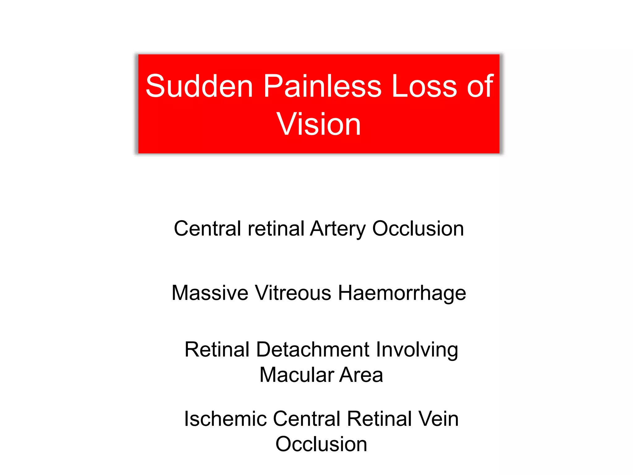
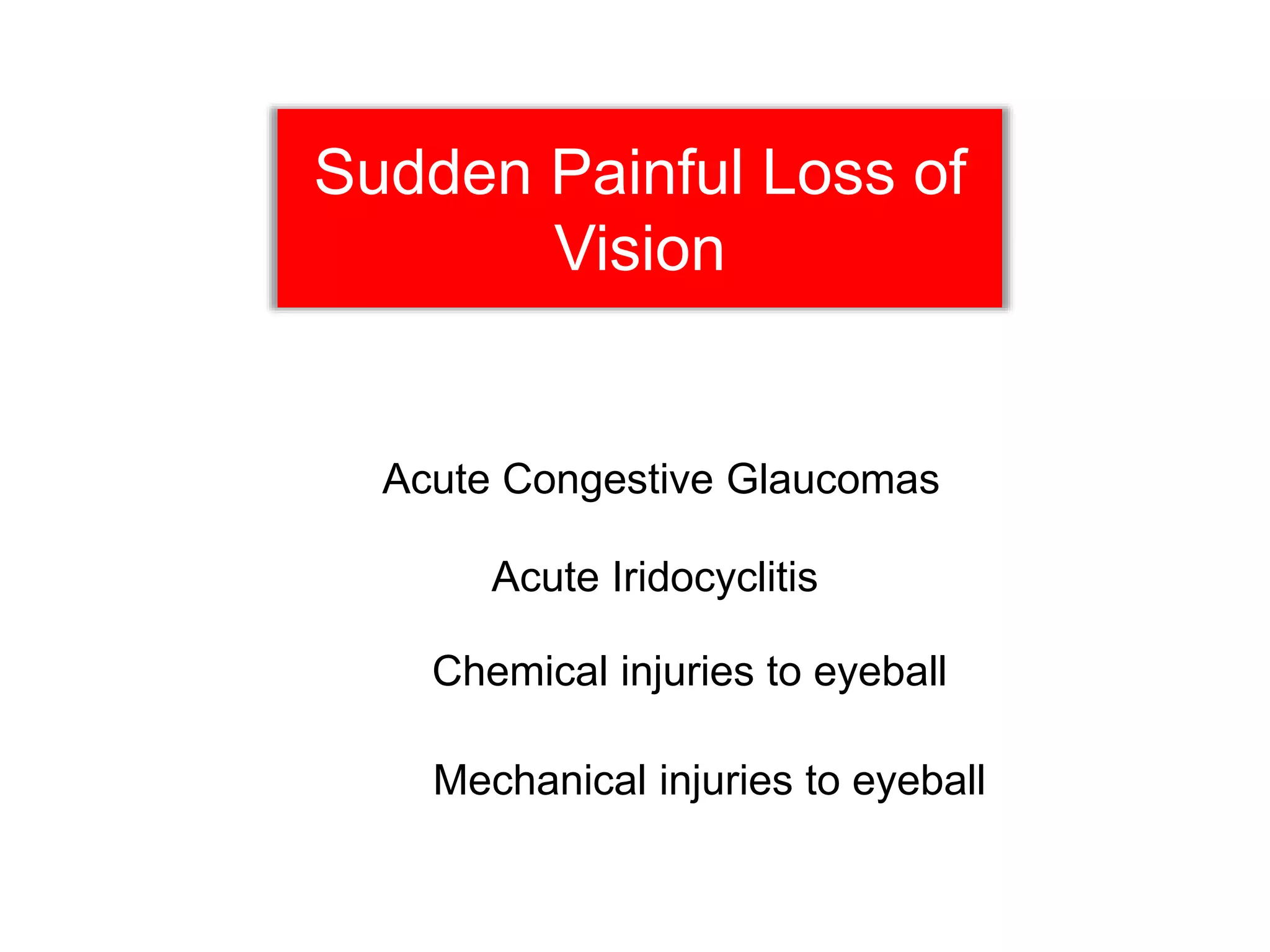
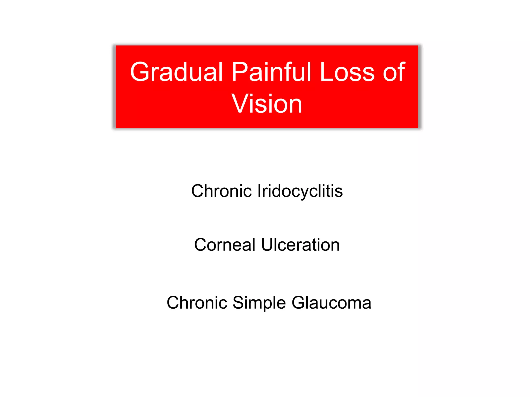
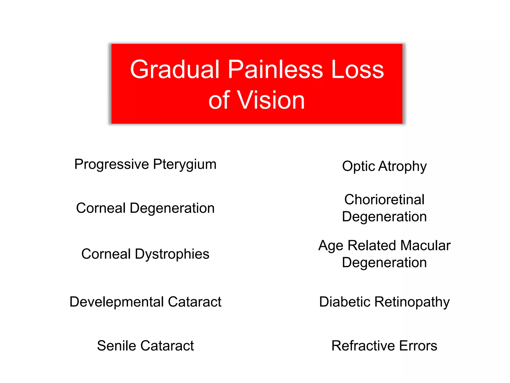
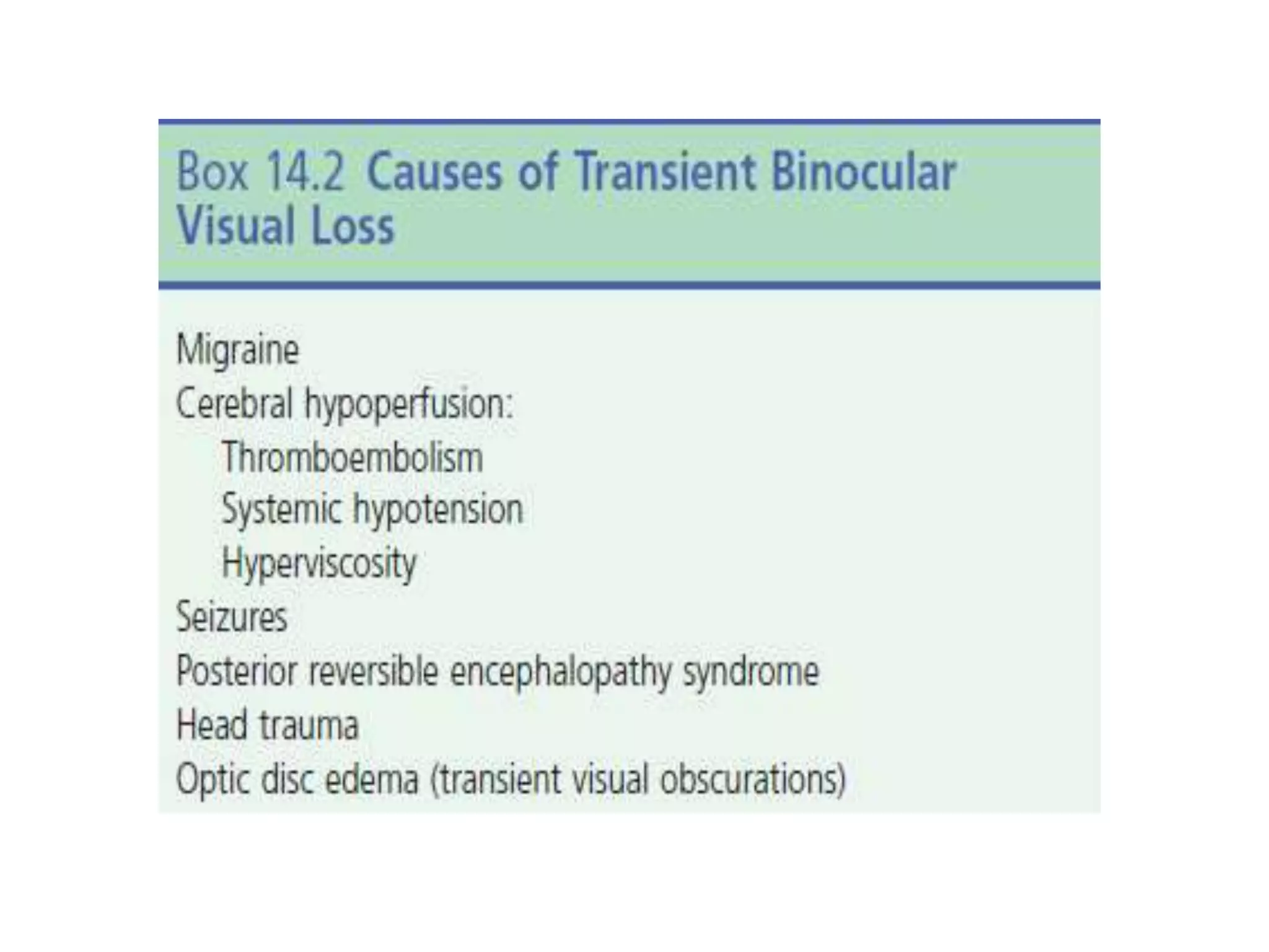
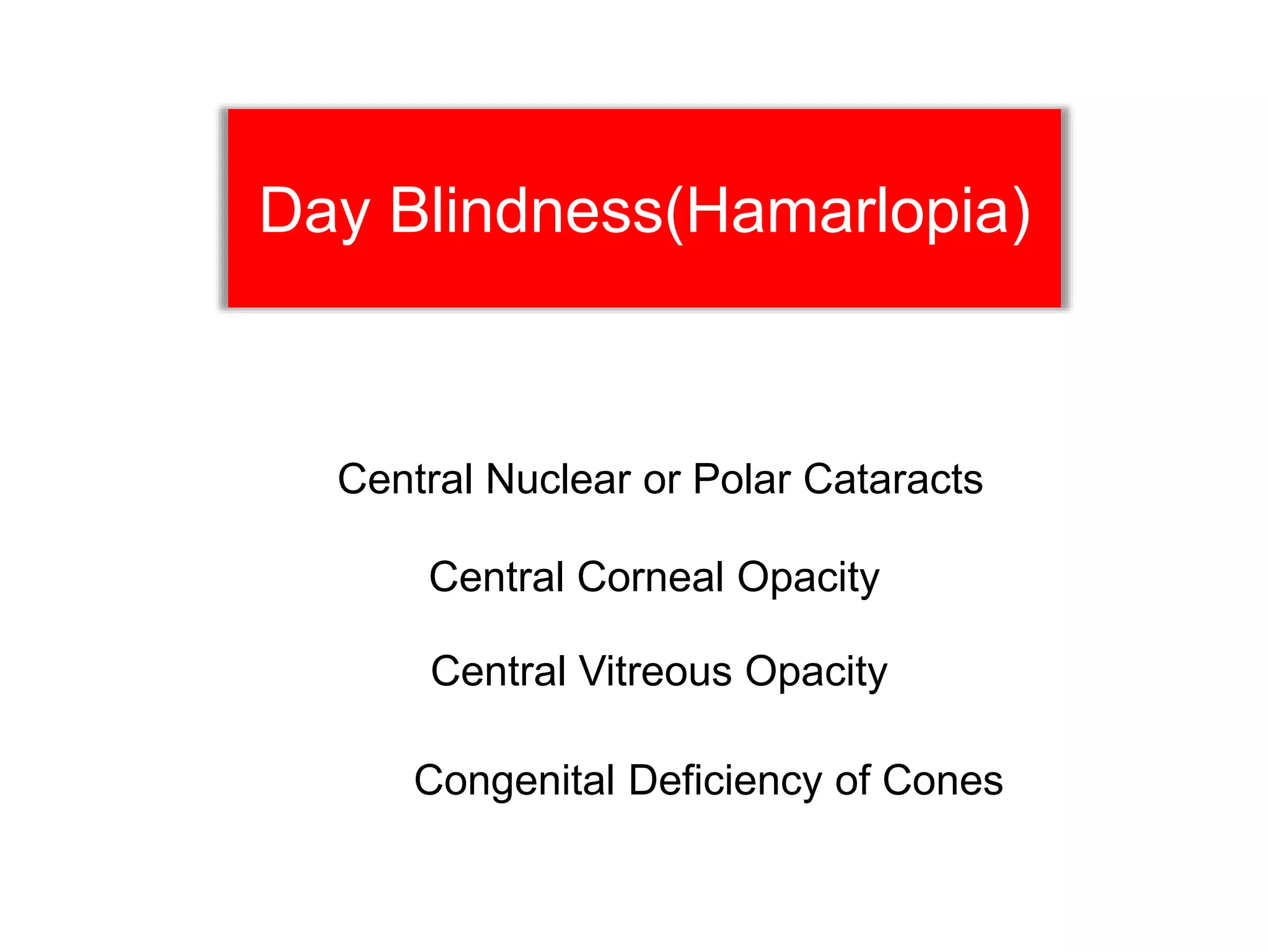
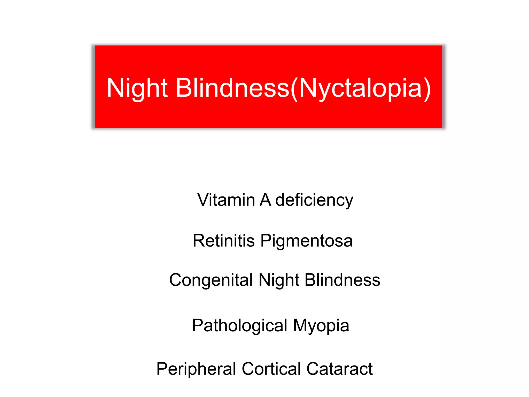
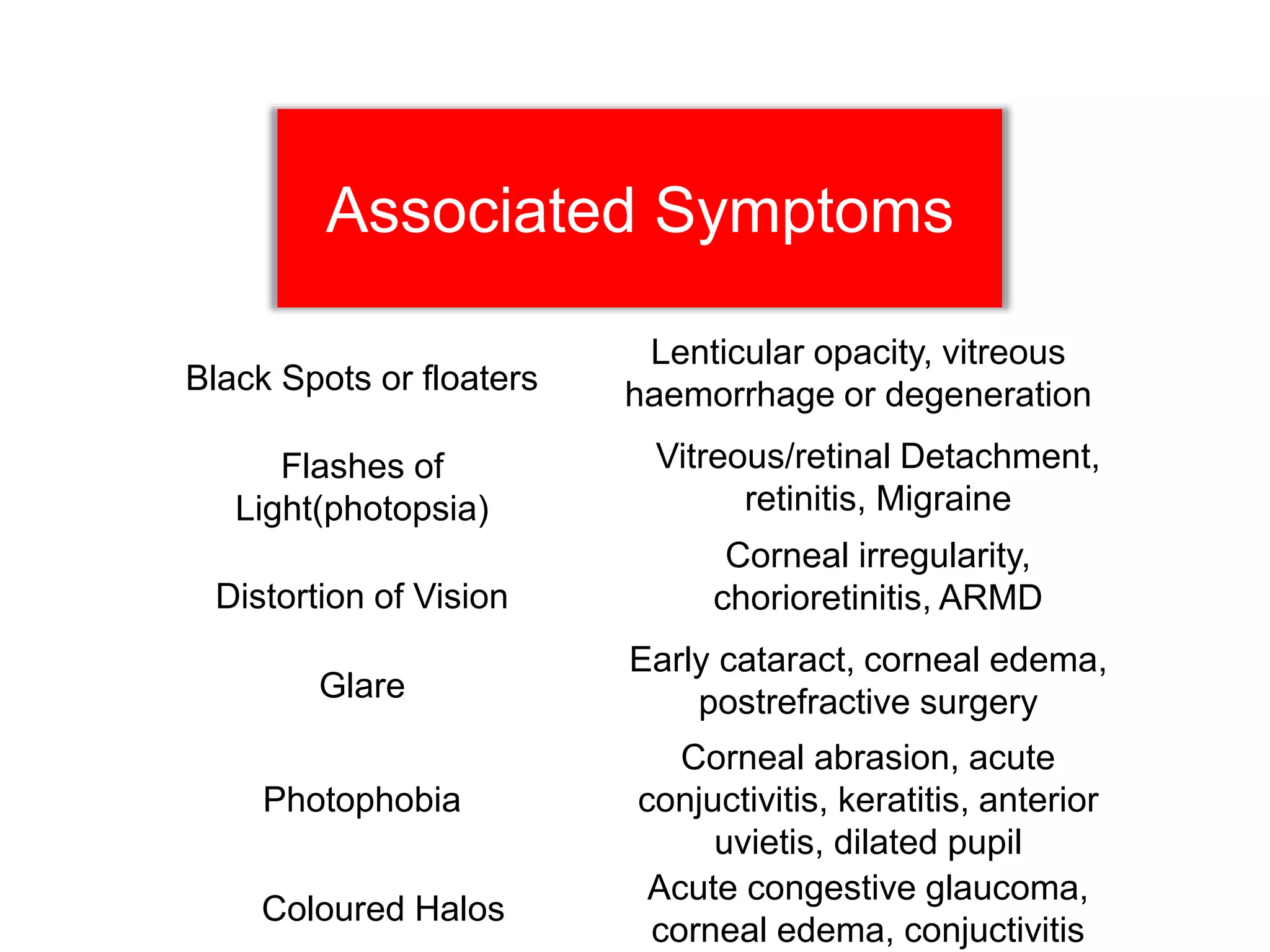
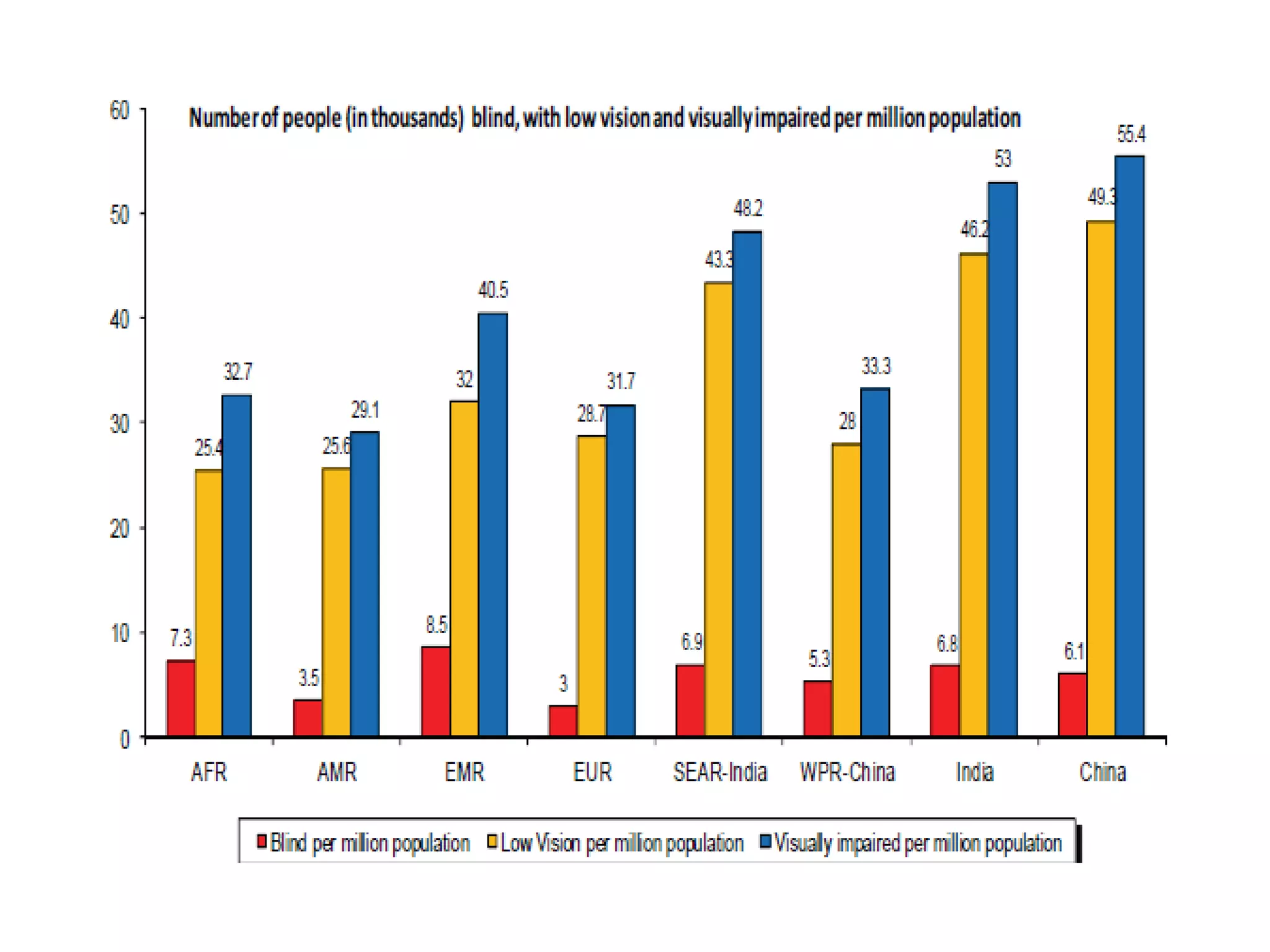
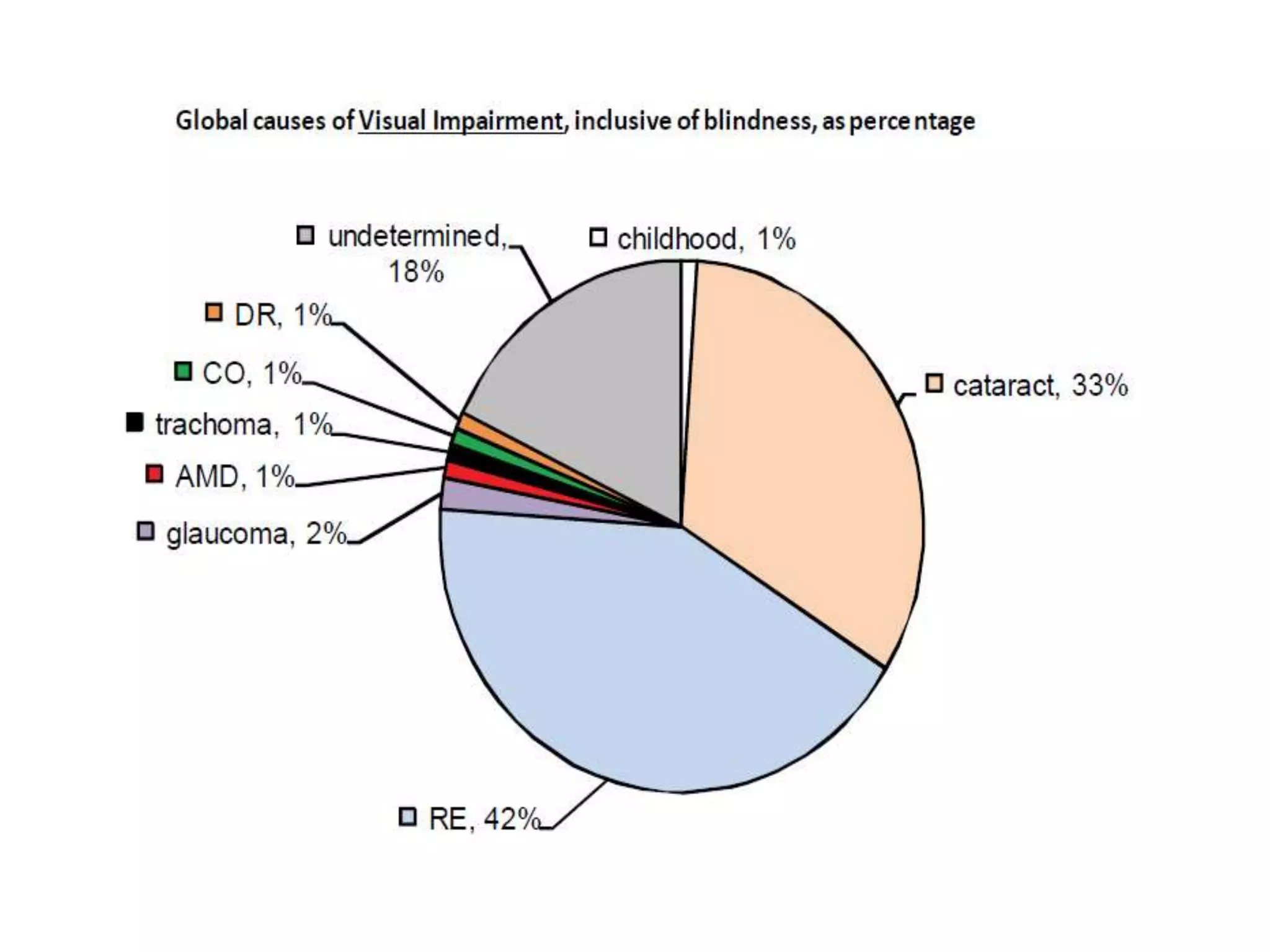
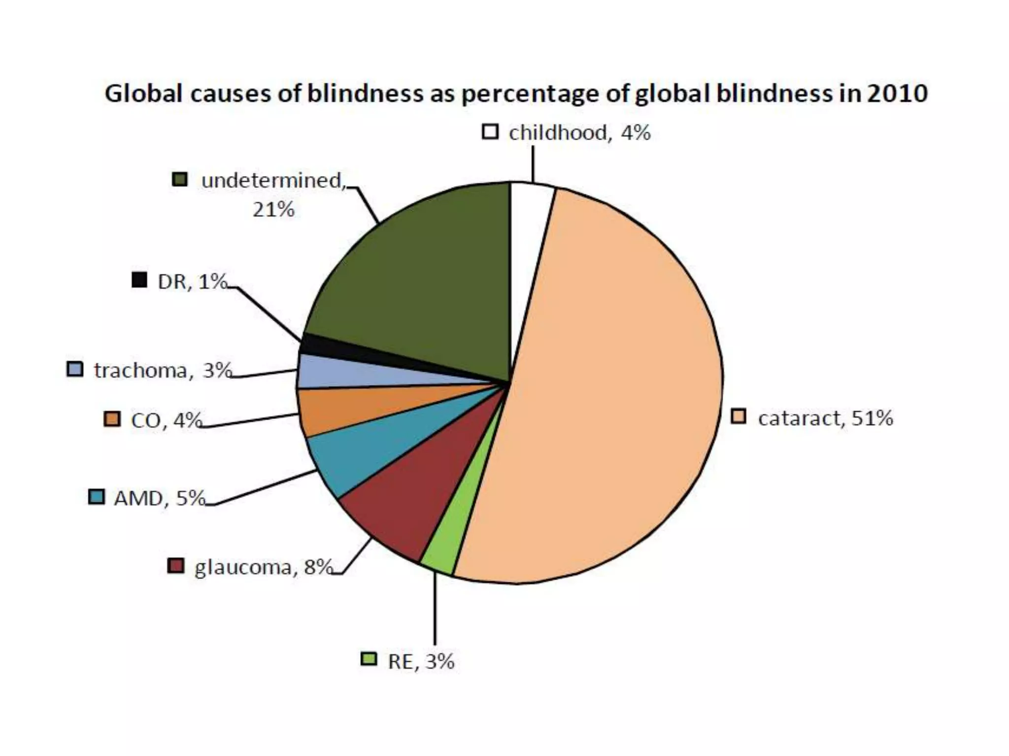
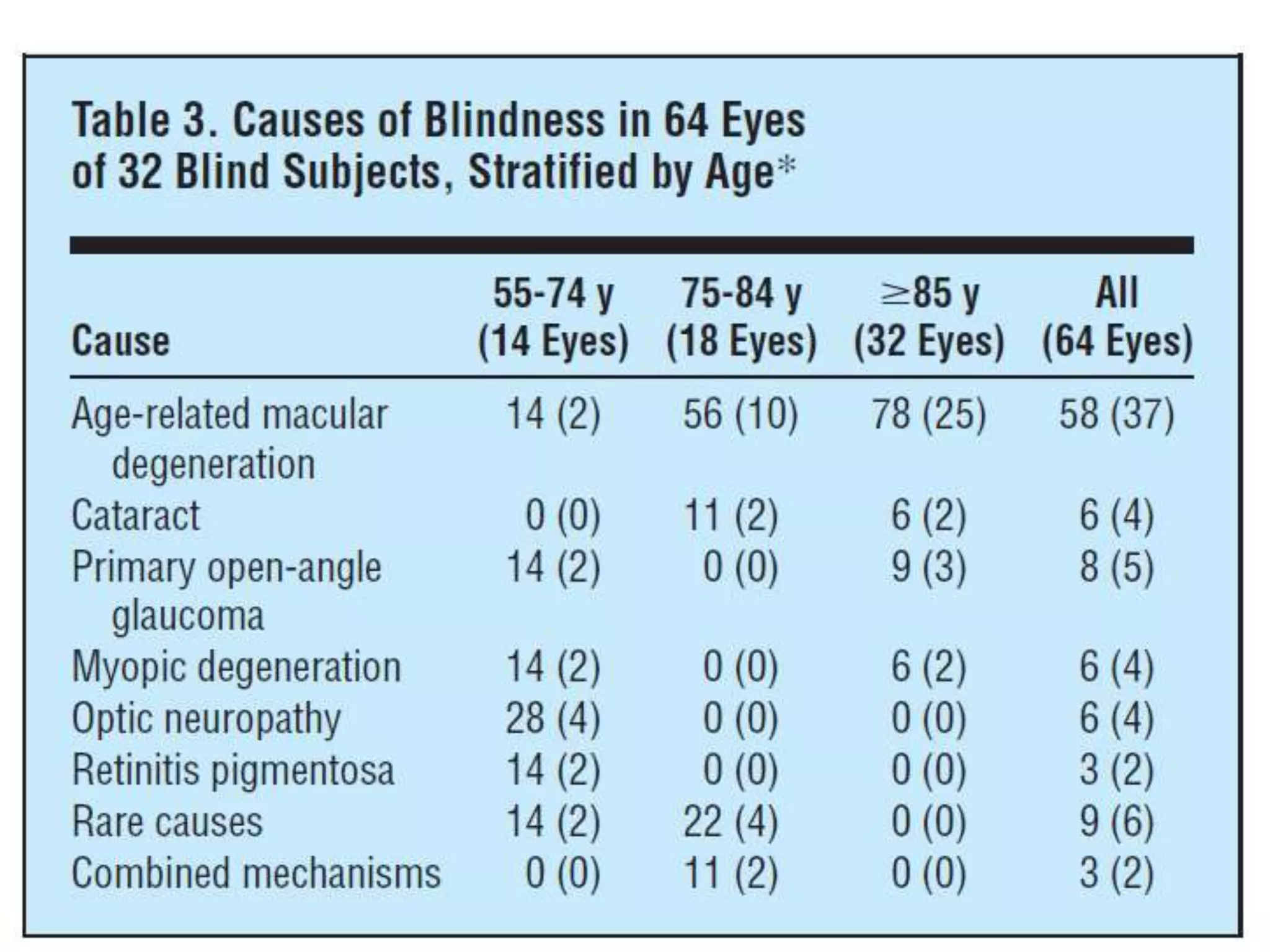
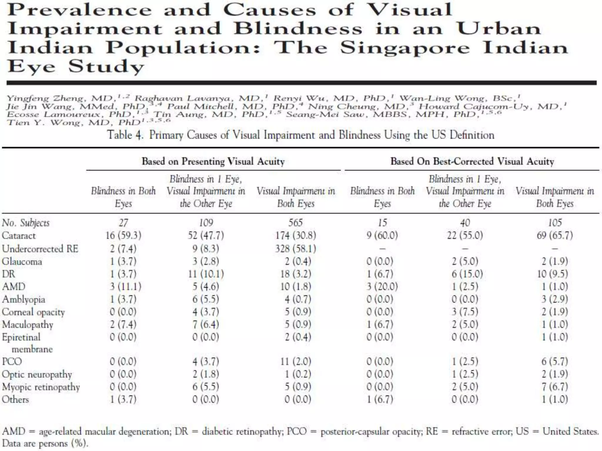
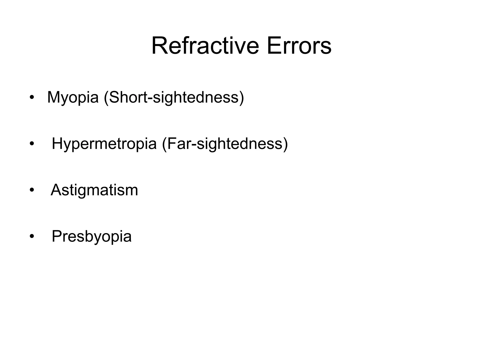
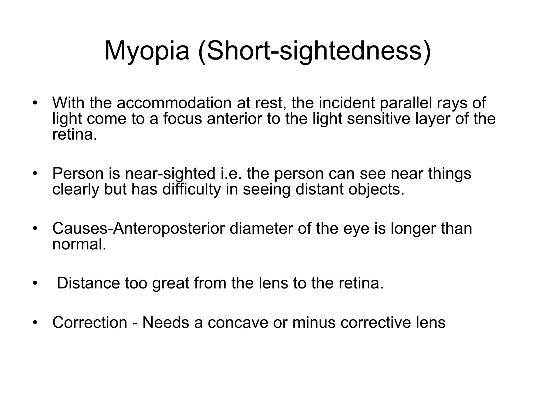
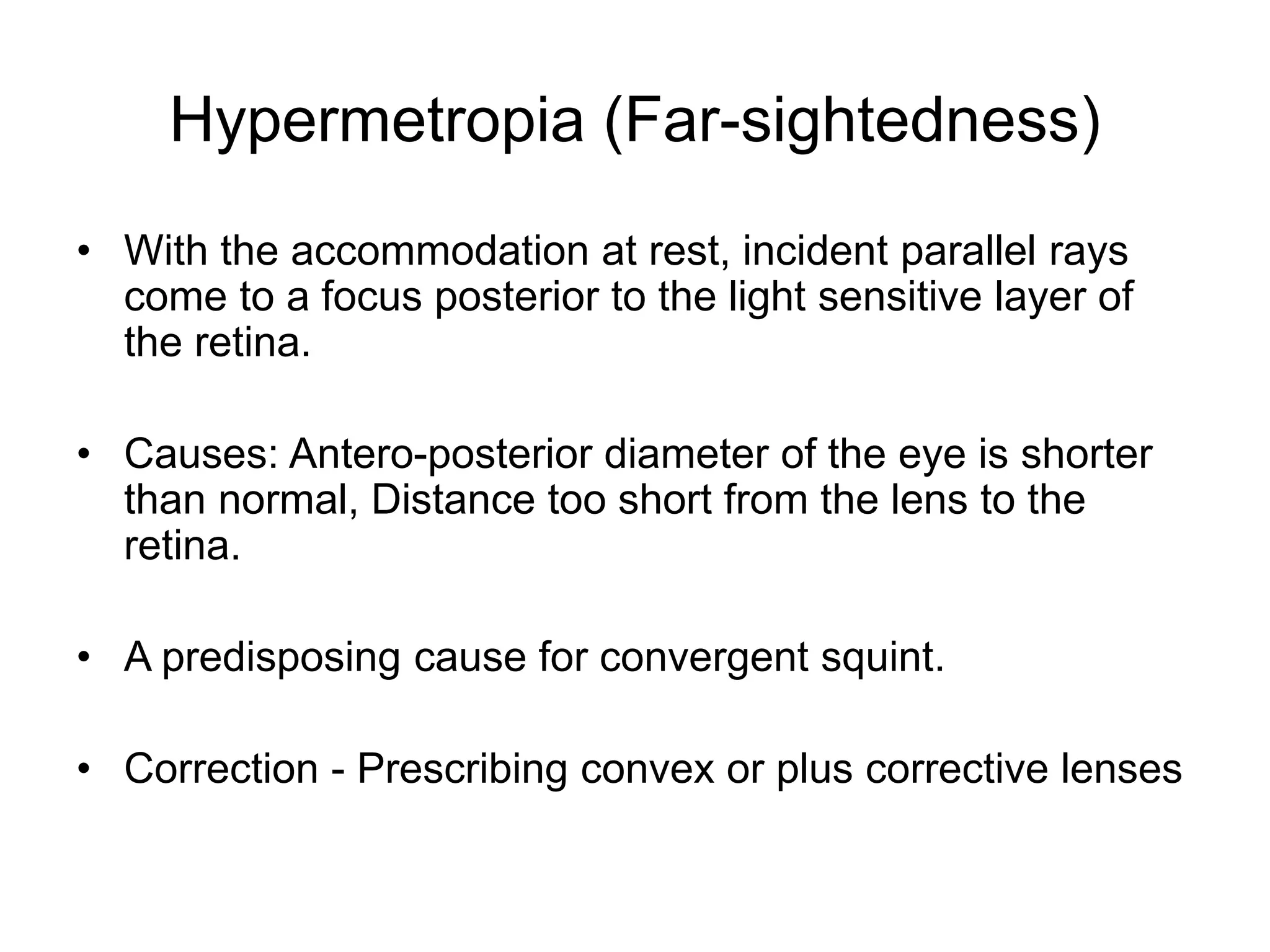
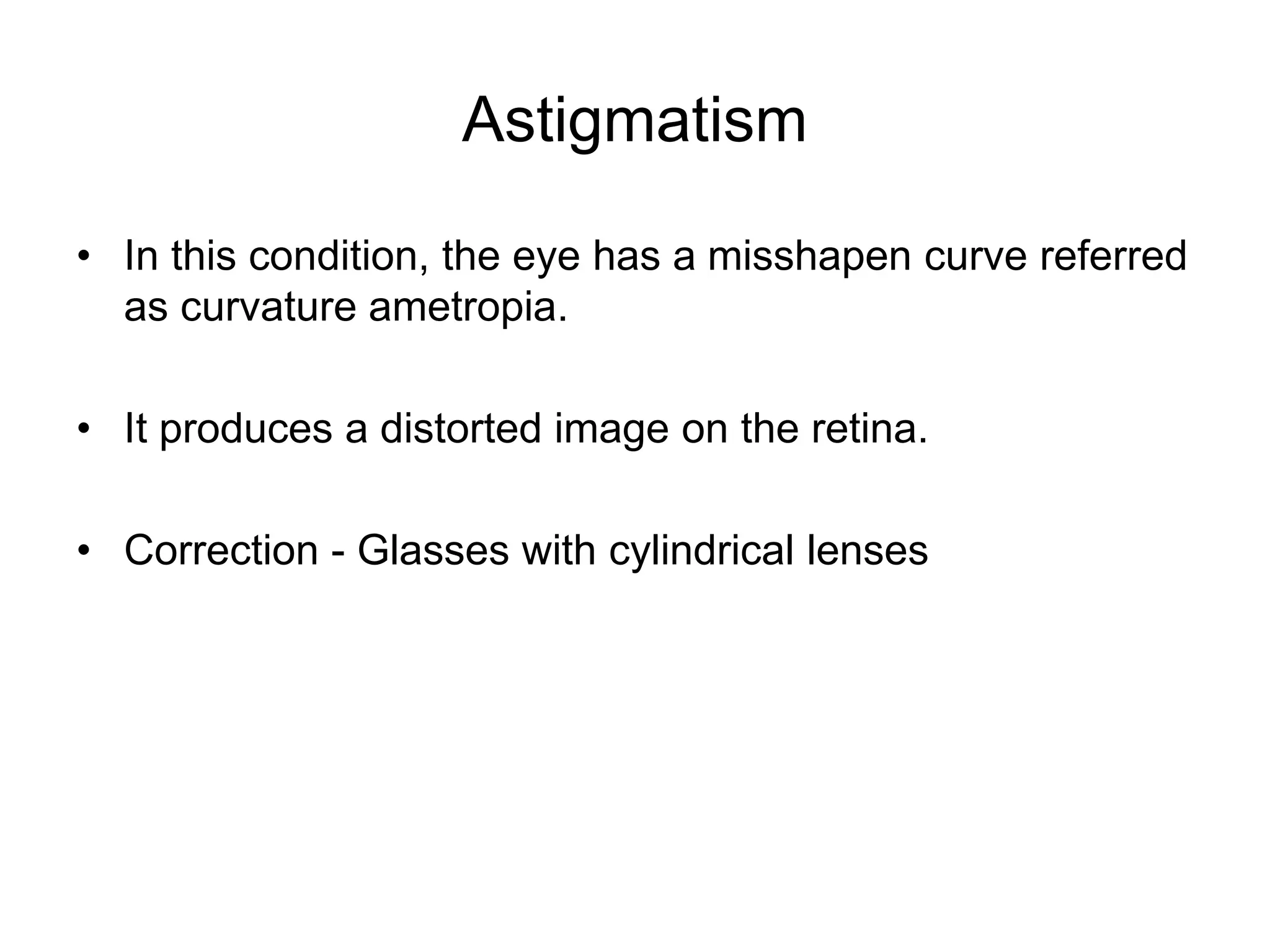
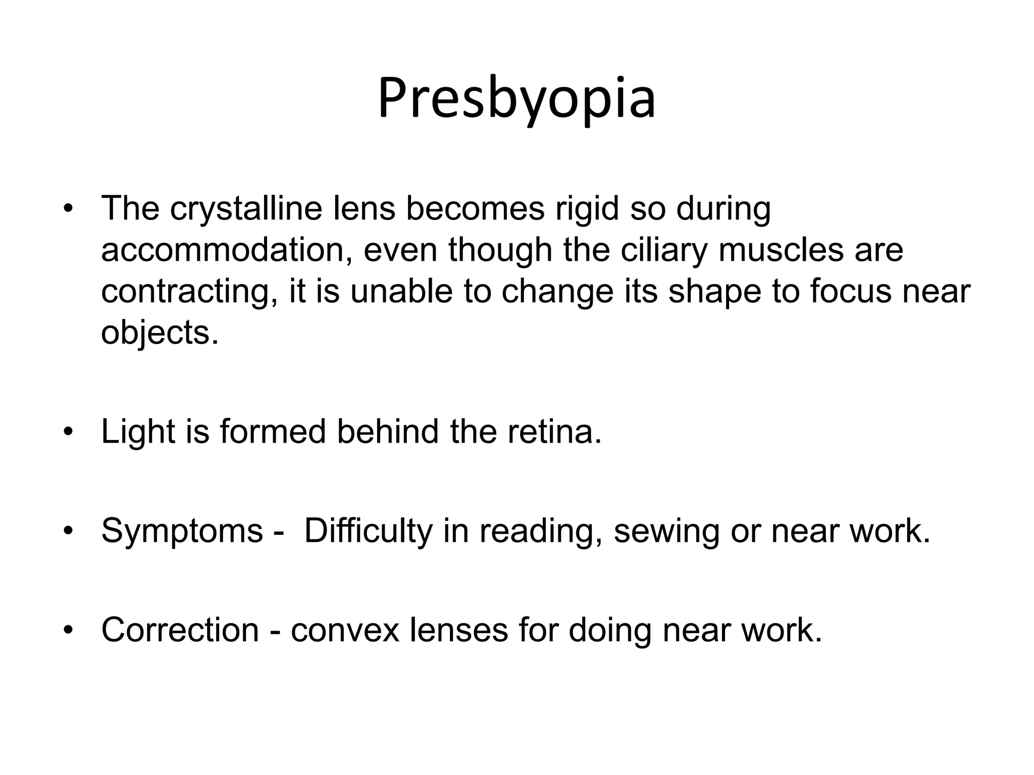
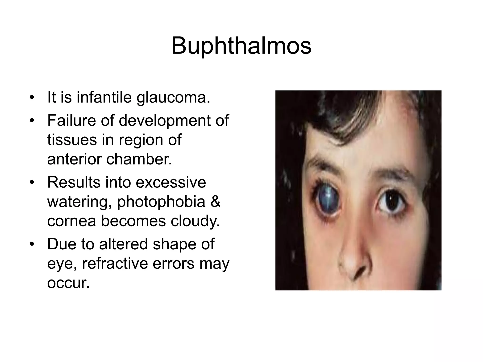
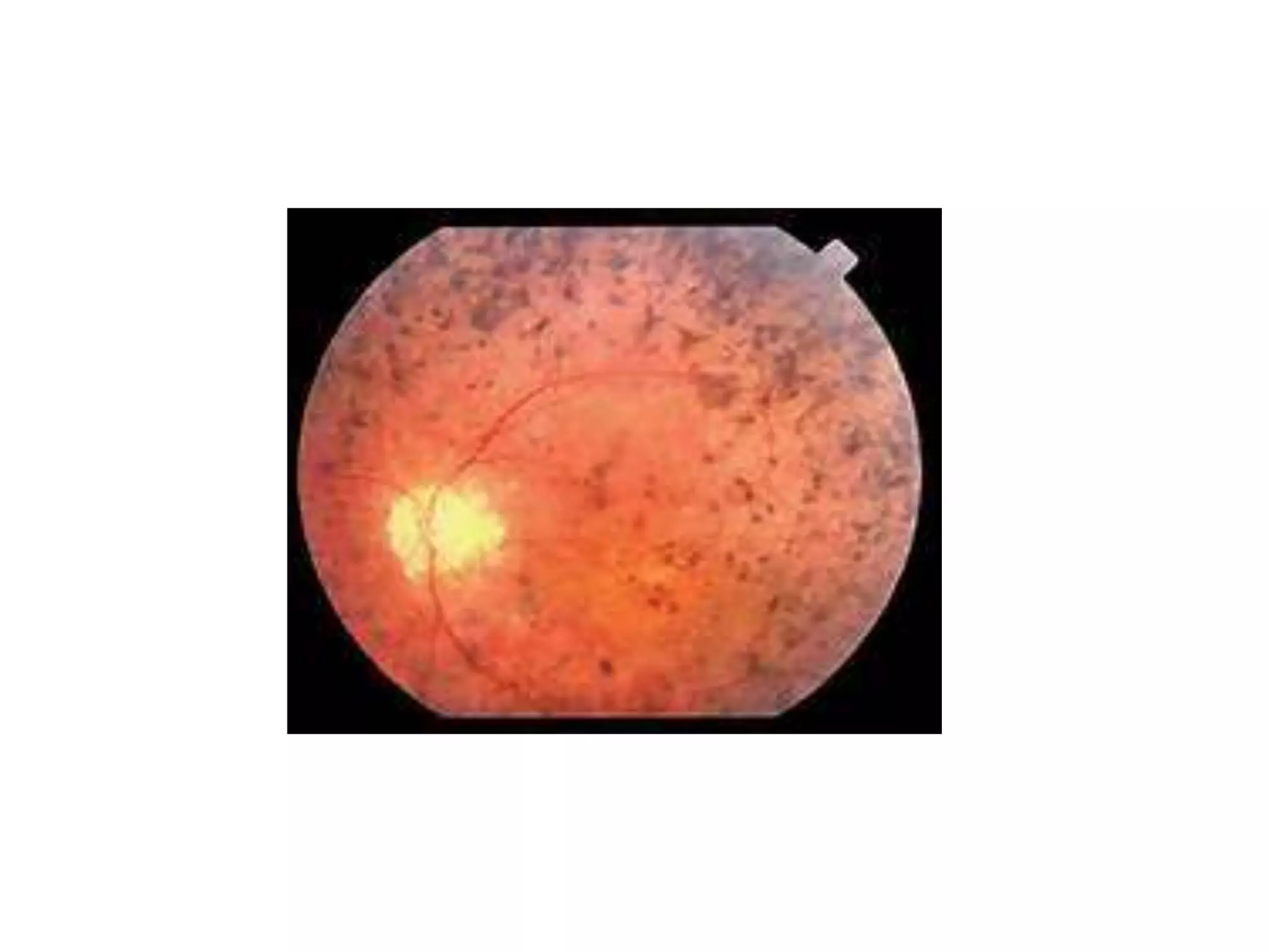
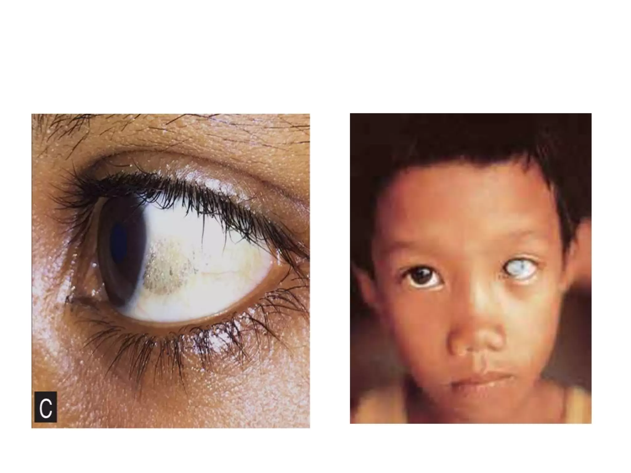
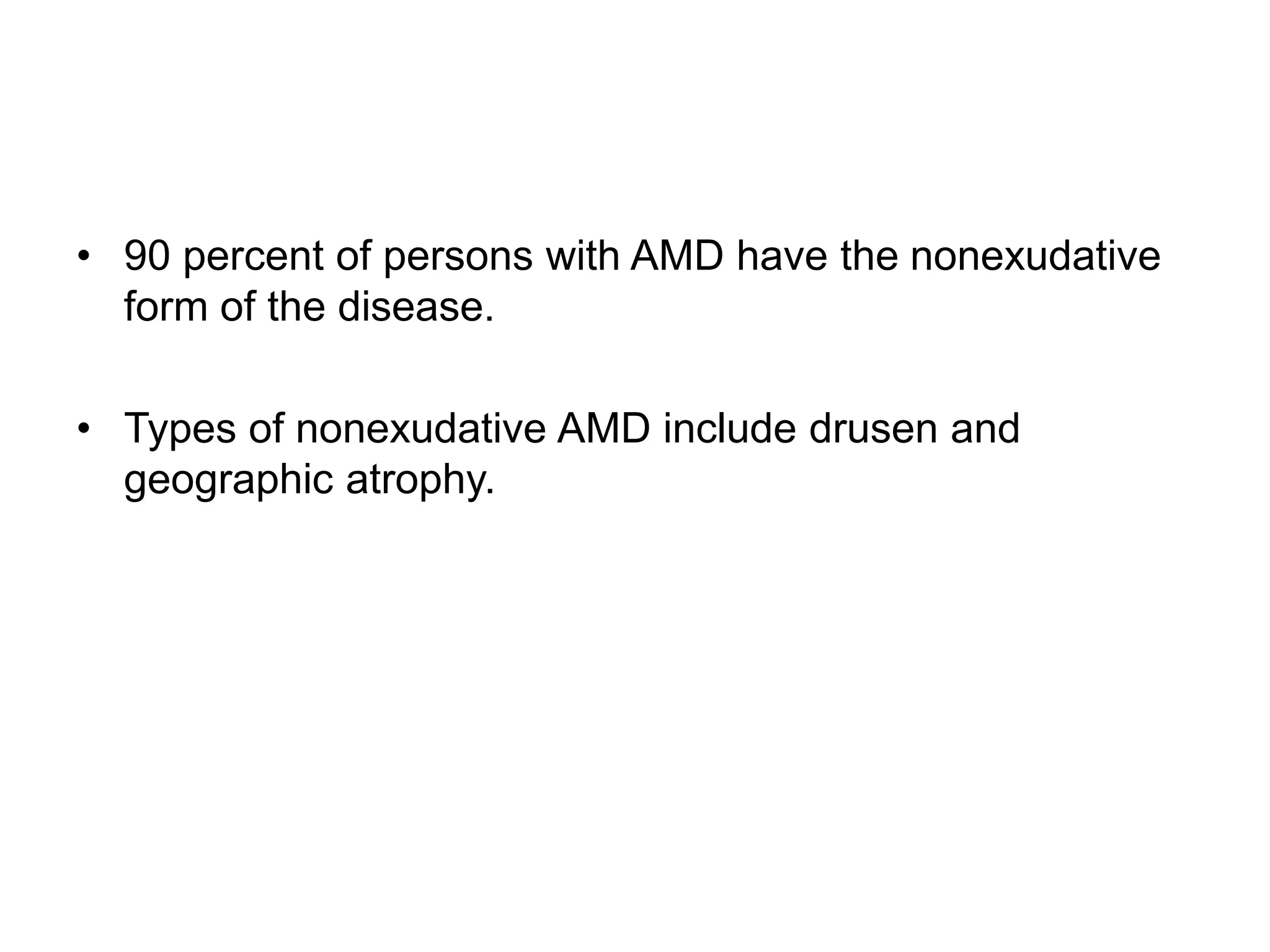
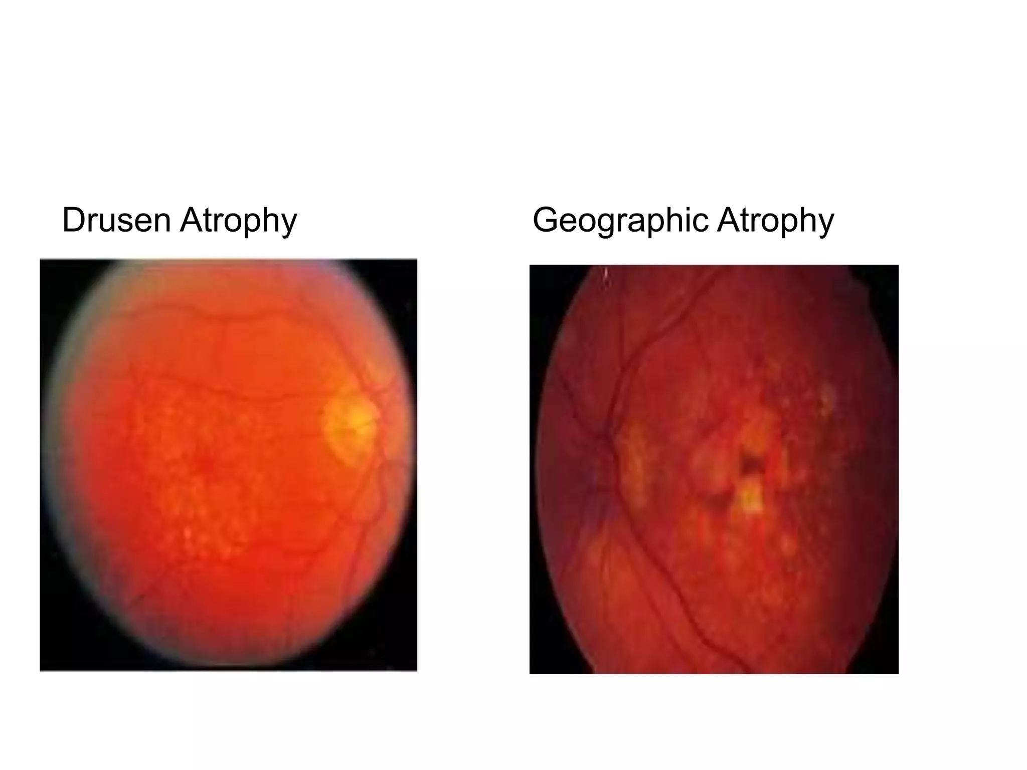
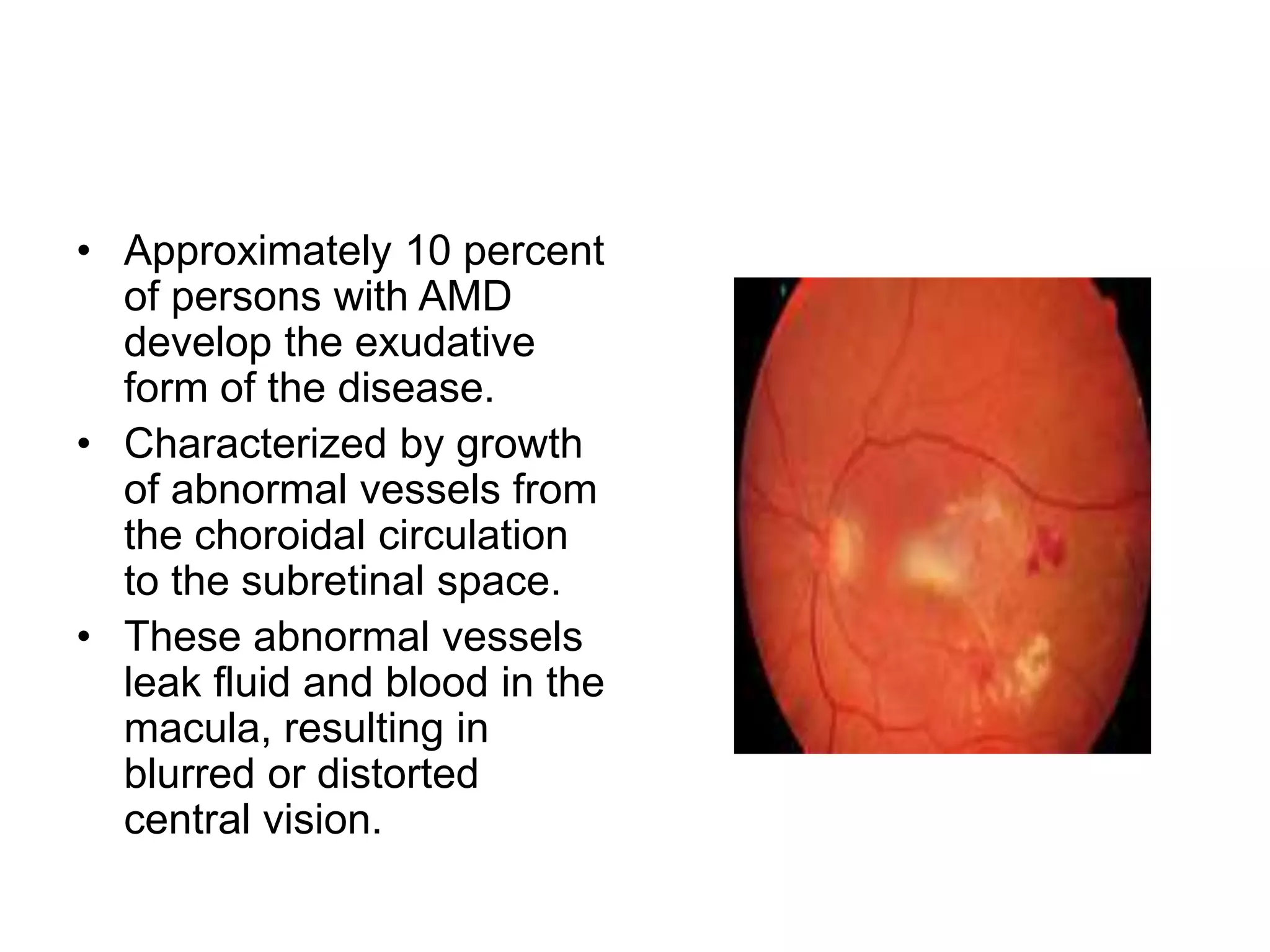
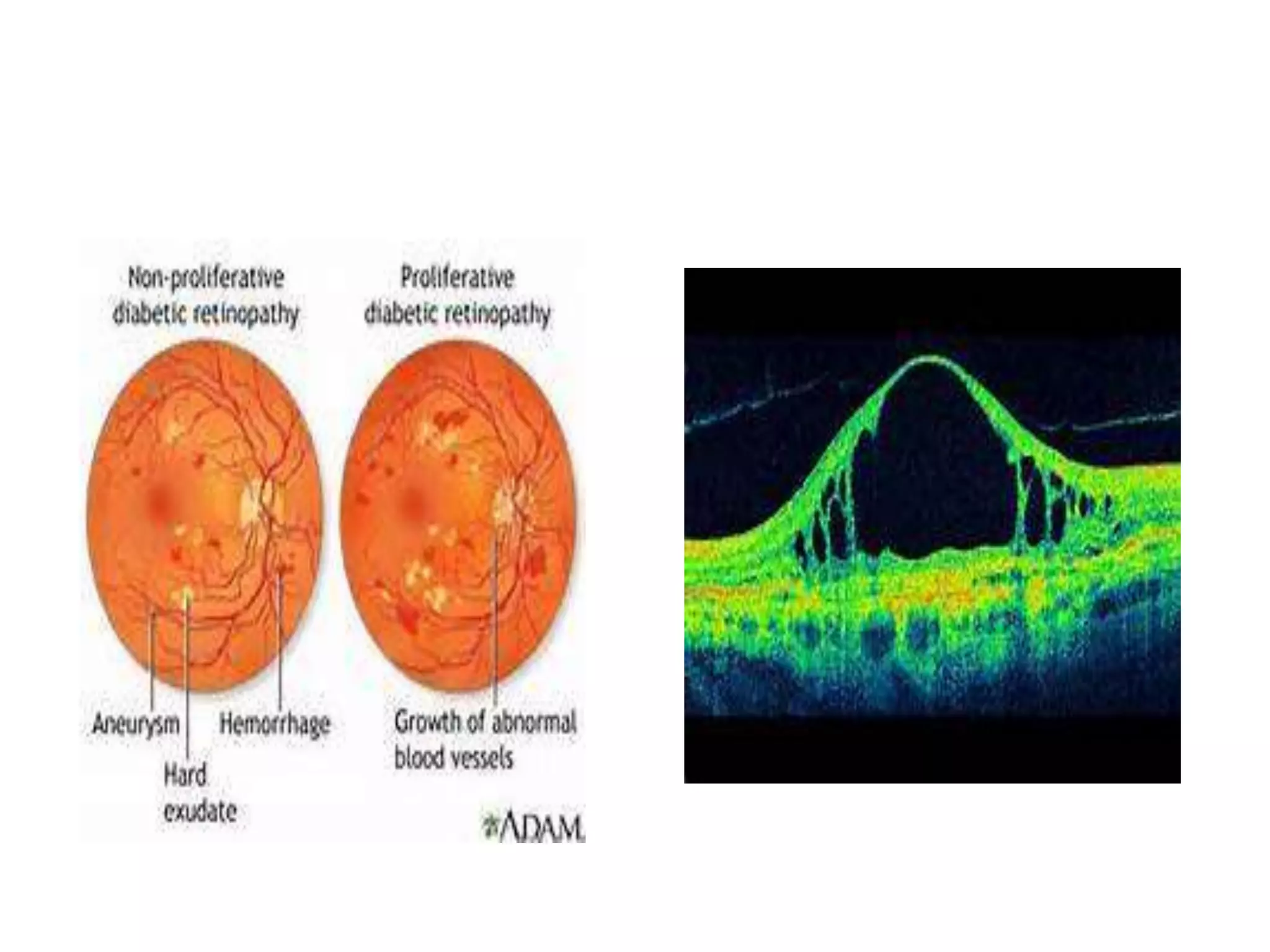
SlideShare a Scribd company logo
Overview of Ophthalmologic Causes
of Visual Impairment
Dr. Nishtha Jain
Senior Resident
Department of Neurology
GMC, Kota.
Visual Impairment
• A functional limitation of the eye(s) or visual system and
can manifest as reduced visual acuity or contrast
sensitivity, visual field loss, photophobia, diplopia, visual
distortion, visual perceptual difficulties, or any
combination.
WHO Grading
Sudden Painless Loss of
Vision
Central retinal Artery Occlusion
Massive Vitreous Haemorrhage
Retinal Detachment Involving
Macular Area
Ischemic Central Retinal Vein
Occlusion
Sudden Painful Loss of
Vision
Acute Iridocyclitis
Acute Congestive Glaucomas
Chemical injuries to eyeball
Mechanical injuries to eyeball
Gradual Painful Loss of
Vision
Chronic Iridocyclitis
Corneal Ulceration
Chronic Simple Glaucoma
Gradual Painless Loss
of Vision
Progressive Pterygium
Corneal Degeneration
Corneal Dystrophies
Develepmental Cataract
Senile Cataract
Optic Atrophy
Chorioretinal
Degeneration
Age Related Macular
Degeneration
Diabetic Retinopathy
Refractive Errors
Day Blindness(Hamarlopia)
Central Corneal Opacity
Central Nuclear or Polar Cataracts
Central Vitreous Opacity
Congenital Deficiency of Cones
Night Blindness(Nyctalopia)
Retinitis Pigmentosa
Vitamin A deficiency
Congenital Night Blindness
Pathological Myopia
Peripheral Cortical Cataract
Associated Symptoms
Black Spots or floaters
Flashes of
Light(photopsia)
Distortion of Vision
Glare
Photophobia
Lenticular opacity, vitreous
haemorrhage or degeneration
Vitreous/retinal Detachment,
retinitis, Migraine
Corneal irregularity,
chorioretinitis, ARMD
Early cataract, corneal edema,
postrefractive surgery
Corneal abrasion, acute
conjuctivitis, keratitis, anterior
uvietis, dilated pupil
Coloured Halos
Acute congestive glaucoma,
corneal edema, conjuctivitis
Refractive Errors
• Myopia (Short-sightedness)
• Hypermetropia (Far-sightedness)
• Astigmatism
• Presbyopia
Myopia (Short-sightedness)
• With the accommodation at rest, the incident parallel rays of
light come to a focus anterior to the light sensitive layer of the
retina.
• Person is near-sighted i.e. the person can see near things
clearly but has difficulty in seeing distant objects.
• Causes-Anteroposterior diameter of the eye is longer than
normal.
• Distance too great from the lens to the retina.
• Correction - Needs a concave or minus corrective lens
Hypermetropia (Far-sightedness)
• With the accommodation at rest, incident parallel rays
come to a focus posterior to the light sensitive layer of
the retina.
• Causes: Antero-posterior diameter of the eye is shorter
than normal, Distance too short from the lens to the
retina.
• A predisposing cause for convergent squint.
• Correction - Prescribing convex or plus corrective lenses
Astigmatism
• In this condition, the eye has a misshapen curve referred
as curvature ametropia.
• It produces a distorted image on the retina.
• Correction - Glasses with cylindrical lenses
Presbyopia
• The crystalline lens becomes rigid so during
accommodation, even though the ciliary muscles are
contracting, it is unable to change its shape to focus near
objects.
• Light is formed behind the retina.
• Symptoms - Difficulty in reading, sewing or near work.
• Correction - convex lenses for doing near work.
Buphthalmos
• It is infantile glaucoma.
• Failure of development of
tissues in region of
anterior chamber.
• Results into excessive
watering, photophobia &
cornea becomes cloudy.
• Due to altered shape of
eye, refractive errors may
occur.
Albinism
• This is a hereditary condition involving defective
development of pigment in hair, skin & eye.
• In ‘ocular albinism’, only eyes are affected.
• Associated with photophobia, decreased visual acuity,
nystagmus and refractive errors.
• Difficult going outside on a bright day
• Any glare would cause difficulty
Precautions
• Such children not to be
seated near windows
• Level of lighting may be
adapted
• Need glasses to help
their distant vision
Retinitis Pigmentosa
• Hereditary slow degenerative disease of the retina.
• Affects the peripheral area of retina.
• Result into night blindness, tunnel vision and inability to
see in dark.
• Progressive and results into blindness in middle or
advanced age.
• The field of vision is so poor that the person falls in the
category of blindness.
Retinoblastoma
• Malignant tumour of the retina.
• Generally confined to infants, probably always congenital
and some cases are heredity.
• Often a bilateral condition.
• Treated by surgery and radiation or photo-coagulation.
• Genetic counselling is desirable.
Retrolental Fibroplasia
• Associated with pre-mature birth children who have been
given high concentration of oxygen.
• Caused due to formation of new vessels and proliferation
of fibrous tissue in the retina.
• Results into formation of a membrane in the back of the
lens of the eye.
• Usually a bilateral condition.
Trachoma
• Caused by an organism chlamydia trachomatis.
• Unhygienic and crowded unhealthy environment
• Primary infection is epithelial and involves both
conjunctiva and cornea.
• Symptoms- Redness, Itching, Tearing, Irritations
• Signs : Diffused conjunctival inflammation characterized
by congestion, formation of follicles on the inner aspect
of the upper lid.
• Vascularisation of the upper margin of cornea and
corneal ulcers
Treatment
• Clean the eyes if there is discharge
• Sulphacetamide eye drops 10% or 20% to be instilled at
least 4 times a day for 6 weeks.
• Advise on personal hygiene and daily washing of face.
• Check other members of the family for trachoma
• Never use medicines containing steroids.
• Trichiasis : distortion of the lids.
• The shape of lids, especially upper lid, is altered and
may be turned inwards, leading to entropion.
• This causes the lids to rub against the cornea.
• Symptoms – Irritation, Photophobia, Tearing, Poor vision
• Remove misdirected eyelashes
• Topical treatment with Erythromycin ointment,
Tetracycline or Rifampicin is far more effective than
Sulfonamides.
• This treatment must be persistent for 5 consecutive days
a month for 12 months.
• Oral Doxycycline 5mg/kg body weight one per month is
easy to administer and is as effective as topical
Tetracycline.
Vitamin A Deficiency
• Also known as: Xerophthalmia, Blinding malnutrition,
Disease of darkness.
• Causes-Insufficient and unbalanced food intake by the
mothers and the children, Low absorption due to
diarrhoea or malnutrition, Increased demand of Vitamin A
during and after measles infection.
• Leads to: Xerosis of the conjunctiva, Keratomalacia and
Night blindness.
Signs
• A line or spot on the conjunctiva
• Thick white spots on both sides of the cornea
• Conjunctiva becomes wrinkled
• Scar forms over cornea
• Immediately- give oral 200,000 IU vitamin A
• Apply antibiotic ointment (twice daily)
• Apply a protective eye shield
• Next day give a further dose of oral vitamin A, 200,000
IU, and again two weeks later (same dose)
Cataract
• Loss of transparency of the lens due to altered physio-
chemical processes within tissues.
• Usually associated with advanced age.
• If present at birth, it is referred as congenital cataract.
• Complaint - Gradual loss of vision over a long period of
time
• Feels as if looking through a dirty window
Examination and findings
• Gray or white pupils
• Usually both eyes are affected
• Usually found in old people
Refer for further examination and possible operation if:
• patient can not perform daily activities
• vision is worse than 6/60 or finger counting at 6 meters
in both eyes
Age-Related Macular Degeneration
• Characterized by degeneration of the macula.
• Risk factors for AMD - Advancing age, family history of
AMD and cardiovascular risk factors such as
hypertension and cigarette smoking.
• AMD can be divided into two categories: nonexudative
(or “dry”) AMD and exudative (or “wet”) AMD.
• 90 percent of persons with AMD have the nonexudative
form of the disease.
• Types of nonexudative AMD include drusen and
geographic atrophy.
Drusen Atrophy Geographic Atrophy
• Approximately 10 percent
of persons with AMD
develop the exudative
form of the disease.
• Characterized by growth
of abnormal vessels from
the choroidal circulation
to the subretinal space.
• These abnormal vessels
leak fluid and blood in the
macula, resulting in
blurred or distorted
central vision.
Glaucoma
• Caused by an obstruction in aqueous outflow channels
at angle of anterior chamber.
• Results in the rise in intraocular pressure which is
detrimental to the eye.
• It is usually a hereditary, symptomatic condition.
• Acute glaucoma strikes suddenly with intense
pain,nausea and blurred vision.
• Treatment is aimed at lowering the internal pressure in
the eye
• Special eye drops used regularly would maintain the
internal pressure at the proper level
• In some instances, surgery may be required.
Risk factors
• age over 35 years of age
• have blood relatives who have Glaucoma
• have diabetes
• asthmatic and patient of arthritis on long term oral
“Corticosteroids”
• have rainbow rings around bulbs, lights or candles
• pain or blurring of sight in the evenings
• children with large eye
Diabetic Retinopathy
• Diabetic macular oedema occurs in approximately 10
percent of all diabetics.
• In patients with diabetes for 20 or more years, incidence
increases to 25 percent.
• Macular oedema is defined as any retinal thickening of
or deposition of hard exudates within one disc diameter
of the centre of the macula.
It is termed significant if any of the following three
characteristics are present:
• Thickening of the retina at or within 500 u of the centre
of fovea
• Deposition of hard exudates associated with the area of
adjacent retinal thickening at or within 500 u of the foveal
centre
• Development of a zone of retinal thickening one disc
diameter or larger
• The decision to treat diabetic macular oedema is based
entirely on clinical and angiographic findings,
independent of patient’s visual acuity.
• Focal treatment consists of directing only green argon
photocoagulation to all leaking microaneurysms.
To conclude
• Developing countries having major burden of visual
impairment.
• Low socioeconomic status/rural population more prone.
• Main cause in adults- cataract, RE, AMD, Glaucoma,
Diabetic Retinopathy
• Main cause in children- Develepmental disorders,
cataract, vitamin A deficiency.
Referrences
• Bradley’s Neurology in Clinical Practice, Sixth Edition.
• Causes, Prevention And Cure Of Visual Impairment. Dr.
Vandana Nath.
• Prevalence and Causes of Visual Impairment and
Blindness in an Urban Indian Population: The Singapore
Indian Eye Study. Zheng et al. Ophthalmology
2011;118:1798–1804.
• Global Data On Visual Impairment. World Health
Organization 2012.

More Related Content

What's hot

Corneal ulcer(bactrial,fungal) 25.02.16, dr.k.n.jha
Corneal ulcer(bactrial,fungal) 25.02.16, dr.k.n.jhaCorneal ulcer(bactrial,fungal) 25.02.16, dr.k.n.jha
Corneal ulcer(bactrial,fungal) 25.02.16, dr.k.n.jha
ophthalmgmcri
 
Ocular trauma
Ocular traumaOcular trauma
Ocular trauma
Md. Nurul Islam
 
The pupillary pathway and its clinical aspects
The pupillary pathway and its clinical aspectsThe pupillary pathway and its clinical aspects
The pupillary pathway and its clinical aspects
Laxmi Eye Institute
 
Primary Angle Closure Glaucoma- Saral
Primary Angle Closure Glaucoma- SaralPrimary Angle Closure Glaucoma- Saral
Primary Angle Closure Glaucoma- Saral
Saral Lamichhane
 
Corneal Dystrophies
Corneal DystrophiesCorneal Dystrophies
Corneal Dystrophies
Puneet Sharma
 
Central retinal artery occlusion
Central retinal artery occlusionCentral retinal artery occlusion
Central retinal artery occlusion
SSSIHMS-PG
 
Differential Diagnosis of Disc Edema
Differential Diagnosis of Disc EdemaDifferential Diagnosis of Disc Edema
Differential Diagnosis of Disc Edema
Sahil Thakur
 
EXTRA OCULAR MUSCLES PHYSIOLOGY
EXTRA OCULAR MUSCLES PHYSIOLOGYEXTRA OCULAR MUSCLES PHYSIOLOGY
EXTRA OCULAR MUSCLES PHYSIOLOGY
Rabia Ammer
 
Pathological myopia 01.03.2014
Pathological myopia 01.03.2014Pathological myopia 01.03.2014
Pathological myopia 01.03.2014
Mohammad Bawtag
 
Anatomy and physiology of cornea
Anatomy and physiology of corneaAnatomy and physiology of cornea
Anatomy and physiology of cornea
SSSIHMS-PG
 
Tarsorrhaphy.pptx
Tarsorrhaphy.pptxTarsorrhaphy.pptx
Tarsorrhaphy.pptx
Gariyashee Lahkar
 
Evaluation of squint
Evaluation of squint Evaluation of squint
Evaluation of squint
Dr.Siddharth Gautam
 
Optic atrophy
Optic atrophyOptic atrophy
Optic atrophy
Angel Das
 
Corneal dystrophies
Corneal dystrophiesCorneal dystrophies
Corneal dystrophies
Najara Thapa
 
Retinitis pigmentosa
Retinitis pigmentosaRetinitis pigmentosa
Retinitis pigmentosa
Othman Al-Abbadi
 
Keratoconus
KeratoconusKeratoconus
Keratoconus
ikramdr01
 
Angle of anterior chamber
Angle of anterior chamberAngle of anterior chamber
Angle of anterior chamber
Kalluri Sahithi Reddy
 
Pterygium and its management
Pterygium and its managementPterygium and its management
Pterygium and its managementDr-Anjali Hiroli
 
Congenital Glaucoma
Congenital GlaucomaCongenital Glaucoma
Congenital Glaucoma
Sahil Thakur
 

What's hot (20)

Corneal ulcer(bactrial,fungal) 25.02.16, dr.k.n.jha
Corneal ulcer(bactrial,fungal) 25.02.16, dr.k.n.jhaCorneal ulcer(bactrial,fungal) 25.02.16, dr.k.n.jha
Corneal ulcer(bactrial,fungal) 25.02.16, dr.k.n.jha
 
Ocular trauma
Ocular traumaOcular trauma
Ocular trauma
 
The pupillary pathway and its clinical aspects
The pupillary pathway and its clinical aspectsThe pupillary pathway and its clinical aspects
The pupillary pathway and its clinical aspects
 
Primary Angle Closure Glaucoma- Saral
Primary Angle Closure Glaucoma- SaralPrimary Angle Closure Glaucoma- Saral
Primary Angle Closure Glaucoma- Saral
 
Corneal Dystrophies
Corneal DystrophiesCorneal Dystrophies
Corneal Dystrophies
 
Central retinal artery occlusion
Central retinal artery occlusionCentral retinal artery occlusion
Central retinal artery occlusion
 
Classification of squint
Classification of squintClassification of squint
Classification of squint
 
Differential Diagnosis of Disc Edema
Differential Diagnosis of Disc EdemaDifferential Diagnosis of Disc Edema
Differential Diagnosis of Disc Edema
 
EXTRA OCULAR MUSCLES PHYSIOLOGY
EXTRA OCULAR MUSCLES PHYSIOLOGYEXTRA OCULAR MUSCLES PHYSIOLOGY
EXTRA OCULAR MUSCLES PHYSIOLOGY
 
Pathological myopia 01.03.2014
Pathological myopia 01.03.2014Pathological myopia 01.03.2014
Pathological myopia 01.03.2014
 
Anatomy and physiology of cornea
Anatomy and physiology of corneaAnatomy and physiology of cornea
Anatomy and physiology of cornea
 
Tarsorrhaphy.pptx
Tarsorrhaphy.pptxTarsorrhaphy.pptx
Tarsorrhaphy.pptx
 
Evaluation of squint
Evaluation of squint Evaluation of squint
Evaluation of squint
 
Optic atrophy
Optic atrophyOptic atrophy
Optic atrophy
 
Corneal dystrophies
Corneal dystrophiesCorneal dystrophies
Corneal dystrophies
 
Retinitis pigmentosa
Retinitis pigmentosaRetinitis pigmentosa
Retinitis pigmentosa
 
Keratoconus
KeratoconusKeratoconus
Keratoconus
 
Angle of anterior chamber
Angle of anterior chamberAngle of anterior chamber
Angle of anterior chamber
 
Pterygium and its management
Pterygium and its managementPterygium and its management
Pterygium and its management
 
Congenital Glaucoma
Congenital GlaucomaCongenital Glaucoma
Congenital Glaucoma
 

Similar to Overview of ophthalmologic causes of visual impairment

opthalmicdisorders-190508204744.pdf
opthalmicdisorders-190508204744.pdfopthalmicdisorders-190508204744.pdf
opthalmicdisorders-190508204744.pdf
RuchikaMaurya4
 
Opthalmic disorders
Opthalmic disorders Opthalmic disorders
Opthalmic disorders
Jamilah AlQahtani
 
Ophthalmic diseases
Ophthalmic diseasesOphthalmic diseases
Ophthalmic diseases
Bahaa Halwany
 
10. 1 disorders of retina
10. 1 disorders of retina10. 1 disorders of retina
10. 1 disorders of retina
SOUMYA SUBRAMANI
 
CATARACTS NEW of the human eye and its management.
CATARACTS NEW of the human eye and its management.CATARACTS NEW of the human eye and its management.
CATARACTS NEW of the human eye and its management.
okumuatanas1
 
Signs and symptoms in ophthalmology.pptx
Signs and symptoms in ophthalmology.pptxSigns and symptoms in ophthalmology.pptx
Signs and symptoms in ophthalmology.pptx
MalaKotadia1
 
Loss of Vision.pptx
Loss of Vision.pptxLoss of Vision.pptx
Loss of Vision.pptx
GracyMatarweh
 
Gradual vision loss
Gradual vision lossGradual vision loss
Gradual vision loss
alijafer99
 
METABOLIC KERATOPATHIES
METABOLIC KERATOPATHIESMETABOLIC KERATOPATHIES
METABOLIC KERATOPATHIES
Marion Kemboi
 
Retinal detachment
Retinal detachmentRetinal detachment
Retinal detachment
JAYDIP NINAMA
 
Sensorineural System Disorders
Sensorineural System DisordersSensorineural System Disorders
Sensorineural System Disorders
Jofred Martinez
 
Glaucoma
GlaucomaGlaucoma
cataract.pptx
cataract.pptxcataract.pptx
cataract.pptx
ROSHAN419311
 
Abc's of vision impairments and communication styles
 Abc's of vision impairments and communication styles Abc's of vision impairments and communication styles
Abc's of vision impairments and communication stylesshelleterp
 
Abc's of vision impairments and communication styles
 Abc's of vision impairments and communication styles Abc's of vision impairments and communication styles
Abc's of vision impairments and communication stylesshelleterp
 
Top Causes of Eye Problems.pptx
Top Causes of Eye Problems.pptxTop Causes of Eye Problems.pptx
Top Causes of Eye Problems.pptx
kradha5
 
Cataract
CataractCataract
Cataract
CataractCataract
Cataract
Abhay Rajpoot
 
retinitis blindness slide about inflammation of retina
retinitis blindness slide about inflammation of retinaretinitis blindness slide about inflammation of retina
retinitis blindness slide about inflammation of retina
TikuSahu6
 
Disorders of the eye
Disorders of the eyeDisorders of the eye
Disorders of the eye
Nikita Sharma
 

Similar to Overview of ophthalmologic causes of visual impairment (20)

opthalmicdisorders-190508204744.pdf
opthalmicdisorders-190508204744.pdfopthalmicdisorders-190508204744.pdf
opthalmicdisorders-190508204744.pdf
 
Opthalmic disorders
Opthalmic disorders Opthalmic disorders
Opthalmic disorders
 
Ophthalmic diseases
Ophthalmic diseasesOphthalmic diseases
Ophthalmic diseases
 
10. 1 disorders of retina
10. 1 disorders of retina10. 1 disorders of retina
10. 1 disorders of retina
 
CATARACTS NEW of the human eye and its management.
CATARACTS NEW of the human eye and its management.CATARACTS NEW of the human eye and its management.
CATARACTS NEW of the human eye and its management.
 
Signs and symptoms in ophthalmology.pptx
Signs and symptoms in ophthalmology.pptxSigns and symptoms in ophthalmology.pptx
Signs and symptoms in ophthalmology.pptx
 
Loss of Vision.pptx
Loss of Vision.pptxLoss of Vision.pptx
Loss of Vision.pptx
 
Gradual vision loss
Gradual vision lossGradual vision loss
Gradual vision loss
 
METABOLIC KERATOPATHIES
METABOLIC KERATOPATHIESMETABOLIC KERATOPATHIES
METABOLIC KERATOPATHIES
 
Retinal detachment
Retinal detachmentRetinal detachment
Retinal detachment
 
Sensorineural System Disorders
Sensorineural System DisordersSensorineural System Disorders
Sensorineural System Disorders
 
Glaucoma
GlaucomaGlaucoma
Glaucoma
 
cataract.pptx
cataract.pptxcataract.pptx
cataract.pptx
 
Abc's of vision impairments and communication styles
 Abc's of vision impairments and communication styles Abc's of vision impairments and communication styles
Abc's of vision impairments and communication styles
 
Abc's of vision impairments and communication styles
 Abc's of vision impairments and communication styles Abc's of vision impairments and communication styles
Abc's of vision impairments and communication styles
 
Top Causes of Eye Problems.pptx
Top Causes of Eye Problems.pptxTop Causes of Eye Problems.pptx
Top Causes of Eye Problems.pptx
 
Cataract
CataractCataract
Cataract
 
Cataract
CataractCataract
Cataract
 
retinitis blindness slide about inflammation of retina
retinitis blindness slide about inflammation of retinaretinitis blindness slide about inflammation of retina
retinitis blindness slide about inflammation of retina
 
Disorders of the eye
Disorders of the eyeDisorders of the eye
Disorders of the eye
 

More from NeurologyKota

CONCEPT OF NODOPATHIES AND PARANODOPATHIES.pptx
CONCEPT OF NODOPATHIES AND PARANODOPATHIES.pptxCONCEPT OF NODOPATHIES AND PARANODOPATHIES.pptx
CONCEPT OF NODOPATHIES AND PARANODOPATHIES.pptx
NeurologyKota
 
NEUROLOGICAL SCALES FOR ASSESSMENT OF CONSCIOUSNESS.pptx
NEUROLOGICAL SCALES FOR ASSESSMENT OF CONSCIOUSNESS.pptxNEUROLOGICAL SCALES FOR ASSESSMENT OF CONSCIOUSNESS.pptx
NEUROLOGICAL SCALES FOR ASSESSMENT OF CONSCIOUSNESS.pptx
NeurologyKota
 
LOCALISATION OF LESION CAUSING COMA.pptx
LOCALISATION OF LESION CAUSING COMA.pptxLOCALISATION OF LESION CAUSING COMA.pptx
LOCALISATION OF LESION CAUSING COMA.pptx
NeurologyKota
 
TREADMILL For_BRAIN_Dr Bharat Bhushan sir.pptx
TREADMILL For_BRAIN_Dr Bharat Bhushan sir.pptxTREADMILL For_BRAIN_Dr Bharat Bhushan sir.pptx
TREADMILL For_BRAIN_Dr Bharat Bhushan sir.pptx
NeurologyKota
 
REMOTE ROBOTIC.pptx
REMOTE ROBOTIC.pptxREMOTE ROBOTIC.pptx
REMOTE ROBOTIC.pptx
NeurologyKota
 
DUAL AND TRIPLE ANTITHROMBOTIC THERAPY FOR SECONDARY STROKE [Autosaved].pptx
DUAL AND TRIPLE ANTITHROMBOTIC THERAPY FOR SECONDARY STROKE [Autosaved].pptxDUAL AND TRIPLE ANTITHROMBOTIC THERAPY FOR SECONDARY STROKE [Autosaved].pptx
DUAL AND TRIPLE ANTITHROMBOTIC THERAPY FOR SECONDARY STROKE [Autosaved].pptx
NeurologyKota
 
SMART WEARABLE DEVICES IN NEUROLOGY new.pptx
SMART WEARABLE DEVICES IN NEUROLOGY new.pptxSMART WEARABLE DEVICES IN NEUROLOGY new.pptx
SMART WEARABLE DEVICES IN NEUROLOGY new.pptx
NeurologyKota
 
ASSESSMENT OF AUTONOMIC FUNCTION TEST.pptx
ASSESSMENT OF AUTONOMIC FUNCTION TEST.pptxASSESSMENT OF AUTONOMIC FUNCTION TEST.pptx
ASSESSMENT OF AUTONOMIC FUNCTION TEST.pptx
NeurologyKota
 
TRANSCRANIAL DOPPLER (1).pptx
TRANSCRANIAL DOPPLER (1).pptxTRANSCRANIAL DOPPLER (1).pptx
TRANSCRANIAL DOPPLER (1).pptx
NeurologyKota
 
INTRACEREBRAL HEMORRHAGE IN YOUNG ADULTS.pptx
INTRACEREBRAL HEMORRHAGE IN YOUNG ADULTS.pptxINTRACEREBRAL HEMORRHAGE IN YOUNG ADULTS.pptx
INTRACEREBRAL HEMORRHAGE IN YOUNG ADULTS.pptx
NeurologyKota
 
CAROTID WEB.pptx
CAROTID WEB.pptxCAROTID WEB.pptx
CAROTID WEB.pptx
NeurologyKota
 
CNS IRIS.pptx
CNS IRIS.pptxCNS IRIS.pptx
CNS IRIS.pptx
NeurologyKota
 
EPILEPTIC ENCEPHALOPATHY
 EPILEPTIC ENCEPHALOPATHY  EPILEPTIC ENCEPHALOPATHY
EPILEPTIC ENCEPHALOPATHY
NeurologyKota
 
Domain Assessment in Dementia.pptx
Domain Assessment in Dementia.pptxDomain Assessment in Dementia.pptx
Domain Assessment in Dementia.pptx
NeurologyKota
 
Young Onset Dementia.pptx
Young Onset Dementia.pptxYoung Onset Dementia.pptx
Young Onset Dementia.pptx
NeurologyKota
 
ENCEPHALOPATHY
ENCEPHALOPATHY ENCEPHALOPATHY
ENCEPHALOPATHY
NeurologyKota
 
NEWER INSIGHT IN FUNCTIONAL NEUROLOGICAL DISORDER
NEWER INSIGHT IN FUNCTIONAL NEUROLOGICAL DISORDER NEWER INSIGHT IN FUNCTIONAL NEUROLOGICAL DISORDER
NEWER INSIGHT IN FUNCTIONAL NEUROLOGICAL DISORDER
NeurologyKota
 
Hyperthermic syndrome in ICU and their management.pptx
Hyperthermic syndrome in ICU and their management.pptxHyperthermic syndrome in ICU and their management.pptx
Hyperthermic syndrome in ICU and their management.pptx
NeurologyKota
 
Entrapment Syndromes of Lower Limb.pptx
Entrapment Syndromes of Lower Limb.pptxEntrapment Syndromes of Lower Limb.pptx
Entrapment Syndromes of Lower Limb.pptx
NeurologyKota
 
MOG and IgG-4 related Neurological manifestation.pptx
MOG and IgG-4 related Neurological manifestation.pptxMOG and IgG-4 related Neurological manifestation.pptx
MOG and IgG-4 related Neurological manifestation.pptx
NeurologyKota
 

More from NeurologyKota (20)

CONCEPT OF NODOPATHIES AND PARANODOPATHIES.pptx
CONCEPT OF NODOPATHIES AND PARANODOPATHIES.pptxCONCEPT OF NODOPATHIES AND PARANODOPATHIES.pptx
CONCEPT OF NODOPATHIES AND PARANODOPATHIES.pptx
 
NEUROLOGICAL SCALES FOR ASSESSMENT OF CONSCIOUSNESS.pptx
NEUROLOGICAL SCALES FOR ASSESSMENT OF CONSCIOUSNESS.pptxNEUROLOGICAL SCALES FOR ASSESSMENT OF CONSCIOUSNESS.pptx
NEUROLOGICAL SCALES FOR ASSESSMENT OF CONSCIOUSNESS.pptx
 
LOCALISATION OF LESION CAUSING COMA.pptx
LOCALISATION OF LESION CAUSING COMA.pptxLOCALISATION OF LESION CAUSING COMA.pptx
LOCALISATION OF LESION CAUSING COMA.pptx
 
TREADMILL For_BRAIN_Dr Bharat Bhushan sir.pptx
TREADMILL For_BRAIN_Dr Bharat Bhushan sir.pptxTREADMILL For_BRAIN_Dr Bharat Bhushan sir.pptx
TREADMILL For_BRAIN_Dr Bharat Bhushan sir.pptx
 
REMOTE ROBOTIC.pptx
REMOTE ROBOTIC.pptxREMOTE ROBOTIC.pptx
REMOTE ROBOTIC.pptx
 
DUAL AND TRIPLE ANTITHROMBOTIC THERAPY FOR SECONDARY STROKE [Autosaved].pptx
DUAL AND TRIPLE ANTITHROMBOTIC THERAPY FOR SECONDARY STROKE [Autosaved].pptxDUAL AND TRIPLE ANTITHROMBOTIC THERAPY FOR SECONDARY STROKE [Autosaved].pptx
DUAL AND TRIPLE ANTITHROMBOTIC THERAPY FOR SECONDARY STROKE [Autosaved].pptx
 
SMART WEARABLE DEVICES IN NEUROLOGY new.pptx
SMART WEARABLE DEVICES IN NEUROLOGY new.pptxSMART WEARABLE DEVICES IN NEUROLOGY new.pptx
SMART WEARABLE DEVICES IN NEUROLOGY new.pptx
 
ASSESSMENT OF AUTONOMIC FUNCTION TEST.pptx
ASSESSMENT OF AUTONOMIC FUNCTION TEST.pptxASSESSMENT OF AUTONOMIC FUNCTION TEST.pptx
ASSESSMENT OF AUTONOMIC FUNCTION TEST.pptx
 
TRANSCRANIAL DOPPLER (1).pptx
TRANSCRANIAL DOPPLER (1).pptxTRANSCRANIAL DOPPLER (1).pptx
TRANSCRANIAL DOPPLER (1).pptx
 
INTRACEREBRAL HEMORRHAGE IN YOUNG ADULTS.pptx
INTRACEREBRAL HEMORRHAGE IN YOUNG ADULTS.pptxINTRACEREBRAL HEMORRHAGE IN YOUNG ADULTS.pptx
INTRACEREBRAL HEMORRHAGE IN YOUNG ADULTS.pptx
 
CAROTID WEB.pptx
CAROTID WEB.pptxCAROTID WEB.pptx
CAROTID WEB.pptx
 
CNS IRIS.pptx
CNS IRIS.pptxCNS IRIS.pptx
CNS IRIS.pptx
 
EPILEPTIC ENCEPHALOPATHY
 EPILEPTIC ENCEPHALOPATHY  EPILEPTIC ENCEPHALOPATHY
EPILEPTIC ENCEPHALOPATHY
 
Domain Assessment in Dementia.pptx
Domain Assessment in Dementia.pptxDomain Assessment in Dementia.pptx
Domain Assessment in Dementia.pptx
 
Young Onset Dementia.pptx
Young Onset Dementia.pptxYoung Onset Dementia.pptx
Young Onset Dementia.pptx
 
ENCEPHALOPATHY
ENCEPHALOPATHY ENCEPHALOPATHY
ENCEPHALOPATHY
 
NEWER INSIGHT IN FUNCTIONAL NEUROLOGICAL DISORDER
NEWER INSIGHT IN FUNCTIONAL NEUROLOGICAL DISORDER NEWER INSIGHT IN FUNCTIONAL NEUROLOGICAL DISORDER
NEWER INSIGHT IN FUNCTIONAL NEUROLOGICAL DISORDER
 
Hyperthermic syndrome in ICU and their management.pptx
Hyperthermic syndrome in ICU and their management.pptxHyperthermic syndrome in ICU and their management.pptx
Hyperthermic syndrome in ICU and their management.pptx
 
Entrapment Syndromes of Lower Limb.pptx
Entrapment Syndromes of Lower Limb.pptxEntrapment Syndromes of Lower Limb.pptx
Entrapment Syndromes of Lower Limb.pptx
 
MOG and IgG-4 related Neurological manifestation.pptx
MOG and IgG-4 related Neurological manifestation.pptxMOG and IgG-4 related Neurological manifestation.pptx
MOG and IgG-4 related Neurological manifestation.pptx
 

Recently uploaded

Gram Stain introduction, principle, Procedure
Gram Stain introduction, principle, ProcedureGram Stain introduction, principle, Procedure
Gram Stain introduction, principle, Procedure
Suraj Goswami
 
Pictures of Superficial & Deep Fascia.ppt.pdf
Pictures of Superficial & Deep Fascia.ppt.pdfPictures of Superficial & Deep Fascia.ppt.pdf
Pictures of Superficial & Deep Fascia.ppt.pdf
Dr. Rabia Inam Gandapore
 
Ophthalmology Clinical Tests for OSCE exam
Ophthalmology Clinical Tests for OSCE examOphthalmology Clinical Tests for OSCE exam
Ophthalmology Clinical Tests for OSCE exam
KafrELShiekh University
 
Physiology of Chemical Sensation of smell.pdf
Physiology of Chemical Sensation of smell.pdfPhysiology of Chemical Sensation of smell.pdf
Physiology of Chemical Sensation of smell.pdf
MedicoseAcademics
 
micro teaching on communication m.sc nursing.pdf
micro teaching on communication m.sc nursing.pdfmicro teaching on communication m.sc nursing.pdf
micro teaching on communication m.sc nursing.pdf
Anurag Sharma
 
Novas diretrizes da OMS para os cuidados perinatais de mais qualidade
Novas diretrizes da OMS para os cuidados perinatais de mais qualidadeNovas diretrizes da OMS para os cuidados perinatais de mais qualidade
Novas diretrizes da OMS para os cuidados perinatais de mais qualidade
Prof. Marcus Renato de Carvalho
 
TEST BANK for Operations Management, 14th Edition by William J. Stevenson, Ve...
TEST BANK for Operations Management, 14th Edition by William J. Stevenson, Ve...TEST BANK for Operations Management, 14th Edition by William J. Stevenson, Ve...
TEST BANK for Operations Management, 14th Edition by William J. Stevenson, Ve...
kevinkariuki227
 
Are There Any Natural Remedies To Treat Syphilis.pdf
Are There Any Natural Remedies To Treat Syphilis.pdfAre There Any Natural Remedies To Treat Syphilis.pdf
Are There Any Natural Remedies To Treat Syphilis.pdf
Little Cross Family Clinic
 
Cervical & Brachial Plexus By Dr. RIG.pptx
Cervical & Brachial Plexus By Dr. RIG.pptxCervical & Brachial Plexus By Dr. RIG.pptx
Cervical & Brachial Plexus By Dr. RIG.pptx
Dr. Rabia Inam Gandapore
 
Pharynx and Clinical Correlations BY Dr.Rabia Inam Gandapore.pptx
Pharynx and Clinical Correlations BY Dr.Rabia Inam Gandapore.pptxPharynx and Clinical Correlations BY Dr.Rabia Inam Gandapore.pptx
Pharynx and Clinical Correlations BY Dr.Rabia Inam Gandapore.pptx
Dr. Rabia Inam Gandapore
 
ANATOMY AND PHYSIOLOGY OF URINARY SYSTEM.pptx
ANATOMY AND PHYSIOLOGY OF URINARY SYSTEM.pptxANATOMY AND PHYSIOLOGY OF URINARY SYSTEM.pptx
ANATOMY AND PHYSIOLOGY OF URINARY SYSTEM.pptx
Swetaba Besh
 
planning for change nursing Management ppt
planning for change nursing Management pptplanning for change nursing Management ppt
planning for change nursing Management ppt
Thangamjayarani
 
Hemodialysis: Chapter 3, Dialysis Water Unit - Dr.Gawad
Hemodialysis: Chapter 3, Dialysis Water Unit - Dr.GawadHemodialysis: Chapter 3, Dialysis Water Unit - Dr.Gawad
Hemodialysis: Chapter 3, Dialysis Water Unit - Dr.Gawad
NephroTube - Dr.Gawad
 
ARTHROLOGY PPT NCISM SYLLABUS AYURVEDA STUDENTS
ARTHROLOGY PPT NCISM SYLLABUS AYURVEDA STUDENTSARTHROLOGY PPT NCISM SYLLABUS AYURVEDA STUDENTS
ARTHROLOGY PPT NCISM SYLLABUS AYURVEDA STUDENTS
Dr. Vinay Pareek
 
Physiology of Special Chemical Sensation of Taste
Physiology of Special Chemical Sensation of TastePhysiology of Special Chemical Sensation of Taste
Physiology of Special Chemical Sensation of Taste
MedicoseAcademics
 
Adv. biopharm. APPLICATION OF PHARMACOKINETICS : TARGETED DRUG DELIVERY SYSTEMS
Adv. biopharm. APPLICATION OF PHARMACOKINETICS : TARGETED DRUG DELIVERY SYSTEMSAdv. biopharm. APPLICATION OF PHARMACOKINETICS : TARGETED DRUG DELIVERY SYSTEMS
Adv. biopharm. APPLICATION OF PHARMACOKINETICS : TARGETED DRUG DELIVERY SYSTEMS
AkankshaAshtankar
 
Superficial & Deep Fascia of the NECK.pptx
Superficial & Deep Fascia of the NECK.pptxSuperficial & Deep Fascia of the NECK.pptx
Superficial & Deep Fascia of the NECK.pptx
Dr. Rabia Inam Gandapore
 
A Classical Text Review on Basavarajeeyam
A Classical Text Review on BasavarajeeyamA Classical Text Review on Basavarajeeyam
A Classical Text Review on Basavarajeeyam
Dr. Jyothirmai Paindla
 
New Drug Discovery and Development .....
New Drug Discovery and Development .....New Drug Discovery and Development .....
New Drug Discovery and Development .....
NEHA GUPTA
 
Dehradun #ℂall #gIRLS Oyo Hotel 9719300533 #ℂall #gIRL in Dehradun
Dehradun #ℂall #gIRLS Oyo Hotel 9719300533 #ℂall #gIRL in DehradunDehradun #ℂall #gIRLS Oyo Hotel 9719300533 #ℂall #gIRL in Dehradun
Dehradun #ℂall #gIRLS Oyo Hotel 9719300533 #ℂall #gIRL in Dehradun
chandankumarsmartiso
 

Recently uploaded (20)

Gram Stain introduction, principle, Procedure
Gram Stain introduction, principle, ProcedureGram Stain introduction, principle, Procedure
Gram Stain introduction, principle, Procedure
 
Pictures of Superficial & Deep Fascia.ppt.pdf
Pictures of Superficial & Deep Fascia.ppt.pdfPictures of Superficial & Deep Fascia.ppt.pdf
Pictures of Superficial & Deep Fascia.ppt.pdf
 
Ophthalmology Clinical Tests for OSCE exam
Ophthalmology Clinical Tests for OSCE examOphthalmology Clinical Tests for OSCE exam
Ophthalmology Clinical Tests for OSCE exam
 
Physiology of Chemical Sensation of smell.pdf
Physiology of Chemical Sensation of smell.pdfPhysiology of Chemical Sensation of smell.pdf
Physiology of Chemical Sensation of smell.pdf
 
micro teaching on communication m.sc nursing.pdf
micro teaching on communication m.sc nursing.pdfmicro teaching on communication m.sc nursing.pdf
micro teaching on communication m.sc nursing.pdf
 
Novas diretrizes da OMS para os cuidados perinatais de mais qualidade
Novas diretrizes da OMS para os cuidados perinatais de mais qualidadeNovas diretrizes da OMS para os cuidados perinatais de mais qualidade
Novas diretrizes da OMS para os cuidados perinatais de mais qualidade
 
TEST BANK for Operations Management, 14th Edition by William J. Stevenson, Ve...
TEST BANK for Operations Management, 14th Edition by William J. Stevenson, Ve...TEST BANK for Operations Management, 14th Edition by William J. Stevenson, Ve...
TEST BANK for Operations Management, 14th Edition by William J. Stevenson, Ve...
 
Are There Any Natural Remedies To Treat Syphilis.pdf
Are There Any Natural Remedies To Treat Syphilis.pdfAre There Any Natural Remedies To Treat Syphilis.pdf
Are There Any Natural Remedies To Treat Syphilis.pdf
 
Cervical & Brachial Plexus By Dr. RIG.pptx
Cervical & Brachial Plexus By Dr. RIG.pptxCervical & Brachial Plexus By Dr. RIG.pptx
Cervical & Brachial Plexus By Dr. RIG.pptx
 
Pharynx and Clinical Correlations BY Dr.Rabia Inam Gandapore.pptx
Pharynx and Clinical Correlations BY Dr.Rabia Inam Gandapore.pptxPharynx and Clinical Correlations BY Dr.Rabia Inam Gandapore.pptx
Pharynx and Clinical Correlations BY Dr.Rabia Inam Gandapore.pptx
 
ANATOMY AND PHYSIOLOGY OF URINARY SYSTEM.pptx
ANATOMY AND PHYSIOLOGY OF URINARY SYSTEM.pptxANATOMY AND PHYSIOLOGY OF URINARY SYSTEM.pptx
ANATOMY AND PHYSIOLOGY OF URINARY SYSTEM.pptx
 
planning for change nursing Management ppt
planning for change nursing Management pptplanning for change nursing Management ppt
planning for change nursing Management ppt
 
Hemodialysis: Chapter 3, Dialysis Water Unit - Dr.Gawad
Hemodialysis: Chapter 3, Dialysis Water Unit - Dr.GawadHemodialysis: Chapter 3, Dialysis Water Unit - Dr.Gawad
Hemodialysis: Chapter 3, Dialysis Water Unit - Dr.Gawad
 
ARTHROLOGY PPT NCISM SYLLABUS AYURVEDA STUDENTS
ARTHROLOGY PPT NCISM SYLLABUS AYURVEDA STUDENTSARTHROLOGY PPT NCISM SYLLABUS AYURVEDA STUDENTS
ARTHROLOGY PPT NCISM SYLLABUS AYURVEDA STUDENTS
 
Physiology of Special Chemical Sensation of Taste
Physiology of Special Chemical Sensation of TastePhysiology of Special Chemical Sensation of Taste
Physiology of Special Chemical Sensation of Taste
 
Adv. biopharm. APPLICATION OF PHARMACOKINETICS : TARGETED DRUG DELIVERY SYSTEMS
Adv. biopharm. APPLICATION OF PHARMACOKINETICS : TARGETED DRUG DELIVERY SYSTEMSAdv. biopharm. APPLICATION OF PHARMACOKINETICS : TARGETED DRUG DELIVERY SYSTEMS
Adv. biopharm. APPLICATION OF PHARMACOKINETICS : TARGETED DRUG DELIVERY SYSTEMS
 
Superficial & Deep Fascia of the NECK.pptx
Superficial & Deep Fascia of the NECK.pptxSuperficial & Deep Fascia of the NECK.pptx
Superficial & Deep Fascia of the NECK.pptx
 
A Classical Text Review on Basavarajeeyam
A Classical Text Review on BasavarajeeyamA Classical Text Review on Basavarajeeyam
A Classical Text Review on Basavarajeeyam
 
New Drug Discovery and Development .....
New Drug Discovery and Development .....New Drug Discovery and Development .....
New Drug Discovery and Development .....
 
Dehradun #ℂall #gIRLS Oyo Hotel 9719300533 #ℂall #gIRL in Dehradun
Dehradun #ℂall #gIRLS Oyo Hotel 9719300533 #ℂall #gIRL in DehradunDehradun #ℂall #gIRLS Oyo Hotel 9719300533 #ℂall #gIRL in Dehradun
Dehradun #ℂall #gIRLS Oyo Hotel 9719300533 #ℂall #gIRL in Dehradun
 

Overview of ophthalmologic causes of visual impairment

  • 1. Overview of Ophthalmologic Causes of Visual Impairment Dr. Nishtha Jain Senior Resident Department of Neurology GMC, Kota.
  • 2. Visual Impairment • A functional limitation of the eye(s) or visual system and can manifest as reduced visual acuity or contrast sensitivity, visual field loss, photophobia, diplopia, visual distortion, visual perceptual difficulties, or any combination.
  • 4. Sudden Painless Loss of Vision Central retinal Artery Occlusion Massive Vitreous Haemorrhage Retinal Detachment Involving Macular Area Ischemic Central Retinal Vein Occlusion
  • 5. Sudden Painful Loss of Vision Acute Iridocyclitis Acute Congestive Glaucomas Chemical injuries to eyeball Mechanical injuries to eyeball
  • 6. Gradual Painful Loss of Vision Chronic Iridocyclitis Corneal Ulceration Chronic Simple Glaucoma
  • 7. Gradual Painless Loss of Vision Progressive Pterygium Corneal Degeneration Corneal Dystrophies Develepmental Cataract Senile Cataract Optic Atrophy Chorioretinal Degeneration Age Related Macular Degeneration Diabetic Retinopathy Refractive Errors
  • 8.
  • 9.
  • 10. Day Blindness(Hamarlopia) Central Corneal Opacity Central Nuclear or Polar Cataracts Central Vitreous Opacity Congenital Deficiency of Cones
  • 11. Night Blindness(Nyctalopia) Retinitis Pigmentosa Vitamin A deficiency Congenital Night Blindness Pathological Myopia Peripheral Cortical Cataract
  • 12. Associated Symptoms Black Spots or floaters Flashes of Light(photopsia) Distortion of Vision Glare Photophobia Lenticular opacity, vitreous haemorrhage or degeneration Vitreous/retinal Detachment, retinitis, Migraine Corneal irregularity, chorioretinitis, ARMD Early cataract, corneal edema, postrefractive surgery Corneal abrasion, acute conjuctivitis, keratitis, anterior uvietis, dilated pupil Coloured Halos Acute congestive glaucoma, corneal edema, conjuctivitis
  • 13.
  • 14.
  • 15.
  • 16.
  • 17.
  • 18.
  • 19.
  • 20. Refractive Errors • Myopia (Short-sightedness) • Hypermetropia (Far-sightedness) • Astigmatism • Presbyopia
  • 21. Myopia (Short-sightedness) • With the accommodation at rest, the incident parallel rays of light come to a focus anterior to the light sensitive layer of the retina. • Person is near-sighted i.e. the person can see near things clearly but has difficulty in seeing distant objects. • Causes-Anteroposterior diameter of the eye is longer than normal. • Distance too great from the lens to the retina. • Correction - Needs a concave or minus corrective lens
  • 22. Hypermetropia (Far-sightedness) • With the accommodation at rest, incident parallel rays come to a focus posterior to the light sensitive layer of the retina. • Causes: Antero-posterior diameter of the eye is shorter than normal, Distance too short from the lens to the retina. • A predisposing cause for convergent squint. • Correction - Prescribing convex or plus corrective lenses
  • 23. Astigmatism • In this condition, the eye has a misshapen curve referred as curvature ametropia. • It produces a distorted image on the retina. • Correction - Glasses with cylindrical lenses
  • 24. Presbyopia • The crystalline lens becomes rigid so during accommodation, even though the ciliary muscles are contracting, it is unable to change its shape to focus near objects. • Light is formed behind the retina. • Symptoms - Difficulty in reading, sewing or near work. • Correction - convex lenses for doing near work.
  • 25. Buphthalmos • It is infantile glaucoma. • Failure of development of tissues in region of anterior chamber. • Results into excessive watering, photophobia & cornea becomes cloudy. • Due to altered shape of eye, refractive errors may occur.
  • 26. Albinism • This is a hereditary condition involving defective development of pigment in hair, skin & eye. • In ‘ocular albinism’, only eyes are affected. • Associated with photophobia, decreased visual acuity, nystagmus and refractive errors. • Difficult going outside on a bright day • Any glare would cause difficulty
  • 27. Precautions • Such children not to be seated near windows • Level of lighting may be adapted • Need glasses to help their distant vision
  • 28. Retinitis Pigmentosa • Hereditary slow degenerative disease of the retina. • Affects the peripheral area of retina. • Result into night blindness, tunnel vision and inability to see in dark. • Progressive and results into blindness in middle or advanced age. • The field of vision is so poor that the person falls in the category of blindness.
  • 29.
  • 30. Retinoblastoma • Malignant tumour of the retina. • Generally confined to infants, probably always congenital and some cases are heredity. • Often a bilateral condition. • Treated by surgery and radiation or photo-coagulation. • Genetic counselling is desirable.
  • 31.
  • 32. Retrolental Fibroplasia • Associated with pre-mature birth children who have been given high concentration of oxygen. • Caused due to formation of new vessels and proliferation of fibrous tissue in the retina. • Results into formation of a membrane in the back of the lens of the eye. • Usually a bilateral condition.
  • 33. Trachoma • Caused by an organism chlamydia trachomatis. • Unhygienic and crowded unhealthy environment • Primary infection is epithelial and involves both conjunctiva and cornea. • Symptoms- Redness, Itching, Tearing, Irritations • Signs : Diffused conjunctival inflammation characterized by congestion, formation of follicles on the inner aspect of the upper lid. • Vascularisation of the upper margin of cornea and corneal ulcers
  • 34. Treatment • Clean the eyes if there is discharge • Sulphacetamide eye drops 10% or 20% to be instilled at least 4 times a day for 6 weeks. • Advise on personal hygiene and daily washing of face. • Check other members of the family for trachoma • Never use medicines containing steroids.
  • 35. • Trichiasis : distortion of the lids. • The shape of lids, especially upper lid, is altered and may be turned inwards, leading to entropion. • This causes the lids to rub against the cornea. • Symptoms – Irritation, Photophobia, Tearing, Poor vision
  • 36. • Remove misdirected eyelashes • Topical treatment with Erythromycin ointment, Tetracycline or Rifampicin is far more effective than Sulfonamides. • This treatment must be persistent for 5 consecutive days a month for 12 months. • Oral Doxycycline 5mg/kg body weight one per month is easy to administer and is as effective as topical Tetracycline.
  • 37.
  • 38. Vitamin A Deficiency • Also known as: Xerophthalmia, Blinding malnutrition, Disease of darkness. • Causes-Insufficient and unbalanced food intake by the mothers and the children, Low absorption due to diarrhoea or malnutrition, Increased demand of Vitamin A during and after measles infection. • Leads to: Xerosis of the conjunctiva, Keratomalacia and Night blindness.
  • 39. Signs • A line or spot on the conjunctiva • Thick white spots on both sides of the cornea • Conjunctiva becomes wrinkled • Scar forms over cornea
  • 40. • Immediately- give oral 200,000 IU vitamin A • Apply antibiotic ointment (twice daily) • Apply a protective eye shield • Next day give a further dose of oral vitamin A, 200,000 IU, and again two weeks later (same dose)
  • 41.
  • 42. Cataract • Loss of transparency of the lens due to altered physio- chemical processes within tissues. • Usually associated with advanced age. • If present at birth, it is referred as congenital cataract. • Complaint - Gradual loss of vision over a long period of time • Feels as if looking through a dirty window
  • 43. Examination and findings • Gray or white pupils • Usually both eyes are affected • Usually found in old people Refer for further examination and possible operation if: • patient can not perform daily activities • vision is worse than 6/60 or finger counting at 6 meters in both eyes
  • 44. Age-Related Macular Degeneration • Characterized by degeneration of the macula. • Risk factors for AMD - Advancing age, family history of AMD and cardiovascular risk factors such as hypertension and cigarette smoking. • AMD can be divided into two categories: nonexudative (or “dry”) AMD and exudative (or “wet”) AMD.
  • 45. • 90 percent of persons with AMD have the nonexudative form of the disease. • Types of nonexudative AMD include drusen and geographic atrophy.
  • 47. • Approximately 10 percent of persons with AMD develop the exudative form of the disease. • Characterized by growth of abnormal vessels from the choroidal circulation to the subretinal space. • These abnormal vessels leak fluid and blood in the macula, resulting in blurred or distorted central vision.
  • 48. Glaucoma • Caused by an obstruction in aqueous outflow channels at angle of anterior chamber. • Results in the rise in intraocular pressure which is detrimental to the eye. • It is usually a hereditary, symptomatic condition. • Acute glaucoma strikes suddenly with intense pain,nausea and blurred vision.
  • 49. • Treatment is aimed at lowering the internal pressure in the eye • Special eye drops used regularly would maintain the internal pressure at the proper level • In some instances, surgery may be required.
  • 50. Risk factors • age over 35 years of age • have blood relatives who have Glaucoma • have diabetes • asthmatic and patient of arthritis on long term oral “Corticosteroids” • have rainbow rings around bulbs, lights or candles • pain or blurring of sight in the evenings • children with large eye
  • 51. Diabetic Retinopathy • Diabetic macular oedema occurs in approximately 10 percent of all diabetics. • In patients with diabetes for 20 or more years, incidence increases to 25 percent. • Macular oedema is defined as any retinal thickening of or deposition of hard exudates within one disc diameter of the centre of the macula.
  • 52. It is termed significant if any of the following three characteristics are present: • Thickening of the retina at or within 500 u of the centre of fovea • Deposition of hard exudates associated with the area of adjacent retinal thickening at or within 500 u of the foveal centre • Development of a zone of retinal thickening one disc diameter or larger
  • 53. • The decision to treat diabetic macular oedema is based entirely on clinical and angiographic findings, independent of patient’s visual acuity. • Focal treatment consists of directing only green argon photocoagulation to all leaking microaneurysms.
  • 54.
  • 55. To conclude • Developing countries having major burden of visual impairment. • Low socioeconomic status/rural population more prone. • Main cause in adults- cataract, RE, AMD, Glaucoma, Diabetic Retinopathy • Main cause in children- Develepmental disorders, cataract, vitamin A deficiency.
  • 56. Referrences • Bradley’s Neurology in Clinical Practice, Sixth Edition. • Causes, Prevention And Cure Of Visual Impairment. Dr. Vandana Nath. • Prevalence and Causes of Visual Impairment and Blindness in an Urban Indian Population: The Singapore Indian Eye Study. Zheng et al. Ophthalmology 2011;118:1798–1804. • Global Data On Visual Impairment. World Health Organization 2012.