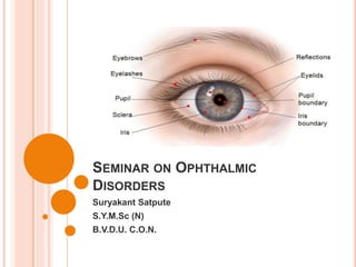
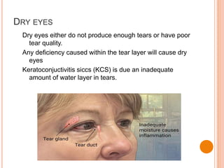
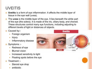
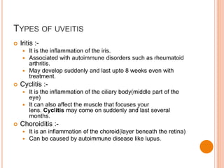
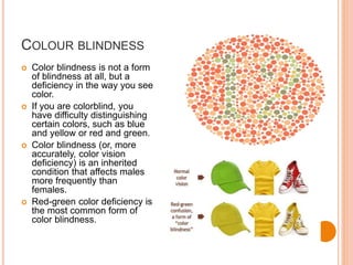
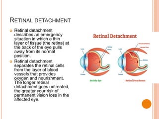
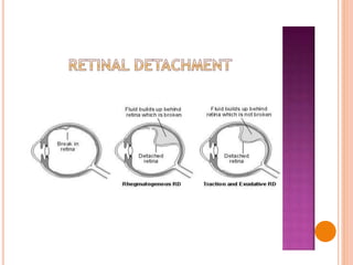
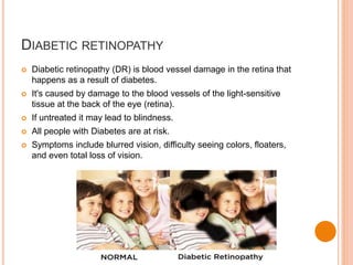
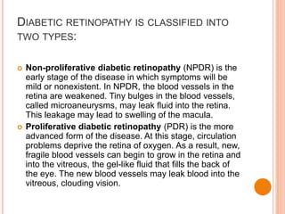
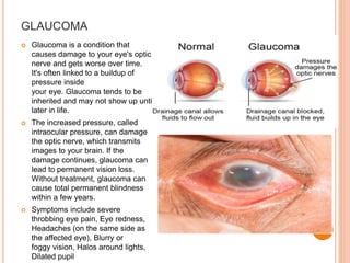
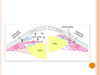
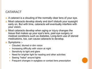
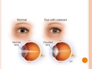
SEMINAR ON OPHTHALMIC
DISORDERS
Suryakant Satpute
S.Y.M.Sc (N)
B.V.D.U. C.O.N.
OUTLINE
 Anatomy and Physiology of the Eye.
 Disorders in the Accessory Structure.
 Dry Eyes
 Conjuctivitis
 Disorders in the Fibrous Tunic
 Keratoconus
 Refractive Errors
 Disorders in the Vascular Tunic
 Colour Blindness
 Retinal Detachment
 Diabetic Retinopathy
 Age related Macular Degenration
 Disorders in the Interior of the Eye
 Glaucoma
 Cataract
 Floaters
ANATOMY AND PHYSIOLOGY OF THE EYE
 Unlike most organs of the body, the eye is available for external
examination.
 The human eye can be distinguished into 3 layers:-
 The OUTER REGION consists of the cornea and the sclera. The cornea
refracts and transmits the light to the lens and the retina and protects the
eye against infection and structural damage to the deeper parts. The
sclera forms a connective tissue coat that protects the eye from internal
and external forces and maintains its shape. The visible part of the sclera
is covered by a transparent mucous membrane, the conjunctiva.
 The MIDDLE LAYER of the eye is composed of the iris, the ciliary body
and the choroid. The iris controls the size of the pupil, and thus the
amount of light reaching the retina; the ciliary body controls the power
and shape of the lens and is the site of aqueous production; and the
choroid is a vascular layer that provides oxygen and nutrients to the
outer retinal layers.
 The INNER LAYER of the eye is the retina, a complex, layered structure
of neurons that capture and process light. The three transparent
structures surrounded by the ocular layers are called the aqueous, the
vitreous and the lens.
ANATOMY AND PHYSIOLOGY
DRY EYES
Dry eyes either do not produce enough tears or have poor
tear quality.
Any deficiency caused within the tear layer will cause dry
eyes
Keratoconjuctivitis siccs (KCS) is due an inadequate
amount of water layer in tears.
CAUSES OF DRY EYE
 Treatment
 Medical treatment:
 Artificial tear substitute
 Anti-inflammatory agents
 Mucolytic agents
 Surgical treatment:
 Punctal plugs
 Permanent punctal occlusion Tarsorhaphy
 Repaire of eye lid malposition.
 Others:
 Warm compress
 Eye lid hygiene
CONJUNCTIVITIS (PINK EYE)
 Infection of conjunctiva
 Common in children and is highly contagious.
 Symptoms :
 Red eye
 Swelling of conjunctiva
 Watering of the eyes
 Itching
 Sensitive to light
TYPES OF CONJUNCTIVITIS
KERATOCONUS
 Keratoconus is a degenerative disorder.
 Structural changes within the cornea causes it to
thin and change into a more conical shape
REFRACTIVE ERRORS
A refractive error is a very common eye disorder.
It occurs when the eye cannot clearly focus the images
from the outside world. The shape of your eye does not
bend light correctly, resulting in a blurred image, which is
sometimes so severe that it causes visual impairment.
Symptoms :-
 Blurred vision.
 Reading problem.
 Inability to see close objects.
 Eye irritation or weakness in the eyes.
 Advanced signs include, headaches due to stress on vascular
eye muscle, squinting of eyes, eye fatigues, different
sensation, or excessive itchiness in your eyes.
UVEITIS
 Uveitis is a form of eye inflammation. It affects the middle layer of
tissue in the eye wall (uvea).
 The uvea is the middle layer of the eye. It lies beneath the white part
of the eye (the sclera). It is made of the iris, ciliary body, and choroid.
These structures control many eye functions, including adjusting to
different levels of light or distances of objects.
 Caused by:-
 Foreign organism
 Injury
 Inflammatory disease
 Symptoms :-
 Redness of eye
 Blurred vision
 Increased sensitivity to light
 Floating spots before the eye
 Treatment :-
 Steroid eye drop
 antibiotic
TYPES OF UVEITIS
 Iritis :-
 It is the inflammation of the iris.
 Associated with autoimmune disorders such as rheumatoid
arthritis.
 May develop suddenly and last upto 8 weeks even with
treatment.
 Cyclitis :-
 It is the inflammation of the ciliary body(middle part of the
eye)
 It can also affect the muscle that focuses your
lens. Cyclitis may come on suddenly and last several
months.
 Choroiditis :-
 It is an inflammation of the choroid(layer beneath the retina)
 Can be caused by autoimmune disease like lupus.
COLOUR BLINDNESS
 Color blindness is not a form
of blindness at all, but a
deficiency in the way you see
color.
 If you are colorblind, you
have difficulty distinguishing
certain colors, such as blue
and yellow or red and green.
 Color blindness (or, more
accurately, color vision
deficiency) is an inherited
condition that affects males
more frequently than
females.
 Red-green color deficiency is
the most common form of
color blindness.
RETINAL DETACHMENT
 Retinal detachment
describes an emergency
situation in which a thin
layer of tissue (the retina) at
the back of the eye pulls
away from its normal
position.
 Retinal detachment
separates the retinal cells
from the layer of blood
vessels that provides
oxygen and nourishment.
The longer retinal
detachment goes untreated,
the greater your risk of
permanent vision loss in the
affected eye.
DIABETIC RETINOPATHY
 Diabetic retinopathy (DR) is blood vessel damage in the retina that
happens as a result of diabetes.
 It's caused by damage to the blood vessels of the light-sensitive
tissue at the back of the eye (retina).
 If untreated it may lead to blindness.
 All people with Diabetes are at risk.
 Symptoms include blurred vision, difficulty seeing colors, floaters,
and even total loss of vision.
DIABETIC RETINOPATHY IS CLASSIFIED INTO
TWO TYPES:
 Non-proliferative diabetic retinopathy (NPDR) is the
early stage of the disease in which symptoms will be
mild or nonexistent. In NPDR, the blood vessels in the
retina are weakened. Tiny bulges in the blood vessels,
called microaneurysms, may leak fluid into the retina.
This leakage may lead to swelling of the macula.
 Proliferative diabetic retinopathy (PDR) is the more
advanced form of the disease. At this stage, circulation
problems deprive the retina of oxygen. As a result, new,
fragile blood vessels can begin to grow in the retina and
into the vitreous, the gel-like fluid that fills the back of
the eye. The new blood vessels may leak blood into the
vitreous, clouding vision.
AGE RELATED MACULAR DEGENERATION
(AMD)
 Macular degeneration is the leading cause of severe,
irreversible vision loss in people over age 60.
 It occurs when the small central portion of the retina, known as the
macula, deteriorates. The retina is the light-sensing nerve tissue at
the back of the eye.
 Because the disease develops as a person ages, it is often referred
to as age-related macular degeneration (AMD). Although macular
degeneration is almost never a totally blinding condition, it can be a
source of significant central visual disability.
GLAUCOMA
 Glaucoma is a condition that
causes damage to your eye's optic
nerve and gets worse over time.
It's often linked to a buildup of
pressure inside
your eye. Glaucoma tends to be
inherited and may not show up until
later in life.
 The increased pressure, called
intraocular pressure, can damage
the optic nerve, which transmits
images to your brain. If the
damage continues, glaucoma can
lead to permanent vision loss.
Without treatment, glaucoma can
cause total permanent blindness
within a few years.
 Symptoms include severe
throbbing eye pain, Eye redness,
Headaches (on the same side as
the affected eye), Blurry or
foggy vision, Halos around lights,
Dilated pupil
TYPES OF GLOUCOMA
 Open-angle glaucoma. It’s the most common type. Your
doctor may also call it wide-angle glaucoma. The drain
structure in your eye -- it’s called the trabecular meshwork --
looks normal, but fluid doesn’t flow out like it should.
 Angle-closure glaucoma. It’s less common in the West than
in Asia. You may also hear it called acute or chronic angle-
closure or narrow-angle glaucoma. Your eye doesn’t drain
right because the drain space between your iris
and cornea becomes too narrow. This can cause a sudden
buildup of pressure in your eye. It’s also linked
to farsightedness and cataracts, a clouding of the lens inside
your eye.
 Treatment :-
 Eye drops
 Iridotomy
 Microsurgery (trabeculectomy)
CATARACT
 A cataract is a clouding of the normally clear lens of your eye.
 Most cataracts develop slowly and don't disturb your eyesight
early on. But with time, cataracts will eventually interfere with
your vision.
 Most cataracts develop when aging or injury changes the
tissue that makes up your eye's lens, past eye surgery or
medical conditions such as diabetes. Long-term use of steroid
medications, too, can cause cataracts to develop.
 Symptoms :-
 Clouded, blurred or dim vision
 Increasing difficulty with vision at night
 Sensitivity to light and glare
 Need for brighter light for reading and other activities
 Seeing "halos" around lights
 Frequent changes in eyeglass or contact lens prescription
Bibliography
 Text book of medical surgical nursing , B.
Venkatessanand Emmess medical publishers,
page-547,548.
 Text book of medical surgical nursing, brunner and
siddharth’s, 12th edition, wolters publication. Page –
1365,1367.
 Future nurses, success in a nut shell second part of
success in nutshell, zodiac publication, page – 68.
Seminar on Ophthalmic Disorders

Seminar on Ophthalmic Disorders

  • 1.
    SEMINAR ON OPHTHALMIC DISORDERS SuryakantSatpute S.Y.M.Sc (N) B.V.D.U. C.O.N.
  • 2.
    OUTLINE  Anatomy andPhysiology of the Eye.  Disorders in the Accessory Structure.  Dry Eyes  Conjuctivitis  Disorders in the Fibrous Tunic  Keratoconus  Refractive Errors  Disorders in the Vascular Tunic  Colour Blindness  Retinal Detachment  Diabetic Retinopathy  Age related Macular Degenration  Disorders in the Interior of the Eye  Glaucoma  Cataract  Floaters
  • 3.
    ANATOMY AND PHYSIOLOGYOF THE EYE  Unlike most organs of the body, the eye is available for external examination.  The human eye can be distinguished into 3 layers:-  The OUTER REGION consists of the cornea and the sclera. The cornea refracts and transmits the light to the lens and the retina and protects the eye against infection and structural damage to the deeper parts. The sclera forms a connective tissue coat that protects the eye from internal and external forces and maintains its shape. The visible part of the sclera is covered by a transparent mucous membrane, the conjunctiva.  The MIDDLE LAYER of the eye is composed of the iris, the ciliary body and the choroid. The iris controls the size of the pupil, and thus the amount of light reaching the retina; the ciliary body controls the power and shape of the lens and is the site of aqueous production; and the choroid is a vascular layer that provides oxygen and nutrients to the outer retinal layers.  The INNER LAYER of the eye is the retina, a complex, layered structure of neurons that capture and process light. The three transparent structures surrounded by the ocular layers are called the aqueous, the vitreous and the lens.
  • 4.
  • 8.
    DRY EYES Dry eyeseither do not produce enough tears or have poor tear quality. Any deficiency caused within the tear layer will cause dry eyes Keratoconjuctivitis siccs (KCS) is due an inadequate amount of water layer in tears.
  • 10.
  • 11.
     Treatment  Medicaltreatment:  Artificial tear substitute  Anti-inflammatory agents  Mucolytic agents  Surgical treatment:  Punctal plugs  Permanent punctal occlusion Tarsorhaphy  Repaire of eye lid malposition.  Others:  Warm compress  Eye lid hygiene
  • 12.
    CONJUNCTIVITIS (PINK EYE) Infection of conjunctiva  Common in children and is highly contagious.  Symptoms :  Red eye  Swelling of conjunctiva  Watering of the eyes  Itching  Sensitive to light
  • 13.
  • 15.
    KERATOCONUS  Keratoconus isa degenerative disorder.  Structural changes within the cornea causes it to thin and change into a more conical shape
  • 17.
    REFRACTIVE ERRORS A refractiveerror is a very common eye disorder. It occurs when the eye cannot clearly focus the images from the outside world. The shape of your eye does not bend light correctly, resulting in a blurred image, which is sometimes so severe that it causes visual impairment. Symptoms :-  Blurred vision.  Reading problem.  Inability to see close objects.  Eye irritation or weakness in the eyes.  Advanced signs include, headaches due to stress on vascular eye muscle, squinting of eyes, eye fatigues, different sensation, or excessive itchiness in your eyes.
  • 21.
    UVEITIS  Uveitis isa form of eye inflammation. It affects the middle layer of tissue in the eye wall (uvea).  The uvea is the middle layer of the eye. It lies beneath the white part of the eye (the sclera). It is made of the iris, ciliary body, and choroid. These structures control many eye functions, including adjusting to different levels of light or distances of objects.  Caused by:-  Foreign organism  Injury  Inflammatory disease  Symptoms :-  Redness of eye  Blurred vision  Increased sensitivity to light  Floating spots before the eye  Treatment :-  Steroid eye drop  antibiotic
  • 22.
    TYPES OF UVEITIS Iritis :-  It is the inflammation of the iris.  Associated with autoimmune disorders such as rheumatoid arthritis.  May develop suddenly and last upto 8 weeks even with treatment.  Cyclitis :-  It is the inflammation of the ciliary body(middle part of the eye)  It can also affect the muscle that focuses your lens. Cyclitis may come on suddenly and last several months.  Choroiditis :-  It is an inflammation of the choroid(layer beneath the retina)  Can be caused by autoimmune disease like lupus.
  • 24.
    COLOUR BLINDNESS  Colorblindness is not a form of blindness at all, but a deficiency in the way you see color.  If you are colorblind, you have difficulty distinguishing certain colors, such as blue and yellow or red and green.  Color blindness (or, more accurately, color vision deficiency) is an inherited condition that affects males more frequently than females.  Red-green color deficiency is the most common form of color blindness.
  • 25.
    RETINAL DETACHMENT  Retinaldetachment describes an emergency situation in which a thin layer of tissue (the retina) at the back of the eye pulls away from its normal position.  Retinal detachment separates the retinal cells from the layer of blood vessels that provides oxygen and nourishment. The longer retinal detachment goes untreated, the greater your risk of permanent vision loss in the affected eye.
  • 27.
    DIABETIC RETINOPATHY  Diabeticretinopathy (DR) is blood vessel damage in the retina that happens as a result of diabetes.  It's caused by damage to the blood vessels of the light-sensitive tissue at the back of the eye (retina).  If untreated it may lead to blindness.  All people with Diabetes are at risk.  Symptoms include blurred vision, difficulty seeing colors, floaters, and even total loss of vision.
  • 28.
    DIABETIC RETINOPATHY ISCLASSIFIED INTO TWO TYPES:  Non-proliferative diabetic retinopathy (NPDR) is the early stage of the disease in which symptoms will be mild or nonexistent. In NPDR, the blood vessels in the retina are weakened. Tiny bulges in the blood vessels, called microaneurysms, may leak fluid into the retina. This leakage may lead to swelling of the macula.  Proliferative diabetic retinopathy (PDR) is the more advanced form of the disease. At this stage, circulation problems deprive the retina of oxygen. As a result, new, fragile blood vessels can begin to grow in the retina and into the vitreous, the gel-like fluid that fills the back of the eye. The new blood vessels may leak blood into the vitreous, clouding vision.
  • 29.
    AGE RELATED MACULARDEGENERATION (AMD)  Macular degeneration is the leading cause of severe, irreversible vision loss in people over age 60.  It occurs when the small central portion of the retina, known as the macula, deteriorates. The retina is the light-sensing nerve tissue at the back of the eye.  Because the disease develops as a person ages, it is often referred to as age-related macular degeneration (AMD). Although macular degeneration is almost never a totally blinding condition, it can be a source of significant central visual disability.
  • 31.
    GLAUCOMA  Glaucoma isa condition that causes damage to your eye's optic nerve and gets worse over time. It's often linked to a buildup of pressure inside your eye. Glaucoma tends to be inherited and may not show up until later in life.  The increased pressure, called intraocular pressure, can damage the optic nerve, which transmits images to your brain. If the damage continues, glaucoma can lead to permanent vision loss. Without treatment, glaucoma can cause total permanent blindness within a few years.  Symptoms include severe throbbing eye pain, Eye redness, Headaches (on the same side as the affected eye), Blurry or foggy vision, Halos around lights, Dilated pupil
  • 32.
    TYPES OF GLOUCOMA Open-angle glaucoma. It’s the most common type. Your doctor may also call it wide-angle glaucoma. The drain structure in your eye -- it’s called the trabecular meshwork -- looks normal, but fluid doesn’t flow out like it should.  Angle-closure glaucoma. It’s less common in the West than in Asia. You may also hear it called acute or chronic angle- closure or narrow-angle glaucoma. Your eye doesn’t drain right because the drain space between your iris and cornea becomes too narrow. This can cause a sudden buildup of pressure in your eye. It’s also linked to farsightedness and cataracts, a clouding of the lens inside your eye.  Treatment :-  Eye drops  Iridotomy  Microsurgery (trabeculectomy)
  • 34.
    CATARACT  A cataractis a clouding of the normally clear lens of your eye.  Most cataracts develop slowly and don't disturb your eyesight early on. But with time, cataracts will eventually interfere with your vision.  Most cataracts develop when aging or injury changes the tissue that makes up your eye's lens, past eye surgery or medical conditions such as diabetes. Long-term use of steroid medications, too, can cause cataracts to develop.  Symptoms :-  Clouded, blurred or dim vision  Increasing difficulty with vision at night  Sensitivity to light and glare  Need for brighter light for reading and other activities  Seeing "halos" around lights  Frequent changes in eyeglass or contact lens prescription
  • 36.
    Bibliography  Text bookof medical surgical nursing , B. Venkatessanand Emmess medical publishers, page-547,548.  Text book of medical surgical nursing, brunner and siddharth’s, 12th edition, wolters publication. Page – 1365,1367.  Future nurses, success in a nut shell second part of success in nutshell, zodiac publication, page – 68.