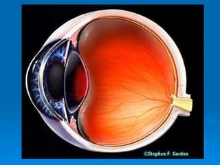
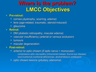
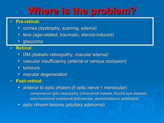
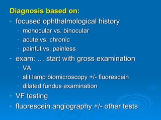
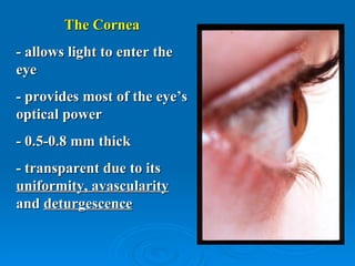
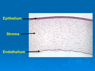
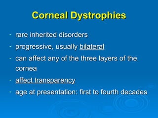
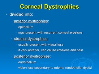
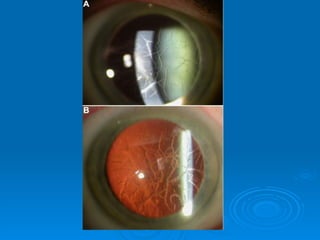
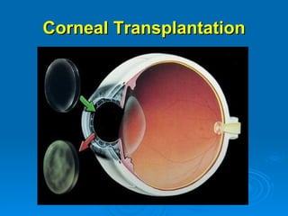
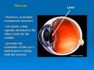
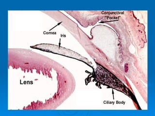
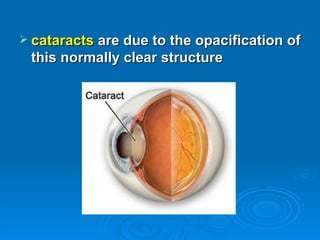
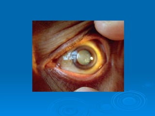
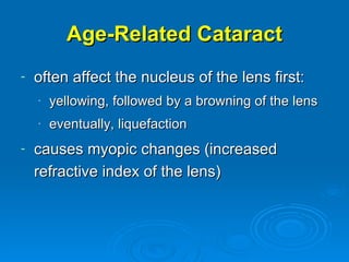
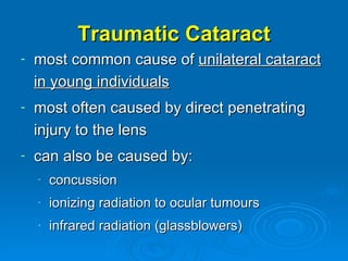
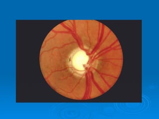
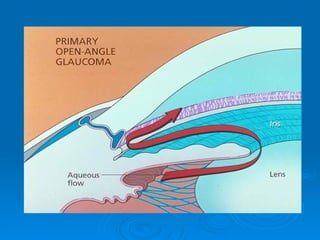
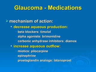
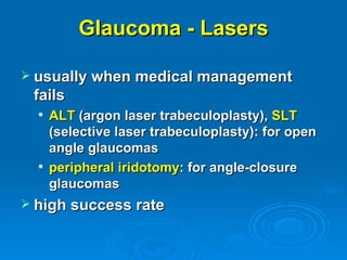
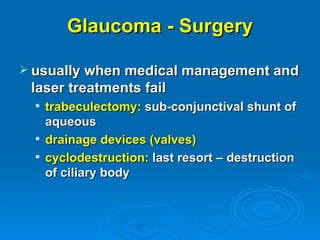
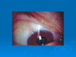
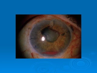
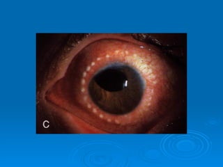
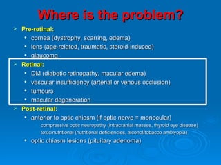
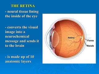
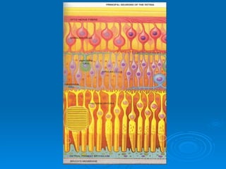
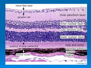
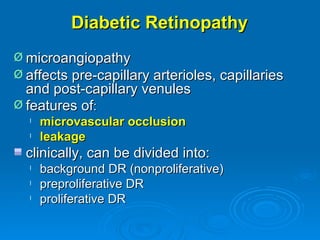
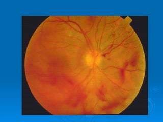
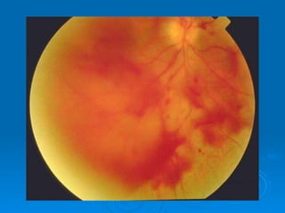
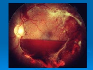
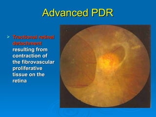
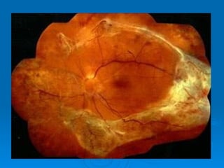
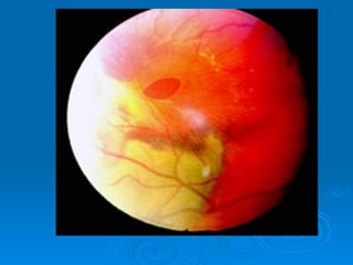
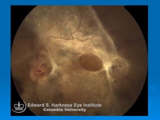
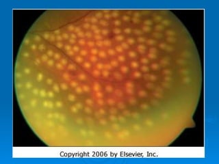
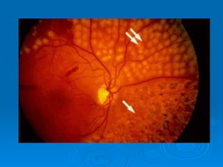
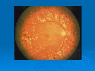
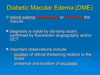
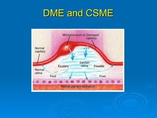
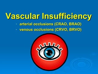
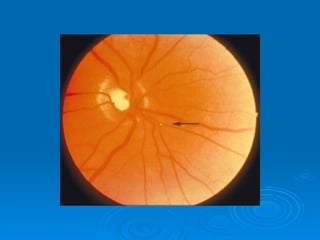
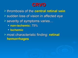
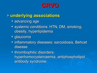
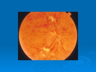
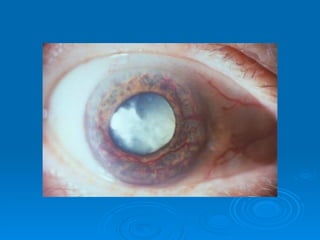
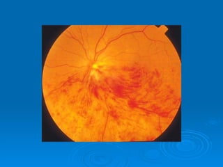
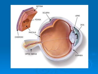
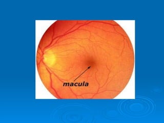
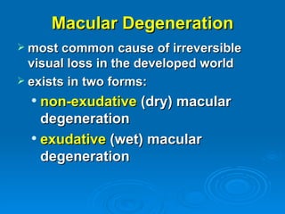
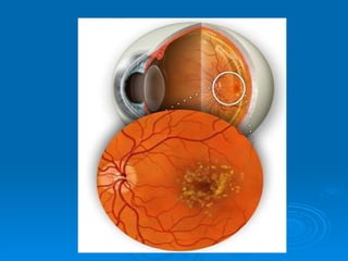
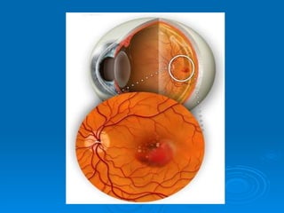
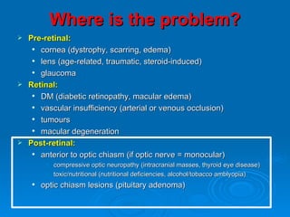
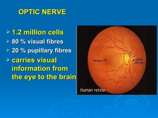
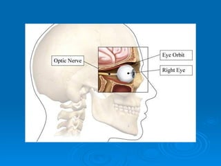
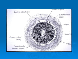
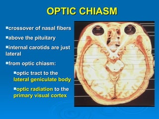
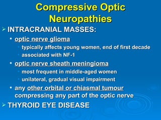
Chronic Visual
Disturbance and Visual
        Loss
          Setareh Ziai
          April 2nd, 2009
    sziai@ottawahospital.on.ca
QUICK review
Basic Anatomy
Where is the problem?
              LMCC Objectives
 Pre-retinal:
    cornea (dystrophy, scarring, edema)
    lens (age-related, traumatic, steroid-induced)
    glaucoma
 Retinal:
    DM (diabetic retinopathy, macular edema)
    vascular insufficiency (arterial or venous occlusion)
    tumours
    macular degeneration
 Post-retinal:
    anterior to optic chiasm (if optic nerve = monocular)
         • compressive optic neuropathy (intracranial masses, thyroid eye disease)
         • toxic/nutritional (nutritional deficiencies, alcohol/tobacco amblyopia)
       optic chiasm lesions (pituitary adenoma)
Where is the problem?
 Pre-retinal:
    cornea (dystrophy, scarring, edema)
    lens (age-related, traumatic, steroid-induced)
    glaucoma
 Retinal:
    DM (diabetic retinopathy, macular edema)
    vascular insufficiency (arterial or venous occlusion)
    tumours
    macular degeneration
 Post-retinal:
    anterior to optic chiasm (if optic nerve = monocular)
         • compressive optic neuropathy (intracranial masses, thyroid eye disease)
         • toxic/nutritional (nutritional deficiencies, alcohol/tobacco amblyopia)
       optic chiasm lesions (pituitary adenoma)
Diagnosis based on:
- focused ophthalmological history
    -   monocular vs. binocular
    -   acute vs. chronic
    -   painful vs. painless
-   exam: … start with gross examination
    -   VA
    -   slit lamp biomicroscopy +/- fluorescein
    -   dilated fundus examination
- VF testing
- fluorescein angiography +/- other tests
 **Remember for exam:
     sometimes, chronic visual loss in ONE eye is
      noted incidentally some time later due to
      occlusion of normal eye…: CHRONIC LOSS
      OF VISION CAN PRESENT ACUTELY!!
Corneal Causes
    -    dystrophy
        - scarring
         - edema
The Cornea
- allows light to enter the
eye
- provides most of the eye’s
optical power
- 0.5-0.8 mm thick
- transparent due to its
uniformity, avascularity
and deturgescence
Epithelium


 Stroma


Endothelium
Corneal Dystrophies
-   rare inherited disorders
-   progressive, usually bilateral
-   can affect any of the three layers of the
    cornea
-   affect transparency
-   age at presentation: first to fourth decades
Corneal Dystrophies
-   divided into:
    -   anterior dystrophies:
         - epithelium
         - may present with recurrent corneal erosions
    -   stromal dystrophies:
         - usually present with visual loss
         - if very anterior, can cause erosions and pain
    -   posterior dystrophies:
         - endothelium
         - vision loss secondary to edema (endothelial dysfx)
Corneal Scarring
-   multiple causes:
    -   trauma
    -   infectious (eg., herpes)
    -   post-surgical
Corneal Edema
-   most often caused by dysfunction of the
    corneal endothelium:
    -   dystrophy
    -   trauma
    -   infectious (eg., herpes)
    -   post-surgical
Corneal
Transplantation
If the corneal stroma opacifies due to
   trauma or infection, or if there is
   swelling or an irregularity of the
   surface of the cornea, light cannot
   properly reach the retina.

In some cases, a cornea from a
  deceased donor can be transplanted.
Corneal Transplantation
Lens-Related Causes
      (cataract)
         - age-related
          - traumatic
     -   steroid induced
The Lens
                           Lens


- biconvex, avascular,
transparent structure
- sits inside a thin
capsule, attached to the
ciliary body by the
zonules
- provides the
remainder of the eye’s
optical power (along
with the cornea)
 cataracts are due to the opacification of
 this normally clear structure
Age-Related Cataract
-   often affect the nucleus of the lens first:
    -   yellowing, followed by a browning of the lens
    -   eventually, liquefaction
-   causes myopic changes (increased
    refractive index of the lens)
Traumatic Cataract
-   most common cause of unilateral cataract
    in young individuals
-   most often caused by direct penetrating
    injury to the lens
-   can also be caused by:
    -   concussion
    -   ionizing radiation to ocular tumours
    -   infrared radiation (glassblowers)
Steroid-Induced Cataract
-   both systemic and topical steroids can be
    the culprits
-   posterior part of lens affected first
-   children may be more susceptible
-   if lens changes develop, dose should be
    reduced to the minimum necessary
-   early opacities may regress with
    discontinuation of therapy
Glaucoma
Glaucoma

 disease of the optic nerve, often
 caused by an increase in intraocular
 pressure due to poor drainage of
 aqueous from the trabecular
 meshwork…
Glaucoma

 if left untreated, glaucoma can lead to
  permanent damage to the optic nerve
  and resultant visual field loss
 can progress to blindness
Glaucoma

 by definition, glaucoma is a trimodal
 disease, characterized by:

             increased IOP
          optic nerve changes

          visual field changes
Goldmann Applanation Tonometer
Glaucoma
 classification:


     primary: open-angle, angle-closure
     secondary: inflammatory, traumatic,
      neovascular, steroid-induced etc…
     congenital
Risk Factors for Glaucoma

 age
 african-american heritage
 high IOP
 family history
 myopia
Symptoms of Glaucoma

 often asymptomatic
 with late disease, constriction of
  peripheral, and later central visual field
 with very high IOP, can have blurry
  vision and halos around lights
Glaucoma: Optic Nerve
          Changes
 increased cup:disc ratio
 thinning of neural rim
 progressive loss of nerve fiber layer
 flame hemorrhages on disc
Primary Open Angle
           Glaucoma
 most common (90%)
 usually bilateral (can be asymmetric)
 prevalence increases with age
 angle is open, eye is quiet
 increased resistance to aqueous drainage
 at the level of the trabecular meshwork is
 thought to be the main pathophysiologic
 feature
Treatment options

 goal is to stabilize the IOP to protect
  the optic nerve against further damage
 options:
     drops
     laser
     surgery
Glaucoma - Medications

 mechanism of action:
     decrease aqueous production:
      • beta blockers: timolol
      • alpha agonists: brimonidine
      • carbonic anhydrase inhibitors: diamox
     increase aqueous outflow:
      •   miotics: pilocarpine
      •   epinephrine
      •   prostaglandin analogs: latanoprost
Glaucoma - Lasers

 usually when medical management
 fails
     ALT (argon laser trabeculoplasty), SLT
      (selective laser trabeculoplasty): for open
      angle glaucomas
     peripheral iridotomy: for angle-closure
      glaucomas
 high success rate
Glaucoma - Surgery

 usually when medical management and
 laser treatments fail
     trabeculectomy: sub-conjunctival shunt of
      aqueous
     drainage devices (valves)
     cyclodestruction: last resort – destruction
      of ciliary body
Where is the problem?
 Pre-retinal:
    cornea (dystrophy, scarring, edema)
    lens (age-related, traumatic, steroid-induced)
    glaucoma
 Retinal:
    DM (diabetic retinopathy, macular edema)
    vascular insufficiency (arterial or venous occlusion)
    tumours
    macular degeneration
 Post-retinal:
    anterior to optic chiasm (if optic nerve = monocular)
         • compressive optic neuropathy (intracranial masses, thyroid eye disease)
         • toxic/nutritional (nutritional deficiencies, alcohol/tobacco amblyopia)
       optic chiasm lesions (pituitary adenoma)
THE RETINA
- neural tissue lining
the inside of the eye

- converts the visual
image into a
neurochemical
message and sends it
to the brain

- is made up of 10
anatomic layers
Diabetes
    - diabetic retinopathy
-   diabetic macular edema
Diabetic Retinopathy
Ø microangiopathy
Ø affects pre-capillary arterioles, capillaries
  and post-capillary venules
Ø features of:
  l   microvascular occlusion
  l   leakage
  clinically, can be divided into:
  l   background DR (nonproliferative)
  l   preproliferative DR
  l   proliferative DR
Diabetic Retinopathy: Epidemiology
    239 million people by 2010
    doubling in prevalence since 1994
       diabetes will affect:
           28 million in western Europe
          18.9 million in North America
          138.2 million in Asia
          1.3 million in Australasia


•   #1 cause of blindness in patients 20-64 yrs
•   prevalence increases with duration of diabetes and
    patient age

       rare to find DR in children < 10 yrs, regardless of duration
       risk of developing DR increases after puberty
Epidemiology
Wisconsin Epidemiologic Study of Diabetic Retinopathy
l Between 1979-1980
l 1210 patients with Type 1
l 1780 patients with Type 2
l predominantly white population


l   After 20 yrs, DR present in:
     • 99% of Type 1
     • 60% of Type 2
WESDR: Frequency of retinopathy in
  subjects with type 1diabetes
WESDR: Frequency of retinopathy in
  subjects with type 2 diabetes
Diabetic Retinopathy: Risk Factors
Ø duration of diabetes: most important risk
  factor
Ø poor metabolic control
Ø pregnancy: can be associated with rapid
  progression
Ø HTN
Ø nephropathy
Ø smoking
Ø obesity
Ø hyperlipidemia
Classification of Diabetic Retinopathy
Ø   Classified into 2 stages

        Nonproliferative Diabetic Retinopathy (NPDR)
          early stage
          also known as background DR (BDR)
          further categorized based upon extent of DR
            l mild, moderate, severe, very severe


    l   Proliferative Diabetic Retinopathy (PDR)
         • more advanced stage

    •   ***Macular edema
         • May be present at any stage of DR
NPDR
Ø typically asymptomatic
  l   fluctuating visual acuity:
        • fluctuating blood sugar
  l   decreased visual acuity:
          CSME
          macular ischemia
 review these patients annually
Mild NPDR
Microaneurysm
Moderate NPDR
Severe NPDR
Proliferative Diabetic Retinopathy
Ø   more likely to become
    symptomatic than early
    NPDR

Ø   may have decreased
    vision, sudden vision loss,
    floaters, cobwebs, flashes,
    dull eye ache

Ø   PDR can also affect visual
    function by affecting the
    macula with resulting
    macular ischemia and/or
    edema
Proliferative DR
Ø   affects 5-10% of the diabetic population
Ø   neovascularization is the hallmark
     l NVD: neovascularization of the disc

     l NVE: neovascularization elsewhere


Ø   new vessels are not only extremely fragile
    (intraretinal or vitreous hemorrhage), but
    often associated with fibrous proliferation,
    leading to an increased risk of tractional retinal
    detachment
Advanced PDR

   Tractional retinal
    detachment
    resulting from
    contraction of
    the fibrovascular
    proliferative
    tissue on the
    retina
Panretinal Photocoagulation for High-risk
                     PDR
 goal is to induce
  involution (or at
  least arrest) of new
  vessels by creating
  areas of retinal
  ischemia
 1200-3000 burns
 4 sessions
Vitrectomy for Vitreous
  Hemorrhage / TRD
Diabetic Macular Edema (DME)
Ø   retinal edema threatening or involving the
    macula

    diagnosis is made by slit-lamp exam,
    confirmed by fluorescein angiography and/or
    OCT

    important observations include:
     l location of retinal thickening relative to the

       fovea
     l presence and location of exudates
DME and CSME
Treatment of CSME
   argon laser application

   intravitreal steroid injection

   intravitreal anti-VEGF injection

   pars plana vitrectomy
Ophthalmological Follow-Up
   Diabetic Screening

    l   Type 1 diabetics:
         Dilated funduscopic exam (DFE) 5 yrs after
          diagnosis
         Newly diagnosed patients with Type 1 diabetes
          rarely have retinopathy during the first 5 yrs

    l   Type 2 diabetics:
          Type 2 diabetics typically diagnosed yrs after initial
          onset
          DFE at the time of diagnosis
          Significant portion of newly diagnosed Type 2
          diabetics have established DR at the time of
          diagnosis
Vascular Insufficiency
  -   arterial occlusions (CRAO, BRAO)
  -   venous occlusions (CRVO, BRVO)
CRAO
CRAO
 most of the retina is supplied by the
  central retinal artery (branch of the
  ophthalmic artery, which is the first branch
  of the ICA)
 if this supply is interrupted (embolus,
  thrombosis, inflammation, vasculitis or
  compression), the retina becomes
  ischemic
 irreversible damage occurs after
  approximately 90 minutes
CRAO
 presentation is with sudden and
  profound loss of vision
 RAPD is present
 orange reflex from the choroid stands
  out at the fovea, and contrasts with the
  surrounding pale retina (cherry-red
  spot)
 must r/o temporal arteritis
CRAO
 most commonly the result of
  atherosclerosis (thrombosis) but may
  also be caused by calcific emboli
 often in older patients, with a hx of
  arteriosclerosis
 may have had a hx of amaurosis fugax
  (transient visual loss)
CRAO
   OPHTHALMOLOGIC EMERGENCY!!
   treatment:
      decrease IOP

      paracentesis

      ocular massage


   goal: to send the embolus distally
   **remember to r/o giant cell arteritis! (ESR, CRP, plt)
   poor prognosis: 60% < 20/400
BRAO
BRAO
 sudden and profound     altitudinal or
  sectoral visual field loss
 similar causes as CRAO
 identify and treat associated medical
  conditions (HTN, DM,
  hypercholesterolemia, smoking,
  vasculitis etc…)
BRAO
 retinal cloudiness in ischemic area
 +/- visible embolus
    also has a poor prognosis, unless the
    obstruction can be dislodged within a
    few hours
CRVO
CRVO
 thrombosis of the central retinal vein
 sudden loss of vision in affected eye
 severity of symptoms varies…
     non-ischemic: 75%
     Ischemic
 most characteristic finding:   retinal
 hemorrhages
CRVO
 underlying associations
     advancing age
     systemic conditions: HTN, DM, smoking,
      obesity, hyperlipidemia
     glaucoma
     inflammatory diseases: sarcoidosis, Behcet
      disease
     thrombophilic disorders:
      hyperhomocysteinaemia, antiphospholipid
      antibody syndrome
CRVO
 Treatment:
     treat associated medical conditions
     decrease IOP if elevated
     pan-retinal photocoagulation
      (laser) if:
       • neovascularization (iris, angle,
         retina)
       • …especially if ischemic CRVO
BRVO
BRVO
 thrombosis of a branch of the        central
  retinal vein
 visual loss depends on the amount of
  macular drainage compromised by the
  occlusion (peripheral occlusions may be
  asymptomatic)
 characteristic findings in one sector of the
  retina:
     dilatation and tortuosity of veins
     retinal hemorrhages
     retinal/macular edema
  
BRVO
 obstruction often at arterio-venous
  crossings: arteries and veins share
  adventitial sheath… thickening of the
  arteriole (arteriosclerosis) compresses
  the vein, eventually causing an occlusion
 often associated with:
    hypertension (75%)

    diabetes (10%)
BRVO
 prognosis: depends on amt of venous
  drainage involved by the occlusion and
  severity of macular ischemia: within 6 mos,
  about 50% of eyes have a VA of 20/30 or
  better
 main complications:
     chronic macular edema
     neovascularization
 laser photocoagulation may be helpful in
 above cases
Retinal Tumours
 ocular tumours:
     ciliary body:
       • melanoma
     choroid:
       •   melanoma
       •   hemangioma
       •   metastases
     primary ocular lymphoma
     retina and optic nerve:
       •   retinoblastoma
       •   astrocytoma
       •   hemangioma
Choroidal Melanoma
 most common primary intraocular
  tumour in adults
 presentation usually in 6th decade:
       asymptomatic vs. visual field defect and/or
        decreased visual acuity
   signs:
      raised, usually pigmented lesion visible at

       the back of the eye
      may be associated with retinal detachment

      optic nerve may be involved
Choroidal Melanoma
 treatment:
     consider size, location, activity of tumour,
      state of fellow eye, general health/age of pt,
      pt’s wishes/fears
       • brachytherapy
       • external radiotherapy
       • transpupillary thermotherapy
       • local resection
       • enucleation
       • exenteration
       • palliative (may include chemo)
Choroidal Metastases
 …with choroidal melanoma, don’t forget
 general medical investigations!
     mets TO the choroid:
      • most frequently from bronchus in both sexes
        and the breast in women, rarely kidney or GI
            CXR, rectal exam, mammography
     mets FROM the choroid:
      • liver
            hepatic u/s, GGT, ALP
      • lungs (rarely affected before liver)
            CXR
Choroidal Metastases
 usually present with visual impairment
  only IF tumour is near the macula
 signs:
     fast-growing, creamy coloured lesion
     most often in posterior pole
     usually not very elevated (infiltrates laterally)
Choroidal Metastases
 treatment:
     observe: if asxic or receiving systemic chemo
     radiation: external beam or brachy
     transpupillary thermotherapy
     systemic therapy for the primary
     enucleation: for painful blind eye
 prognosis is poor…
     median survival: 8-12 mos for all pts, 15-17
      mos for those with breast ca
Retinoblastoma
 most common malignant tumour of the eye
  in childhood (1:20 000)
 mean age of presentation: 8 mos if
  inherited, 25 mos if sporadic
      60% present with leukocoria (white pupillary
      reflex)
     strabismus (20%)
     occasionally: painful, red eye
     if inherited: often bilateral
Retinoblastoma
 malignant transformation of primitive
 retinal cells before their final differentiation

 can be caused by germinal mutations (can
 be passed on to the next generation), or
 can be sporadic (66% of cases)
Retinoblastoma
 this is a clinical diagnosis, but CSF and
  bone marrow should be examined to check
  for metastatic disease if ON involved or if
  there is evidence of extraocular extension
 rx:
     small: cryotherapy, photocoagulation
     medium: brachytherapy, external beam, chemo
     large/advanced cases: chemoreduction + local
      treatment, enucleation
     metastatic disease: chemo (intrathecal if cells
      in CSF)
Retinoblastoma
 prognosis:
     depends on extent of disease at diagnosis
     overall mortality ~ 5-15%
     ~ 50% of children with the germinal mutation
      will eventually develop a second primary
      tumour (eg., osteosarcoma of the femur or
      pinealoblastoma)
Macular Degeneration
Macula
 1.5 mm in diameter
 central vision: BEST VISUAL ACUITY
 colour vision
 progressive destruction of the macular
 area:
   MACULAR DEGENERATION
Macular Degeneration
 most common cause of irreversible
  visual loss in the developed world
 exists in two forms:
   non-exudative (dry) macular
    degeneration
   exudative (wet) macular

    degeneration
Non-exudative Macular
           Degeneration
 lipid products arising from
 photoreceptor outer segments are
 found under retina
     can be seen with ophthalmoscope!
     called « drusen »
Exudative Macular
         Degeneration
 new vessels from the choroid grow into
 the sub-retinal space; form a sub-
 retinal neovascular membrane

 subsequent hemorrhage into the sub-
 retinal space or even through the retina
 into the vitreous is associated with
 profound loss of vision
Macular Degeneration
 symptoms:
    since fovea is responsible for fine visual
     resolution, any disruption will cause
     severe visual impairment
      • blurry/reduced vision
      • distorted vision (metamorphopsia)
      • reduction (micropsia) or enlargement
        (macropsia) of objects
      • VF loss (scotoma)
Macular Degeneration
 rx:
     non-exudative (usually slowly
      progressive):
       • no actual medical treatment
       • use low vision aids
       • high dose antioxidants MAY be
         beneficial (eg., vitalux)
Macular Degeneration
 rx:
     exudative (can be rapidly progressive and
      devastating):
       • intravitreal injections of anti-VEGF
         factors: bevacizumab, ranibizumab
       • photodynamic therapy (injection of
         photosensitizer into systemic circulation
         followed immediately by laser targeting
         new vessels in macular area)
       • combination of above treatments
Where is the problem?
 Pre-retinal:
    cornea (dystrophy, scarring, edema)
    lens (age-related, traumatic, steroid-induced)
    glaucoma
 Retinal:
    DM (diabetic retinopathy, macular edema)
    vascular insufficiency (arterial or venous occlusion)
    tumours
    macular degeneration
 Post-retinal:
    anterior to optic chiasm (if optic nerve = monocular)
         • compressive optic neuropathy (intracranial masses, thyroid eye disease)
         • toxic/nutritional (nutritional deficiencies, alcohol/tobacco amblyopia)
       optic chiasm lesions (pituitary adenoma)
OPTIC NERVE

 1.2 million cells
   80 % visual fibres
   20 % pupillary fibres
 carries visual
    information from
    the eye to the brain
OPTIC CHIASM
crossover     of nasal fibers
above     the pituitary
internal   carotids are just
lateral
from     optic chiasm:
   optic  tract to the
   lateral geniculate body
   opticradiation to the
   primary visual cortex
Anterior to Optic
        Chiasm
    -     compressive optic neuropathies
-       toxic/nutritional optic neuropathies
Compressive Optic
              Neuropathies
 INTRACRANIAL MASSES:
    optic nerve glioma
     • typically affects young women, end of first decade
     • associated with NF-1
    optic nerve sheath meningioma
     • most frequent in middle-aged women
     • unilateral, gradual visual impairment
    any other orbital or chiasmal tumour
     compressing any part of the optic nerve
 THYROID EYE DISEASE
Thyroid Eye Disease
 may occur in the absence of biochemical
  evidence of thyroid dysfx
 autoimmune reaction (IgG Abs) causing:
     inflammation of EOMs: pleiomorphic cellular
      infiltration associated with increased secretion
      of GAGs and osmotic imbibition of water
       • muscles can become up to 8 times their original
         size!!
 no relation to severity of thyroid dysfx!
Thyroid Eye Disease
 main findings: (not all are always present!)
     soft tissue involvement
     lid retraction
     proptosis
     optic neuropathy
     restrictive myopathy
Thyroid Eye Disease
 vision loss from:
     exposure keratopathy
      • due to severe proptosis resulting in incomplete lid
        closure → chronically exposed cornea → corneal
        ulceration & exposure keratopathy
     optic neuropathy
      • affects 5% of pts
      • compression of ON or its blood supply by
        congested (enlarged) EOMs
      • can lead to severe, permanent visual impairment
      • rx with steroids, surgery if needed
Toxic/Nutritional Optic
            Neuropathies
 nutritional deficiencies
 alcohol-tobacco amblyopia
Nutritional Deficiencies
   pts with extremely poor diets, often in association
    with alcohol-tobacco amblyopia
   usually due to B12 deficiency in combination with
    cyanide toxicity
   symmetrical VF loss
   if early, can be treated with high-dose vitamins
    and restoration of « well-balanced diet »
   eventually leads to optic atrophy and permanent
    vision loss
Alcohol-Tobacco Amblyopia
   affects heavy drinkers, cigar and pipe smokers: deficient in
    protein and the B vitamins
   symptoms: insidious, bilateral, progressive visual
    impairment + dyschromatopsia
   signs: symmetrical VF defect, may have pale (or normal)
    discs
   rx: 1000 units of hydroxocobalamin qweekly X 10 wks +
    multivitamins + « well-balanced diet »
   px:
      good in early cases if comply with rx

      advanced cases: optic atrophy and permanent visual

       loss
Optic Chiasm Lesions
     -   pituitary adenoma
Pituitary Adenoma
 presentation usually in early adult life or
  middle age
 symptoms:
     h/a
     visual symptoms: very gradual onset (often
      not noticed by pt until very well-established)
       • VF defect: usually, bitemporal hemianopia, worst in
         the superior field, and extending inferiorly
       • colour desaturation across vertical midline
       • optic atrophy: in 50% of cases with field defects
         caused by pituitary lesions
Pituitary Adenoma
 investigations:
     MRI: coronal, axial and sagittal sections before
      and after gadolinium injection
     CT: demonstrates enlargement or erosion of
      the sella
     endocrinological investigation: PRL, FSH, TSH,
      GH
Pituitary Adenoma
 treatment options:
     observation
     medical: dopamine agonists (bromocriptine)
     surgery
     radiotherapy: often used as an adjunct
     gamma knife stereotactic radiotherapy
Visual Field Defects
Merci

visual loss compressed

  • 1.
    Chronic Visual Disturbance andVisual Loss Setareh Ziai April 2nd, 2009 sziai@ottawahospital.on.ca
  • 2.
  • 4.
  • 5.
    Where is theproblem? LMCC Objectives  Pre-retinal:  cornea (dystrophy, scarring, edema)  lens (age-related, traumatic, steroid-induced)  glaucoma  Retinal:  DM (diabetic retinopathy, macular edema)  vascular insufficiency (arterial or venous occlusion)  tumours  macular degeneration  Post-retinal:  anterior to optic chiasm (if optic nerve = monocular) • compressive optic neuropathy (intracranial masses, thyroid eye disease) • toxic/nutritional (nutritional deficiencies, alcohol/tobacco amblyopia)  optic chiasm lesions (pituitary adenoma)
  • 6.
    Where is theproblem?  Pre-retinal:  cornea (dystrophy, scarring, edema)  lens (age-related, traumatic, steroid-induced)  glaucoma  Retinal:  DM (diabetic retinopathy, macular edema)  vascular insufficiency (arterial or venous occlusion)  tumours  macular degeneration  Post-retinal:  anterior to optic chiasm (if optic nerve = monocular) • compressive optic neuropathy (intracranial masses, thyroid eye disease) • toxic/nutritional (nutritional deficiencies, alcohol/tobacco amblyopia)  optic chiasm lesions (pituitary adenoma)
  • 7.
    Diagnosis based on: -focused ophthalmological history - monocular vs. binocular - acute vs. chronic - painful vs. painless - exam: … start with gross examination - VA - slit lamp biomicroscopy +/- fluorescein - dilated fundus examination - VF testing - fluorescein angiography +/- other tests
  • 8.
     **Remember forexam:  sometimes, chronic visual loss in ONE eye is noted incidentally some time later due to occlusion of normal eye…: CHRONIC LOSS OF VISION CAN PRESENT ACUTELY!!
  • 9.
    Corneal Causes - dystrophy - scarring - edema
  • 10.
    The Cornea - allowslight to enter the eye - provides most of the eye’s optical power - 0.5-0.8 mm thick - transparent due to its uniformity, avascularity and deturgescence
  • 11.
  • 12.
    Corneal Dystrophies - rare inherited disorders - progressive, usually bilateral - can affect any of the three layers of the cornea - affect transparency - age at presentation: first to fourth decades
  • 13.
    Corneal Dystrophies - divided into: - anterior dystrophies: - epithelium - may present with recurrent corneal erosions - stromal dystrophies: - usually present with visual loss - if very anterior, can cause erosions and pain - posterior dystrophies: - endothelium - vision loss secondary to edema (endothelial dysfx)
  • 17.
    Corneal Scarring - multiple causes: - trauma - infectious (eg., herpes) - post-surgical
  • 18.
    Corneal Edema - most often caused by dysfunction of the corneal endothelium: - dystrophy - trauma - infectious (eg., herpes) - post-surgical
  • 19.
  • 20.
    If the cornealstroma opacifies due to trauma or infection, or if there is swelling or an irregularity of the surface of the cornea, light cannot properly reach the retina. In some cases, a cornea from a deceased donor can be transplanted.
  • 21.
  • 23.
    Lens-Related Causes (cataract) - age-related - traumatic - steroid induced
  • 24.
    The Lens Lens - biconvex, avascular, transparent structure - sits inside a thin capsule, attached to the ciliary body by the zonules - provides the remainder of the eye’s optical power (along with the cornea)
  • 26.
     cataracts aredue to the opacification of this normally clear structure
  • 28.
    Age-Related Cataract - often affect the nucleus of the lens first: - yellowing, followed by a browning of the lens - eventually, liquefaction - causes myopic changes (increased refractive index of the lens)
  • 29.
    Traumatic Cataract - most common cause of unilateral cataract in young individuals - most often caused by direct penetrating injury to the lens - can also be caused by: - concussion - ionizing radiation to ocular tumours - infrared radiation (glassblowers)
  • 30.
    Steroid-Induced Cataract - both systemic and topical steroids can be the culprits - posterior part of lens affected first - children may be more susceptible - if lens changes develop, dose should be reduced to the minimum necessary - early opacities may regress with discontinuation of therapy
  • 31.
  • 32.
    Glaucoma  disease ofthe optic nerve, often caused by an increase in intraocular pressure due to poor drainage of aqueous from the trabecular meshwork…
  • 33.
    Glaucoma  if leftuntreated, glaucoma can lead to permanent damage to the optic nerve and resultant visual field loss  can progress to blindness
  • 34.
    Glaucoma  by definition,glaucoma is a trimodal disease, characterized by: increased IOP  optic nerve changes  visual field changes
  • 35.
  • 36.
    Glaucoma  classification:  primary: open-angle, angle-closure  secondary: inflammatory, traumatic, neovascular, steroid-induced etc…  congenital
  • 37.
    Risk Factors forGlaucoma  age  african-american heritage  high IOP  family history  myopia
  • 38.
    Symptoms of Glaucoma often asymptomatic  with late disease, constriction of peripheral, and later central visual field  with very high IOP, can have blurry vision and halos around lights
  • 39.
    Glaucoma: Optic Nerve Changes  increased cup:disc ratio  thinning of neural rim  progressive loss of nerve fiber layer  flame hemorrhages on disc
  • 43.
    Primary Open Angle Glaucoma  most common (90%)  usually bilateral (can be asymmetric)  prevalence increases with age  angle is open, eye is quiet  increased resistance to aqueous drainage at the level of the trabecular meshwork is thought to be the main pathophysiologic feature
  • 46.
    Treatment options  goalis to stabilize the IOP to protect the optic nerve against further damage  options:  drops  laser  surgery
  • 48.
    Glaucoma - Medications mechanism of action:  decrease aqueous production: • beta blockers: timolol • alpha agonists: brimonidine • carbonic anhydrase inhibitors: diamox  increase aqueous outflow: • miotics: pilocarpine • epinephrine • prostaglandin analogs: latanoprost
  • 49.
    Glaucoma - Lasers usually when medical management fails  ALT (argon laser trabeculoplasty), SLT (selective laser trabeculoplasty): for open angle glaucomas  peripheral iridotomy: for angle-closure glaucomas  high success rate
  • 50.
    Glaucoma - Surgery usually when medical management and laser treatments fail  trabeculectomy: sub-conjunctival shunt of aqueous  drainage devices (valves)  cyclodestruction: last resort – destruction of ciliary body
  • 55.
    Where is theproblem?  Pre-retinal:  cornea (dystrophy, scarring, edema)  lens (age-related, traumatic, steroid-induced)  glaucoma  Retinal:  DM (diabetic retinopathy, macular edema)  vascular insufficiency (arterial or venous occlusion)  tumours  macular degeneration  Post-retinal:  anterior to optic chiasm (if optic nerve = monocular) • compressive optic neuropathy (intracranial masses, thyroid eye disease) • toxic/nutritional (nutritional deficiencies, alcohol/tobacco amblyopia)  optic chiasm lesions (pituitary adenoma)
  • 56.
    THE RETINA - neuraltissue lining the inside of the eye - converts the visual image into a neurochemical message and sends it to the brain - is made up of 10 anatomic layers
  • 59.
    Diabetes - diabetic retinopathy - diabetic macular edema
  • 60.
    Diabetic Retinopathy Ø microangiopathy Øaffects pre-capillary arterioles, capillaries and post-capillary venules Ø features of: l microvascular occlusion l leakage clinically, can be divided into: l background DR (nonproliferative) l preproliferative DR l proliferative DR
  • 61.
    Diabetic Retinopathy: Epidemiology 239 million people by 2010 doubling in prevalence since 1994 diabetes will affect: 28 million in western Europe 18.9 million in North America 138.2 million in Asia 1.3 million in Australasia • #1 cause of blindness in patients 20-64 yrs • prevalence increases with duration of diabetes and patient age rare to find DR in children < 10 yrs, regardless of duration risk of developing DR increases after puberty
  • 62.
    Epidemiology Wisconsin Epidemiologic Studyof Diabetic Retinopathy l Between 1979-1980 l 1210 patients with Type 1 l 1780 patients with Type 2 l predominantly white population l After 20 yrs, DR present in: • 99% of Type 1 • 60% of Type 2
  • 63.
    WESDR: Frequency ofretinopathy in subjects with type 1diabetes
  • 64.
    WESDR: Frequency ofretinopathy in subjects with type 2 diabetes
  • 65.
    Diabetic Retinopathy: RiskFactors Ø duration of diabetes: most important risk factor Ø poor metabolic control Ø pregnancy: can be associated with rapid progression Ø HTN Ø nephropathy Ø smoking Ø obesity Ø hyperlipidemia
  • 66.
    Classification of DiabeticRetinopathy Ø Classified into 2 stages Nonproliferative Diabetic Retinopathy (NPDR) early stage also known as background DR (BDR) further categorized based upon extent of DR l mild, moderate, severe, very severe l Proliferative Diabetic Retinopathy (PDR) • more advanced stage • ***Macular edema • May be present at any stage of DR
  • 67.
    NPDR Ø typically asymptomatic l fluctuating visual acuity: • fluctuating blood sugar l decreased visual acuity: CSME macular ischemia review these patients annually
  • 68.
  • 69.
  • 70.
  • 71.
  • 72.
    Proliferative Diabetic Retinopathy Ø more likely to become symptomatic than early NPDR Ø may have decreased vision, sudden vision loss, floaters, cobwebs, flashes, dull eye ache Ø PDR can also affect visual function by affecting the macula with resulting macular ischemia and/or edema
  • 73.
    Proliferative DR Ø affects 5-10% of the diabetic population Ø neovascularization is the hallmark l NVD: neovascularization of the disc l NVE: neovascularization elsewhere Ø new vessels are not only extremely fragile (intraretinal or vitreous hemorrhage), but often associated with fibrous proliferation, leading to an increased risk of tractional retinal detachment
  • 80.
    Advanced PDR  Tractional retinal detachment resulting from contraction of the fibrovascular proliferative tissue on the retina
  • 85.
    Panretinal Photocoagulation forHigh-risk PDR  goal is to induce involution (or at least arrest) of new vessels by creating areas of retinal ischemia  1200-3000 burns  4 sessions
  • 89.
    Vitrectomy for Vitreous Hemorrhage / TRD
  • 90.
    Diabetic Macular Edema(DME) Ø retinal edema threatening or involving the macula diagnosis is made by slit-lamp exam, confirmed by fluorescein angiography and/or OCT important observations include: l location of retinal thickening relative to the fovea l presence and location of exudates
  • 91.
  • 92.
    Treatment of CSME  argon laser application  intravitreal steroid injection  intravitreal anti-VEGF injection  pars plana vitrectomy
  • 93.
    Ophthalmological Follow-Up  Diabetic Screening l Type 1 diabetics: Dilated funduscopic exam (DFE) 5 yrs after diagnosis Newly diagnosed patients with Type 1 diabetes rarely have retinopathy during the first 5 yrs l Type 2 diabetics: Type 2 diabetics typically diagnosed yrs after initial onset DFE at the time of diagnosis Significant portion of newly diagnosed Type 2 diabetics have established DR at the time of diagnosis
  • 95.
    Vascular Insufficiency - arterial occlusions (CRAO, BRAO) - venous occlusions (CRVO, BRVO)
  • 96.
  • 97.
    CRAO  most ofthe retina is supplied by the central retinal artery (branch of the ophthalmic artery, which is the first branch of the ICA)  if this supply is interrupted (embolus, thrombosis, inflammation, vasculitis or compression), the retina becomes ischemic  irreversible damage occurs after approximately 90 minutes
  • 98.
    CRAO  presentation iswith sudden and profound loss of vision  RAPD is present  orange reflex from the choroid stands out at the fovea, and contrasts with the surrounding pale retina (cherry-red spot)  must r/o temporal arteritis
  • 99.
    CRAO  most commonlythe result of atherosclerosis (thrombosis) but may also be caused by calcific emboli  often in older patients, with a hx of arteriosclerosis  may have had a hx of amaurosis fugax (transient visual loss)
  • 100.
    CRAO  OPHTHALMOLOGIC EMERGENCY!!  treatment:  decrease IOP  paracentesis  ocular massage  goal: to send the embolus distally  **remember to r/o giant cell arteritis! (ESR, CRP, plt)  poor prognosis: 60% < 20/400
  • 103.
  • 104.
    BRAO  sudden andprofound altitudinal or sectoral visual field loss  similar causes as CRAO  identify and treat associated medical conditions (HTN, DM, hypercholesterolemia, smoking, vasculitis etc…)
  • 105.
    BRAO  retinal cloudinessin ischemic area  +/- visible embolus  also has a poor prognosis, unless the obstruction can be dislodged within a few hours
  • 108.
  • 109.
    CRVO  thrombosis ofthe central retinal vein  sudden loss of vision in affected eye  severity of symptoms varies…  non-ischemic: 75%  Ischemic  most characteristic finding: retinal hemorrhages
  • 110.
    CRVO  underlying associations  advancing age  systemic conditions: HTN, DM, smoking, obesity, hyperlipidemia  glaucoma  inflammatory diseases: sarcoidosis, Behcet disease  thrombophilic disorders: hyperhomocysteinaemia, antiphospholipid antibody syndrome
  • 112.
    CRVO  Treatment:  treat associated medical conditions  decrease IOP if elevated  pan-retinal photocoagulation (laser) if: • neovascularization (iris, angle, retina) • …especially if ischemic CRVO
  • 114.
  • 115.
    BRVO  thrombosis ofa branch of the central retinal vein  visual loss depends on the amount of macular drainage compromised by the occlusion (peripheral occlusions may be asymptomatic)  characteristic findings in one sector of the retina:  dilatation and tortuosity of veins  retinal hemorrhages  retinal/macular edema 
  • 116.
    BRVO  obstruction oftenat arterio-venous crossings: arteries and veins share adventitial sheath… thickening of the arteriole (arteriosclerosis) compresses the vein, eventually causing an occlusion  often associated with:  hypertension (75%)  diabetes (10%)
  • 117.
    BRVO  prognosis: dependson amt of venous drainage involved by the occlusion and severity of macular ischemia: within 6 mos, about 50% of eyes have a VA of 20/30 or better  main complications:  chronic macular edema  neovascularization  laser photocoagulation may be helpful in above cases
  • 119.
  • 121.
     ocular tumours:  ciliary body: • melanoma  choroid: • melanoma • hemangioma • metastases  primary ocular lymphoma  retina and optic nerve: • retinoblastoma • astrocytoma • hemangioma
  • 122.
    Choroidal Melanoma  mostcommon primary intraocular tumour in adults  presentation usually in 6th decade:  asymptomatic vs. visual field defect and/or decreased visual acuity  signs:  raised, usually pigmented lesion visible at the back of the eye  may be associated with retinal detachment  optic nerve may be involved
  • 123.
    Choroidal Melanoma  treatment:  consider size, location, activity of tumour, state of fellow eye, general health/age of pt, pt’s wishes/fears • brachytherapy • external radiotherapy • transpupillary thermotherapy • local resection • enucleation • exenteration • palliative (may include chemo)
  • 126.
    Choroidal Metastases  …withchoroidal melanoma, don’t forget general medical investigations!  mets TO the choroid: • most frequently from bronchus in both sexes and the breast in women, rarely kidney or GI  CXR, rectal exam, mammography  mets FROM the choroid: • liver  hepatic u/s, GGT, ALP • lungs (rarely affected before liver)  CXR
  • 127.
    Choroidal Metastases  usuallypresent with visual impairment only IF tumour is near the macula  signs:  fast-growing, creamy coloured lesion  most often in posterior pole  usually not very elevated (infiltrates laterally)
  • 128.
    Choroidal Metastases  treatment:  observe: if asxic or receiving systemic chemo  radiation: external beam or brachy  transpupillary thermotherapy  systemic therapy for the primary  enucleation: for painful blind eye  prognosis is poor…  median survival: 8-12 mos for all pts, 15-17 mos for those with breast ca
  • 130.
    Retinoblastoma  most commonmalignant tumour of the eye in childhood (1:20 000)  mean age of presentation: 8 mos if inherited, 25 mos if sporadic  60% present with leukocoria (white pupillary reflex)  strabismus (20%)  occasionally: painful, red eye  if inherited: often bilateral
  • 131.
    Retinoblastoma  malignant transformationof primitive retinal cells before their final differentiation  can be caused by germinal mutations (can be passed on to the next generation), or can be sporadic (66% of cases)
  • 132.
    Retinoblastoma  this isa clinical diagnosis, but CSF and bone marrow should be examined to check for metastatic disease if ON involved or if there is evidence of extraocular extension  rx:  small: cryotherapy, photocoagulation  medium: brachytherapy, external beam, chemo  large/advanced cases: chemoreduction + local treatment, enucleation  metastatic disease: chemo (intrathecal if cells in CSF)
  • 133.
    Retinoblastoma  prognosis:  depends on extent of disease at diagnosis  overall mortality ~ 5-15%  ~ 50% of children with the germinal mutation will eventually develop a second primary tumour (eg., osteosarcoma of the femur or pinealoblastoma)
  • 135.
  • 136.
    Macula  1.5 mmin diameter  central vision: BEST VISUAL ACUITY  colour vision  progressive destruction of the macular area: MACULAR DEGENERATION
  • 138.
    Macular Degeneration  mostcommon cause of irreversible visual loss in the developed world  exists in two forms:  non-exudative (dry) macular degeneration  exudative (wet) macular degeneration
  • 139.
    Non-exudative Macular Degeneration  lipid products arising from photoreceptor outer segments are found under retina  can be seen with ophthalmoscope!  called « drusen »
  • 141.
    Exudative Macular Degeneration  new vessels from the choroid grow into the sub-retinal space; form a sub- retinal neovascular membrane  subsequent hemorrhage into the sub- retinal space or even through the retina into the vitreous is associated with profound loss of vision
  • 143.
    Macular Degeneration  symptoms:  since fovea is responsible for fine visual resolution, any disruption will cause severe visual impairment • blurry/reduced vision • distorted vision (metamorphopsia) • reduction (micropsia) or enlargement (macropsia) of objects • VF loss (scotoma)
  • 144.
    Macular Degeneration  rx:  non-exudative (usually slowly progressive): • no actual medical treatment • use low vision aids • high dose antioxidants MAY be beneficial (eg., vitalux)
  • 145.
    Macular Degeneration  rx:  exudative (can be rapidly progressive and devastating): • intravitreal injections of anti-VEGF factors: bevacizumab, ranibizumab • photodynamic therapy (injection of photosensitizer into systemic circulation followed immediately by laser targeting new vessels in macular area) • combination of above treatments
  • 147.
    Where is theproblem?  Pre-retinal:  cornea (dystrophy, scarring, edema)  lens (age-related, traumatic, steroid-induced)  glaucoma  Retinal:  DM (diabetic retinopathy, macular edema)  vascular insufficiency (arterial or venous occlusion)  tumours  macular degeneration  Post-retinal:  anterior to optic chiasm (if optic nerve = monocular) • compressive optic neuropathy (intracranial masses, thyroid eye disease) • toxic/nutritional (nutritional deficiencies, alcohol/tobacco amblyopia)  optic chiasm lesions (pituitary adenoma)
  • 148.
    OPTIC NERVE  1.2million cells  80 % visual fibres  20 % pupillary fibres  carries visual information from the eye to the brain
  • 151.
    OPTIC CHIASM crossover of nasal fibers above the pituitary internal carotids are just lateral from optic chiasm: optic tract to the lateral geniculate body opticradiation to the primary visual cortex
  • 152.
    Anterior to Optic Chiasm - compressive optic neuropathies - toxic/nutritional optic neuropathies
  • 153.
    Compressive Optic Neuropathies  INTRACRANIAL MASSES:  optic nerve glioma • typically affects young women, end of first decade • associated with NF-1  optic nerve sheath meningioma • most frequent in middle-aged women • unilateral, gradual visual impairment  any other orbital or chiasmal tumour compressing any part of the optic nerve  THYROID EYE DISEASE
  • 154.
    Thyroid Eye Disease may occur in the absence of biochemical evidence of thyroid dysfx  autoimmune reaction (IgG Abs) causing:  inflammation of EOMs: pleiomorphic cellular infiltration associated with increased secretion of GAGs and osmotic imbibition of water • muscles can become up to 8 times their original size!!  no relation to severity of thyroid dysfx!
  • 155.
    Thyroid Eye Disease main findings: (not all are always present!)  soft tissue involvement  lid retraction  proptosis  optic neuropathy  restrictive myopathy
  • 160.
    Thyroid Eye Disease vision loss from:  exposure keratopathy • due to severe proptosis resulting in incomplete lid closure → chronically exposed cornea → corneal ulceration & exposure keratopathy  optic neuropathy • affects 5% of pts • compression of ON or its blood supply by congested (enlarged) EOMs • can lead to severe, permanent visual impairment • rx with steroids, surgery if needed
  • 161.
    Toxic/Nutritional Optic Neuropathies  nutritional deficiencies  alcohol-tobacco amblyopia
  • 162.
    Nutritional Deficiencies  pts with extremely poor diets, often in association with alcohol-tobacco amblyopia  usually due to B12 deficiency in combination with cyanide toxicity  symmetrical VF loss  if early, can be treated with high-dose vitamins and restoration of « well-balanced diet »  eventually leads to optic atrophy and permanent vision loss
  • 163.
    Alcohol-Tobacco Amblyopia  affects heavy drinkers, cigar and pipe smokers: deficient in protein and the B vitamins  symptoms: insidious, bilateral, progressive visual impairment + dyschromatopsia  signs: symmetrical VF defect, may have pale (or normal) discs  rx: 1000 units of hydroxocobalamin qweekly X 10 wks + multivitamins + « well-balanced diet »  px:  good in early cases if comply with rx  advanced cases: optic atrophy and permanent visual loss
  • 164.
    Optic Chiasm Lesions - pituitary adenoma
  • 165.
    Pituitary Adenoma  presentationusually in early adult life or middle age  symptoms:  h/a  visual symptoms: very gradual onset (often not noticed by pt until very well-established) • VF defect: usually, bitemporal hemianopia, worst in the superior field, and extending inferiorly • colour desaturation across vertical midline • optic atrophy: in 50% of cases with field defects caused by pituitary lesions
  • 166.
    Pituitary Adenoma  investigations:  MRI: coronal, axial and sagittal sections before and after gadolinium injection  CT: demonstrates enlargement or erosion of the sella  endocrinological investigation: PRL, FSH, TSH, GH
  • 167.
    Pituitary Adenoma  treatmentoptions:  observation  medical: dopamine agonists (bromocriptine)  surgery  radiotherapy: often used as an adjunct  gamma knife stereotactic radiotherapy
  • 168.
  • 169.