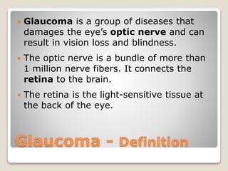
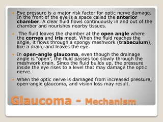
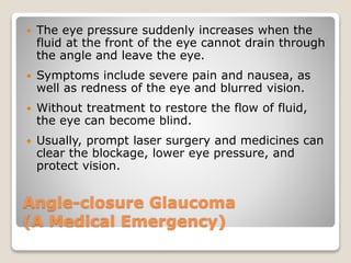
This was a lecture in the course "Significant Medical Conditions in Seniors" presented at Peer Learning in Chapel Hill, NC, USA in 2016 by Michael C. Joseph, MD, MPH.
4. Glaucoma - Definition
Glaucoma is a group of diseases that
damages the eye’s optic nerve and can
result in vision loss and blindness.
The optic nerve is a bundle of more than
1 million nerve fibers. It connects the
retina to the brain.
The retina is the light-sensitive tissue at
the back of the eye.
5. Glaucoma - Mechanism
Eye pressure is a major risk factor for optic nerve damage.
In the front of the eye is a space called the anterior
chamber. A clear fluid flows continuously in and out of the
chamber and nourishes nearby tissues.
The fluid leaves the chamber at the open angle where
the cornea and iris meet. When the fluid reaches the
angle, it flows through a spongy meshwork (trabeculum),
like a drain, and leaves the eye.
In open-angle glaucoma, even though the drainage
angle is “open”, the fluid passes too slowly through the
meshwork drain. Since the fluid builds up, the pressure
inside the eye rises to a level that may damage the optic
nerve.
When the optic nerve is damaged from increased pressure,
open-angle glaucoma, and vision loss may result.
6. Glaucoma - Risk Factors
Anyone can develop glaucoma.
Some people are at higher risk than others:
− African Americans over age 40
− Everyone over age 60, especially Mexican-Americans
− People with a family history of glaucoma
− Chronic use of corticosteroids, e.g. prednisone, cortisone
7. Angle-closure Glaucoma
(A Medical Emergency)
The eye pressure suddenly increases when the
fluid at the front of the eye cannot drain through
the angle and leave the eye.
Symptoms include severe pain and nausea, as
well as redness of the eye and blurred vision.
Without treatment to restore the flow of fluid,
the eye can become blind.
Usually, prompt laser surgery and medicines can
clear the blockage, lower eye pressure, and
protect vision.
8. Glaucoma - Symptoms
At first, open-angle glaucoma has no
symptoms. It causes no pain. Vision stays
normal.
Glaucoma can develop in one or both eyes.
Without treatment, people with glaucoma will
slowly lose their peripheral (side) vision.
As glaucoma remains untreated, people may
miss objects to the side and out of the corner
of their eye. They seem to be looking through
a tunnel.
Over time, straight-ahead (central) vision
may decrease until no vision remains.
9. Glaucoma - Diagnosis
Visual acuity test. This eye chart test measures
how well you see at various distances.
Visual field test. This test measures your peripheral
(side vision). It helps your eye care professional tell if
you have lost peripheral vision, a sign of glaucoma.
Dilated eye exam. In this exam, drops are placed in
your eyes to widen, or dilate, the pupils. Your eye
care professional uses a magnifying lens
(ophthalmoscope) to examine your retina and optic
nerve for signs of damage and other eye problems.
Tonometry is the measurement of pressure inside
the eye by using an instrument called a tonometer.
Numbing drops are applied to your eye for this test.
10. Glaucoma - Treatment
Drug Therapy
Medicines, in the form of eyedrops or pills, are the most
common early treatment for glaucoma. Taken regularly,
these eyedrops lower eye pressure. Some medicines cause
the eye to make less fluid, while others lower pressure by
helping fluid drain from the eye.
Surgery
Laser trabeculoplasty helps fluid drain out of the eye.
The laser makes several evenly spaced burns that stretch
the drainage holes in the meshwork. In many cases, you
will need to keep taking glaucoma medicines after
this procedure.
11. Cataract - Definition
A cataract is a clouding of the lens of the eye
that affects vision. A cataract can occur in one or
both eyes. It cannot spread from one eye to
the other.
Most cataracts are related to aging. Before age
60 most cataracts are small and do not affect
vision.
By age 80, more than half of all Americans either
have a cataract or have had cataract surgery.
12. Cataract - Lens Functions
The lens is a clear part of the eye that helps to focus
light, or an image, on the retina. The lens also
adjusts the eye’s focus, letting us see things clearly
both up close and far away. As you age, the lens
hardens, and far vision is better than close vision
(presbyopia).
In a normal eye, light passes through the transparent
lens to the retina. Once it reaches the retina, light is
changed into nerve signals that are sent to the brain.
The lens must be clear for the retina to receive a
sharp image. If the lens is cloudy from a cataract, the
image you see will be blurred.
13. Cataract - Risk Factors
Age
Diabetes
Excessive exposure to sunlight
Smoking
Obesity
High blood pressure
Previous eye injury or inflammation
Previous eye surgery
Prolonged use of corticosteroid medications
Drinking excessive amounts of alcohol
14. Cataract - Common Symptoms
Cloudy or blurry vision.
Colors seem faded.
Glare. Headlights, lamps, or sunlight may
appear too bright. A halo may appear
around lights.
Poor night vision.
Double vision, or multiple images in one eye.
(This symptom may clear as the cataract
gets larger.)
Frequent prescription changes in your
eyeglasses or contact lenses.
15. Cataract - Treatment
The symptoms of early cataract may be
improved with new eyeglasses, brighter
lighting, anti-glare sunglasses, or magnifying
lenses. If these measures do not help,
surgery is the only effective treatment.
Surgery involves removing the cloudy lens
and replacing it with an artificial lens.
Sometimes a cataract should be removed
even if it does not cause problems with
your vision. For example, a cataract should
be removed if it prevents examination or
treatment of another eye problem, such as
age-related macular degeneration or
diabetic retinopathy.
16. Age-Related Macular Degeneration
(AMD) - Definition
AMD is a disease that blurs the sharp, central
vision you need for “straight-ahead” activities
such as reading, sewing, and driving.
AMD affects the macula, the part of the retina
that allows you to see fine detail.
In some people, AMD advances so slowly that
vision loss does not occur for a long time. In
others, the disease progresses faster and may
lead to a loss of vision in one or both eyes.
AMD causes no pain.
17. Age-Related Macular Degeneration
(AMD) - Progression
As AMD progresses, a blurred area near the
center of vision is a common symptom. Over
time, the blurred area may grow larger, or you
may develop blank spots in your central vision.
Objects also may not appear to be as bright as
they used to be.
AMD by itself does not lead to complete
blindness.
VIDEO
https://www.youtube.com/watch?v=hZe7VxtuQB0
&feature=youtu.be
18. Age-Related Macular Degeneration
(AMD) - Epidemiology
AMD is the number one cause of severe
vision loss and legal blindness in adults over
60 in the U.S.
It escalates with age. It affects 14%-24% of
the U.S. population aged 65-74 years and 35
– 40% of people aged 74 years or more.
More than one person in three can
develop signs of age-related macular
degeneration, with over 200,000 new cases
diagnosed every year.
19. Age-Related
Macular Degeneration (AMD)
Risk Factors
Age is a major risk factor for AMD. The disease is most
likely to occur after age 60, but it can occur earlier.
Other risk factors for AMD include:
Smoking: Research shows that smoking doubles the
risk of AMD.
Race: AMD is more common among Caucasians than
among African-Americans or Hispanics/Latinos.
Family history and Genetics: People with a family
history of AMD are at higher risk. At last count,
researchers had identified nearly 20 genes that can
affect the risk of developing AMD.
20. Age-Related Macular Degeneration
(AMD) Prevention and Treatment
Preventive supplements tested in clinical studies
(Age-Related Eye Disease Study – AREDS; AREDS2):
− 500 milligrams (mg) of vitamin C
− 400 international units of vitamin E
− 80 mg zinc as zinc oxide (25 mg in AREDS2)
− 2 mg copper as cupric oxide
− 15 mg beta-carotene (10 mg lutein and 2 mg zeaxanthin in AREDS2)
− A number of manufacturers offer nutritional supplements that were formulated based
on these 5-year studies. The label may refer to “AREDS” or “AREDS2.”
− AREDS Results (25% decrease in progression of early and moderate AMD)
− AREDS2 Results (10-25% decrease in progression of early AMD)
For moderate or advanced AMD, treatments include injections into the eye,
light therapy to the retina, or laser surgery.
21. RESOURCES
National Eye Institute (NEI) (NIH)
https://nei.nih.gov
Mayo Clinic
http://www.mayoclinic.org
Macular Degeneration Partnership
http://www.amd.org
22. CREDITS
National Eye Institute (NEI) (NIH)
https://nei.nih.gov/health/glaucoma/glaucoma_facts
https://nei.nih.gov/health/cataract/cataract_facts
https://nei.nih.gov/health/maculardegen/armd_facts
Mayo Clinic
http://www.mayoclinic.org/diseases-conditions/cataracts/symptoms-
causes/dxc-20215129
Macular Degeneration Partnership
http://www.amd.org/what-is-macular-degeneration