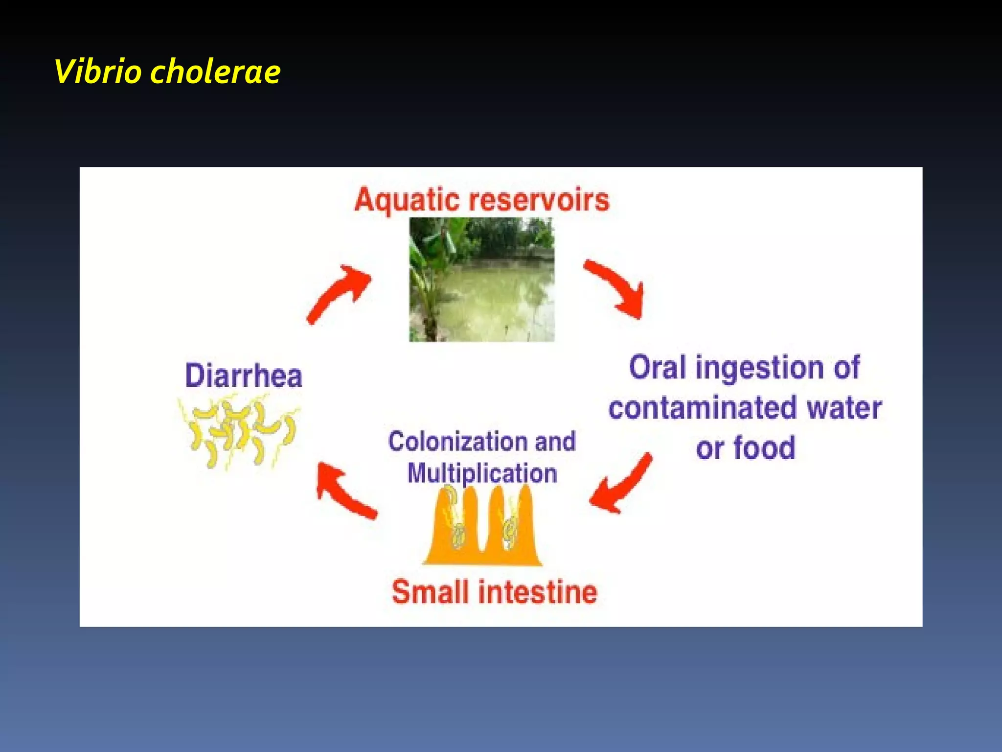
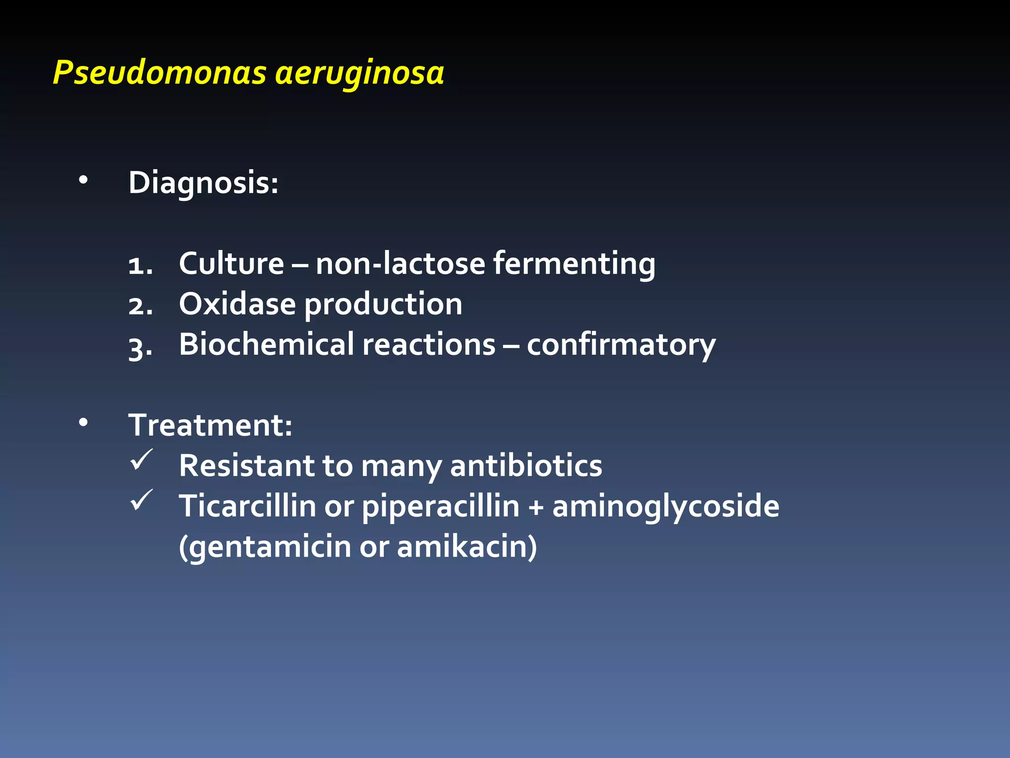
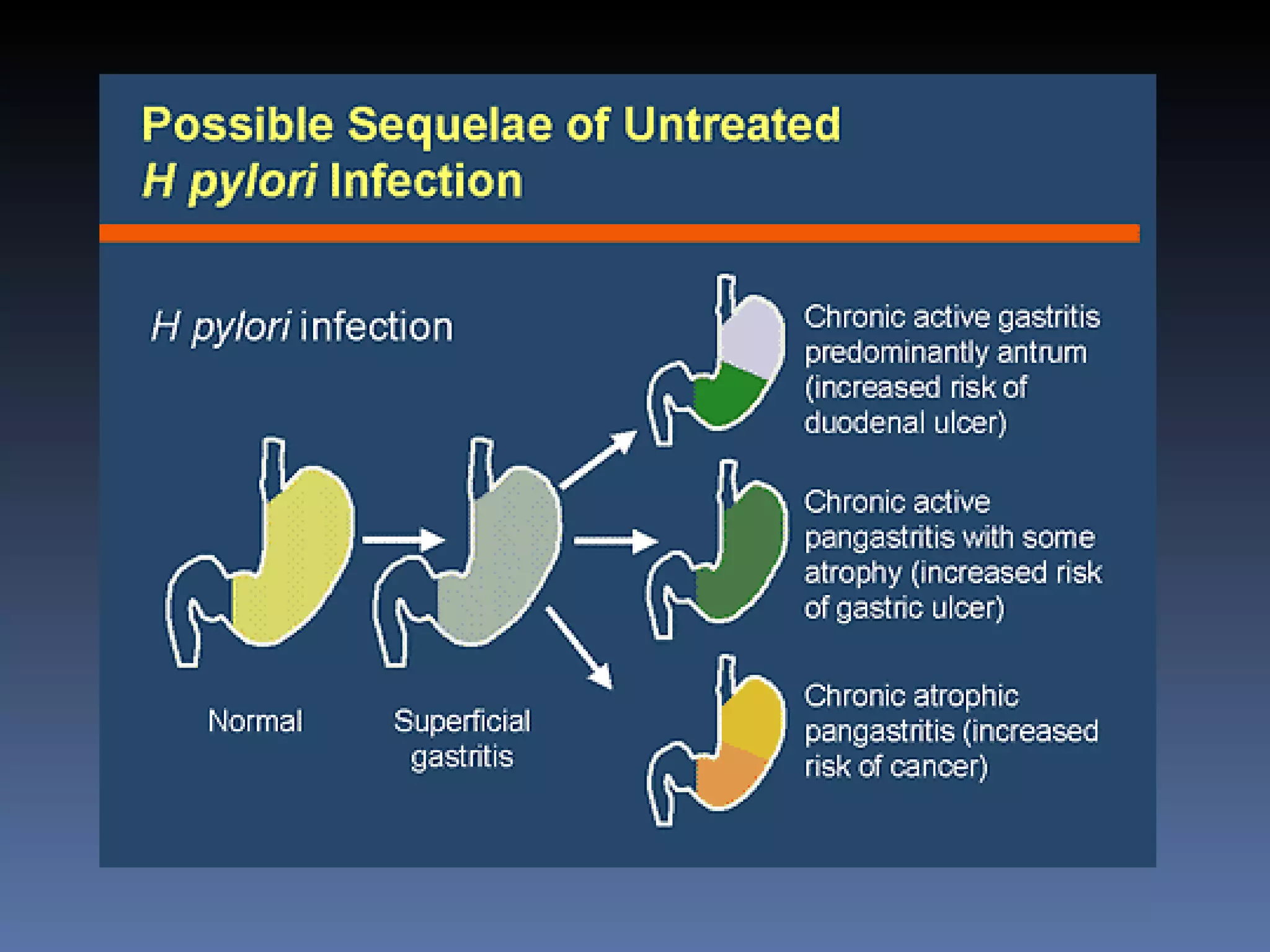
Vibrios, Pseudomonas, Campylobacter, Helicobacter Other gram-negative bacilli
VIBRIOS Among the most common bacteria in surface waters worldwide Curved, aerobic rods; motile with polar flagella oxidase (+) grow best on alkaline media Often found in brackish water
Vibrio cholerae Classified based on somatic O antigen: V. cholerae O1 – most common cause of epidemic & pandemic cholera V. cholerae O139 (Bengal strain) – epidemic Non-O1 group Two biotypes (based on differences in biochemical reactions): El Tor – most common cause of epidemics and cause of 7 th  pandemic Cholerae or classical Three serotypes (based on antigenic differences): Ogawa, Inaba, Hikojima
Vibrio cholerae MOT: fecal-oral Sources of infection: humans, marine shellfish (shrimp and oysters) Sensitive to gastric acid    high infective dose Virulence factors: Mucinase – cause adherence to cells of the brush border of the gut Choleragen – stimulate adenylyl cyclase
Vibrio cholerae
Vibrio cholerae Cholera Toxin B subunit A subunit Binds GM1 ganglioside (surface of epithelial cells) Carried to ER (retrograde transport) Endocytosis  Reduced by protein disulfide isomerase in ER Cytosol  Unfolding  Refolding  Interact with cytosolic ADP ribosylation factors Activate G protein Stimulate adenylate cyclase Inc. cAMP Open CFTR Cl released in lumen; secretion of HCO 3  , Na +  & water
Vibrio cholerae Clinical findings: Voluminous watery diarrhea – hallmark; “rice water” stool Loss of fluid and electrolytes – marked dehydration “ washerwoman” hands Cardiac and renal failure Hypovolemic shock
Vibrio cholerae Diagnosis: Culture on MacConkey’s agar (slow lactose fermenter) or TCBS (Thiosulfate citrate bile salt sucrose) agar plate for selective isolation Oxidase (+) – distinguished from the family Enterobacteriaceae Serology Treatment: Prompt and rapid fluid and electrolyte replacement Tetracycline – shorten duration
Growth on TCBS agar plate
Vibrio parahaemolyticus Halophilic – grows on 8% NaCl solution MOT: ingestion of raw or undercooked seafood, especially shellfish such as oysters Mild to severe watery diarrhea, nausea and vomiting, abdominal cramps, fever    self-limited No specific treatment indicated
 
Vibrio vulnificus Found in warm salt waters Causes severe skin and soft tissue infections (cellulitis), especially in shellfish handlers Rapid, fatal septicemia in immunocompromised people who have eaten raw shellfish Treatment: doxycycline
 
Pseudomonas aeruginosa Gram-negative rods that resemble the Enterobacteriaceae but are strict aerobes Derive energy by oxidation of sugars rather than fermentation Oxidase (+) Able to grow in water containing only traces of nutrients
Pseudomonas aeruginosa Produce two pigments: Pyocyanin (blue) Pyoverdin (fluorescein) – yellow green pigments that fluoresce under UVL Found chiefly in soil and water Found on the skin in moist areas Primarily opportunistic
Pseudomonas aeruginosa Virulence factors: Endotoxins – cause sepsis and shock Exotoxin A – cause tissue necrosis; inhibits eukaryotic protein synthesis similar to diphtheria exotoxin Enzymes Elastases and proteases Pyocyanin – damages the cilia and mucosal cells of respiratory tract
Pseudomonas aeruginosa Clinical: Urinary tract infections Pneumonia in cystic fibrosis patients Burn wound infection Sepsis Ecthyma gangrenosum – black necrotic lesions on skin Malignant external otitis Folliculitis  Osteochondritis of the foot due to punctured wounds through the soles of gym shoes -  most common cause
 
Ecthyma gangrenosum
 
Osteochondritis of foot
 
Pseudomonas aeruginosa Diagnosis: Culture – non-lactose fermenting Oxidase production Biochemical reactions – confirmatory Treatment: Resistant to many antibiotics Ticarcillin or piperacillin + aminoglycoside (gentamicin or amikacin)
Production of pyocyanin, water-soluble green pigment of Pseudomonas aeruginosa. (left tube)
Campylobacter jejuni Comma- or S-shaped rods Microaerophilic (5% oxygen) Grows well at 42 0 C Sources of infection: domestic animals MOT: fecal-oral (poultry, meat, unpasteurized milk common sources)
 
Campylobacter jejuni Clinical: Enterocolitis – begins as watery, foul-smelling diarrhea    bloody stools Associated with  Guillain-Barre syndrome Most common cause of acute neuro-muscular paralysis Autoimmune disease Reactive arthritis Reiter’s syndrome
Campylobacter jejuni Diagnosis: Culture Oxidase (+); sensitive to nalidixic acid Treatment: erythromycin or ciprofloxacin
Helicobacter pylori Curved; gram-negative; similar in appearance to Campylobacter Strongly urease positive    convert urea to ammonia    neutralize gastric acid Rapidly motile – allow organism to penetrate protective mucus layer


Helicobacter pylori Most common cause of chronic gastritis Most common cause of duodenal ulcers Second most common cause of gastric ulcer Associated with development of gastric CA and gastric lymphoma
Helicobacter pylori Diagnosis: Culture Urease production    urea breath test
 
 
Helicobacter pylori Treatment: Proton pump inhibitors (Omeprazole) Antibiotics – amoxicillin, metronidazole, tetracycline Bismuth salts
HAEMOPHILUS Family Pasteurellaceae Small, gram negative, pleomorphic Require enriched media containing blood or its derivatives Facultative anaerobes Obligate parasites
Haemophilus influenzae (Pfeiffer’s Bacilli) Found on mucus membrane of URT in humans (non-capsular form)    encapsulated species uncommon members of normal flora Short, coccoid bacilli in pairs or chains
Haemophilus influenzae Classification: Serotype – based on capsular antigen Biotype – based on biochemical properties a. indole production b. urease activity c. ornithine decarboxylase activity Biogroup – useful for clinical purposes
Haemophilus influenzae: Culture Chocolate agar – flat, grayish brown colonies after 24 hrs incubation Does not grow on sheep blood agar except around colonies of Staphylococci    “satellite phenomenon”
Haemophilus influenzae Growth Characteristics: Requires X factor (hemin) and V factor (NAD) Ferments carbohydrates poorly and irregularly
Haemophilus influenzae Characteristics & Growth Requirements: Species X Factor V Factor Hemolysis H. influenzae H. parainfluenzae H. ducreyi H. haemolyticus H. parahaemolyticus H. aprophilus + - + + - - + + - + + - - - - + + -
Haemophilus influenzae: Virulence Factors Capsule Antiphagocytic; impair ciliary function Main virulence factor With capsular polysaccharides (a to f) Type b – polyribose-ribitol phosphate (PRP)
Somatic antigen Outer membrane proteins    lipooligo-saccharides (endotoxin) IgA1 proteases Haemophilus influenzae: Virulence Factors
Haemophilus influenzae: Clinical Features H. influenzae type b Most common serotype causing systemic disease Meningitis Pneumonia & empyema Epiglottitis Cellulitis Septic arthritis
Non-typable (non-encapsulated) H. influenzae opportunistic Chronic bronchitis Otitis media Sinusitis Conjunctivitis  Haemophilus influenzae: Clinical Features
Meningitis 2 0  to bacteremic spread from nasopharynx Peak incidence: 3 – 18 mos. old Epiglottitis Cellulitis & swelling of supraglottic tissues Pharyngitis, fever & dyspnea    complete airway obstruction    death
Cellulitis Reddish blue patches on cheeks or periorbital areas
Arthritis Infection of a single large joint Children < 2 y/o or immunocompromised patients or those with previously damaged joints Conjunctivitis Epidemic and endemic H. influenzae biogroup aegypticus
Haemophilus influenzae Clinical Features: Sepsis with gangrene
Haemophilus influenzae: Prevention Chemoprophylaxis with Rifampicin for non-immune children < 4 y/o who are close contacts Hib conjugate vaccine >  2 mos. old    Hib conjugated with C. diphtheriae toxin protein or N. meningitidis outer membrane complex >  15 mos. old    Hib conjugated with diphtheria toxoid
Haemophilus aegypticus H. influenzae biotype III Koch-Weeks bacillus Resembles H. influenzae closely Diseases: Conjunctivitis – highly communicable Brazilian purpuric fever – fever, purpura, shock and death
Haemophilus ducreyi Causes chancroid (soft chancre) Ragged ulcer on genitalia with marked swelling and tenderness Lymph nodes enlarged and painful Organism grows best on chocolate agar incubated in 10% CO 2 No permanent immunity
Haemophilus ducreyi
Bordetella pertussis Small, coccobacillary, encapsulated, gram (-) With bipolar metachromatic granules (toluidine blue stain) Non-motile; strict aerobe Forms acid from glucose and lactose
Bordetella pertussis Requires enriched media Bordet-Gengou medium (potato-blood-glycerol agar) Contains Pen G 0.5 ug/mL Virulence genes –  bvgA and bvgS
Bordetella pertussis Gram stain Culture on chocolate agar
Bordetella pertussis: Virulence Factors Filamentous hemagglutinin Protein on pili; adhesion to ciliated epithelial cells Pertussis toxin promote lymphocytosis via inhibition of signal transduction by chemokine receptors    lymphocytes do not enter lymphoid tissues promote sensitization to histamine enhance insulin secretion stimulate adenylate cyclase via ADP-ribosylation
Adenylyl cyclase toxin – inhibit phagocytosis Tracheal cytotoxin Fragment of bacterial peptidoglycan Induce nitric oxide    destroy ciliated epithelium Dermonecrotic toxin Hemolysin Bordetella pertussis: Virulence Factors
Adheres to and multiplies rapidly on epithelial surface of trachea and bronchi    interfere with ciliary action No invasion of blood Bordetella pertussis: Pathogenesis
MOT: airborne droplets Source of infection: patients in early catarrhal stage Disease: Pertussis or Whooping Cough    acute tracheobronchitis Incubation period: approx. 2 weeks Bordetella pertussis: Pathogenesis
Clinical: Stages of Disease Catarrhal Mild coughing and sneezing Highly infectious but not very ill Paroxysmal (1-4 weeks) Series of hacking coughs, accompanied by copious amts. of mucus, ending with inspiratory “whoop”    exhaustion, vomiting, cyanosis and convulsions High wbc count (16,000-30,000/uL) with absolute lymphocytosis Convalescence - slow
Specimen: saline nasal wash (preferred) or nasopharyngeal swab Direct fluorescence antibody test – 50% sensitivity Culture of saline nasal wash fluid PCR – most sensitive Serology – (+) only on third week of illness    of little diagnostic value Bordetella pertussis: Diagnosis
First defense is antibody that prevents attachment Recovery from disease or immunization is followed by immunity Second infection may occur but is mild Re-infection occurring years later in adults may be severe Vaccine-induced immunity not completely protective
Chemoprophylaxis –  Erythromycin     for exposed, unimmunized individuals OR exposed, immunized children < 4 years old Vaccine – two vaccines available: acellular vaccine – contains 5 purified antigens    main immunogen is inactivated pertussis toxin; first vaccine to contain a genetically inactivated toxoid    ADP-ribosylating activity removed DPT x 3 doses
BRUCELLA Zoonotic    obligate parasite of animals & humans Intracellular organism Gram negative coccobacilli Aerobic; non-motile; non-spore-forming
BRUCELLA Catalase (+); oxidase (+) Produces H 2 S Culture: trypticase soy agar OR blood culture media;  B. abortus  requires 5-10% CO 2  for growth
Route of infection in humans: Intestinal tract – ingestion of infected milk & contaminated dairy products (cheese from unpasteurized goat’s milk) Mucous membranes – droplets Skin – contact with infected tissues of animals Pathogenesis:  endotoxin – O antigen polysaccharide
Species Animal Pathology  B. melitensis Goats Acute and severe infection B. suis Swine Chronic with suppurative lesions; caseating granulomas B. abortus Cattle Mild disease without suppuration; Non-caseating granulomas of the RES (LN, liver, spleen, BM) B. canis Dogs  Mild disease
Clinical:  Brucellosis (Undulant or Malta Fever) Acute Malaise, fever, weakness, aches & sweats Fever rises in the afternoon    fall during the night with drenching sweats (+) lymphadenopathy w/ palpable spleen;  +  hepatitis with jaundice Chronic With psychoneurotic symptoms Weakness, aches & pains, low grade fever
Diagnosis: Culture BM & blood – commonly used specimen Brucella agar, trypticase soy medium, brain heart infusion medium, chocolate agar Serology – inc. IgM during 1 st  week of illness; peak at 3 months

Other Gram Negative Bacilli

  • 1.
    Vibrios, Pseudomonas, Campylobacter,Helicobacter Other gram-negative bacilli
  • 2.
    VIBRIOS Among themost common bacteria in surface waters worldwide Curved, aerobic rods; motile with polar flagella oxidase (+) grow best on alkaline media Often found in brackish water
  • 3.
    Vibrio cholerae Classifiedbased on somatic O antigen: V. cholerae O1 – most common cause of epidemic & pandemic cholera V. cholerae O139 (Bengal strain) – epidemic Non-O1 group Two biotypes (based on differences in biochemical reactions): El Tor – most common cause of epidemics and cause of 7 th pandemic Cholerae or classical Three serotypes (based on antigenic differences): Ogawa, Inaba, Hikojima
  • 4.
    Vibrio cholerae MOT:fecal-oral Sources of infection: humans, marine shellfish (shrimp and oysters) Sensitive to gastric acid  high infective dose Virulence factors: Mucinase – cause adherence to cells of the brush border of the gut Choleragen – stimulate adenylyl cyclase
  • 5.
  • 6.
    Vibrio cholerae CholeraToxin B subunit A subunit Binds GM1 ganglioside (surface of epithelial cells) Carried to ER (retrograde transport) Endocytosis Reduced by protein disulfide isomerase in ER Cytosol Unfolding Refolding Interact with cytosolic ADP ribosylation factors Activate G protein Stimulate adenylate cyclase Inc. cAMP Open CFTR Cl released in lumen; secretion of HCO 3 , Na + & water
  • 7.
    Vibrio cholerae Clinicalfindings: Voluminous watery diarrhea – hallmark; “rice water” stool Loss of fluid and electrolytes – marked dehydration “ washerwoman” hands Cardiac and renal failure Hypovolemic shock
  • 8.
    Vibrio cholerae Diagnosis:Culture on MacConkey’s agar (slow lactose fermenter) or TCBS (Thiosulfate citrate bile salt sucrose) agar plate for selective isolation Oxidase (+) – distinguished from the family Enterobacteriaceae Serology Treatment: Prompt and rapid fluid and electrolyte replacement Tetracycline – shorten duration
  • 9.
    Growth on TCBSagar plate
  • 10.
    Vibrio parahaemolyticus Halophilic– grows on 8% NaCl solution MOT: ingestion of raw or undercooked seafood, especially shellfish such as oysters Mild to severe watery diarrhea, nausea and vomiting, abdominal cramps, fever  self-limited No specific treatment indicated
  • 11.
  • 12.
    Vibrio vulnificus Foundin warm salt waters Causes severe skin and soft tissue infections (cellulitis), especially in shellfish handlers Rapid, fatal septicemia in immunocompromised people who have eaten raw shellfish Treatment: doxycycline
  • 13.
  • 14.
    Pseudomonas aeruginosa Gram-negativerods that resemble the Enterobacteriaceae but are strict aerobes Derive energy by oxidation of sugars rather than fermentation Oxidase (+) Able to grow in water containing only traces of nutrients
  • 15.
    Pseudomonas aeruginosa Producetwo pigments: Pyocyanin (blue) Pyoverdin (fluorescein) – yellow green pigments that fluoresce under UVL Found chiefly in soil and water Found on the skin in moist areas Primarily opportunistic
  • 16.
    Pseudomonas aeruginosa Virulencefactors: Endotoxins – cause sepsis and shock Exotoxin A – cause tissue necrosis; inhibits eukaryotic protein synthesis similar to diphtheria exotoxin Enzymes Elastases and proteases Pyocyanin – damages the cilia and mucosal cells of respiratory tract
  • 17.
    Pseudomonas aeruginosa Clinical:Urinary tract infections Pneumonia in cystic fibrosis patients Burn wound infection Sepsis Ecthyma gangrenosum – black necrotic lesions on skin Malignant external otitis Folliculitis Osteochondritis of the foot due to punctured wounds through the soles of gym shoes - most common cause
  • 18.
  • 19.
  • 20.
  • 21.
  • 22.
  • 23.
    Pseudomonas aeruginosa Diagnosis:Culture – non-lactose fermenting Oxidase production Biochemical reactions – confirmatory Treatment: Resistant to many antibiotics Ticarcillin or piperacillin + aminoglycoside (gentamicin or amikacin)
  • 24.
    Production of pyocyanin,water-soluble green pigment of Pseudomonas aeruginosa. (left tube)
  • 25.
    Campylobacter jejuni Comma-or S-shaped rods Microaerophilic (5% oxygen) Grows well at 42 0 C Sources of infection: domestic animals MOT: fecal-oral (poultry, meat, unpasteurized milk common sources)
  • 26.
  • 27.
    Campylobacter jejuni Clinical:Enterocolitis – begins as watery, foul-smelling diarrhea  bloody stools Associated with Guillain-Barre syndrome Most common cause of acute neuro-muscular paralysis Autoimmune disease Reactive arthritis Reiter’s syndrome
  • 28.
    Campylobacter jejuni Diagnosis:Culture Oxidase (+); sensitive to nalidixic acid Treatment: erythromycin or ciprofloxacin
  • 29.
    Helicobacter pylori Curved;gram-negative; similar in appearance to Campylobacter Strongly urease positive  convert urea to ammonia  neutralize gastric acid Rapidly motile – allow organism to penetrate protective mucus layer
  • 30.
  • 31.
  • 32.
  • 33.
  • 34.
    Helicobacter pylori Mostcommon cause of chronic gastritis Most common cause of duodenal ulcers Second most common cause of gastric ulcer Associated with development of gastric CA and gastric lymphoma
  • 35.
    Helicobacter pylori Diagnosis:Culture Urease production  urea breath test
  • 36.
  • 37.
  • 38.
    Helicobacter pylori Treatment:Proton pump inhibitors (Omeprazole) Antibiotics – amoxicillin, metronidazole, tetracycline Bismuth salts
  • 39.
    HAEMOPHILUS Family PasteurellaceaeSmall, gram negative, pleomorphic Require enriched media containing blood or its derivatives Facultative anaerobes Obligate parasites
  • 40.
    Haemophilus influenzae (Pfeiffer’sBacilli) Found on mucus membrane of URT in humans (non-capsular form)  encapsulated species uncommon members of normal flora Short, coccoid bacilli in pairs or chains
  • 41.
    Haemophilus influenzae Classification:Serotype – based on capsular antigen Biotype – based on biochemical properties a. indole production b. urease activity c. ornithine decarboxylase activity Biogroup – useful for clinical purposes
  • 42.
    Haemophilus influenzae: CultureChocolate agar – flat, grayish brown colonies after 24 hrs incubation Does not grow on sheep blood agar except around colonies of Staphylococci  “satellite phenomenon”
  • 43.
    Haemophilus influenzae GrowthCharacteristics: Requires X factor (hemin) and V factor (NAD) Ferments carbohydrates poorly and irregularly
  • 44.
    Haemophilus influenzae Characteristics& Growth Requirements: Species X Factor V Factor Hemolysis H. influenzae H. parainfluenzae H. ducreyi H. haemolyticus H. parahaemolyticus H. aprophilus + - + + - - + + - + + - - - - + + -
  • 45.
    Haemophilus influenzae: VirulenceFactors Capsule Antiphagocytic; impair ciliary function Main virulence factor With capsular polysaccharides (a to f) Type b – polyribose-ribitol phosphate (PRP)
  • 46.
    Somatic antigen Outermembrane proteins  lipooligo-saccharides (endotoxin) IgA1 proteases Haemophilus influenzae: Virulence Factors
  • 47.
    Haemophilus influenzae: ClinicalFeatures H. influenzae type b Most common serotype causing systemic disease Meningitis Pneumonia & empyema Epiglottitis Cellulitis Septic arthritis
  • 48.
    Non-typable (non-encapsulated) H.influenzae opportunistic Chronic bronchitis Otitis media Sinusitis Conjunctivitis Haemophilus influenzae: Clinical Features
  • 49.
    Meningitis 2 0 to bacteremic spread from nasopharynx Peak incidence: 3 – 18 mos. old Epiglottitis Cellulitis & swelling of supraglottic tissues Pharyngitis, fever & dyspnea  complete airway obstruction  death
  • 50.
    Cellulitis Reddish bluepatches on cheeks or periorbital areas
  • 51.
    Arthritis Infection ofa single large joint Children < 2 y/o or immunocompromised patients or those with previously damaged joints Conjunctivitis Epidemic and endemic H. influenzae biogroup aegypticus
  • 52.
    Haemophilus influenzae ClinicalFeatures: Sepsis with gangrene
  • 53.
    Haemophilus influenzae: PreventionChemoprophylaxis with Rifampicin for non-immune children < 4 y/o who are close contacts Hib conjugate vaccine > 2 mos. old  Hib conjugated with C. diphtheriae toxin protein or N. meningitidis outer membrane complex > 15 mos. old  Hib conjugated with diphtheria toxoid
  • 54.
    Haemophilus aegypticus H.influenzae biotype III Koch-Weeks bacillus Resembles H. influenzae closely Diseases: Conjunctivitis – highly communicable Brazilian purpuric fever – fever, purpura, shock and death
  • 55.
    Haemophilus ducreyi Causeschancroid (soft chancre) Ragged ulcer on genitalia with marked swelling and tenderness Lymph nodes enlarged and painful Organism grows best on chocolate agar incubated in 10% CO 2 No permanent immunity
  • 56.
  • 57.
    Bordetella pertussis Small,coccobacillary, encapsulated, gram (-) With bipolar metachromatic granules (toluidine blue stain) Non-motile; strict aerobe Forms acid from glucose and lactose
  • 58.
    Bordetella pertussis Requiresenriched media Bordet-Gengou medium (potato-blood-glycerol agar) Contains Pen G 0.5 ug/mL Virulence genes – bvgA and bvgS
  • 59.
    Bordetella pertussis Gramstain Culture on chocolate agar
  • 60.
    Bordetella pertussis: VirulenceFactors Filamentous hemagglutinin Protein on pili; adhesion to ciliated epithelial cells Pertussis toxin promote lymphocytosis via inhibition of signal transduction by chemokine receptors  lymphocytes do not enter lymphoid tissues promote sensitization to histamine enhance insulin secretion stimulate adenylate cyclase via ADP-ribosylation
  • 61.
    Adenylyl cyclase toxin– inhibit phagocytosis Tracheal cytotoxin Fragment of bacterial peptidoglycan Induce nitric oxide  destroy ciliated epithelium Dermonecrotic toxin Hemolysin Bordetella pertussis: Virulence Factors
  • 62.
    Adheres to andmultiplies rapidly on epithelial surface of trachea and bronchi  interfere with ciliary action No invasion of blood Bordetella pertussis: Pathogenesis
  • 63.
    MOT: airborne dropletsSource of infection: patients in early catarrhal stage Disease: Pertussis or Whooping Cough  acute tracheobronchitis Incubation period: approx. 2 weeks Bordetella pertussis: Pathogenesis
  • 64.
    Clinical: Stages ofDisease Catarrhal Mild coughing and sneezing Highly infectious but not very ill Paroxysmal (1-4 weeks) Series of hacking coughs, accompanied by copious amts. of mucus, ending with inspiratory “whoop”  exhaustion, vomiting, cyanosis and convulsions High wbc count (16,000-30,000/uL) with absolute lymphocytosis Convalescence - slow
  • 65.
    Specimen: saline nasalwash (preferred) or nasopharyngeal swab Direct fluorescence antibody test – 50% sensitivity Culture of saline nasal wash fluid PCR – most sensitive Serology – (+) only on third week of illness  of little diagnostic value Bordetella pertussis: Diagnosis
  • 66.
    First defense isantibody that prevents attachment Recovery from disease or immunization is followed by immunity Second infection may occur but is mild Re-infection occurring years later in adults may be severe Vaccine-induced immunity not completely protective
  • 67.
    Chemoprophylaxis – Erythromycin  for exposed, unimmunized individuals OR exposed, immunized children < 4 years old Vaccine – two vaccines available: acellular vaccine – contains 5 purified antigens  main immunogen is inactivated pertussis toxin; first vaccine to contain a genetically inactivated toxoid  ADP-ribosylating activity removed DPT x 3 doses
  • 68.
    BRUCELLA Zoonotic  obligate parasite of animals & humans Intracellular organism Gram negative coccobacilli Aerobic; non-motile; non-spore-forming
  • 69.
    BRUCELLA Catalase (+);oxidase (+) Produces H 2 S Culture: trypticase soy agar OR blood culture media; B. abortus requires 5-10% CO 2 for growth
  • 70.
    Route of infectionin humans: Intestinal tract – ingestion of infected milk & contaminated dairy products (cheese from unpasteurized goat’s milk) Mucous membranes – droplets Skin – contact with infected tissues of animals Pathogenesis: endotoxin – O antigen polysaccharide
  • 71.
    Species Animal Pathology B. melitensis Goats Acute and severe infection B. suis Swine Chronic with suppurative lesions; caseating granulomas B. abortus Cattle Mild disease without suppuration; Non-caseating granulomas of the RES (LN, liver, spleen, BM) B. canis Dogs Mild disease
  • 72.
    Clinical: Brucellosis(Undulant or Malta Fever) Acute Malaise, fever, weakness, aches & sweats Fever rises in the afternoon  fall during the night with drenching sweats (+) lymphadenopathy w/ palpable spleen; + hepatitis with jaundice Chronic With psychoneurotic symptoms Weakness, aches & pains, low grade fever
  • 73.
    Diagnosis: Culture BM& blood – commonly used specimen Brucella agar, trypticase soy medium, brain heart infusion medium, chocolate agar Serology – inc. IgM during 1 st week of illness; peak at 3 months