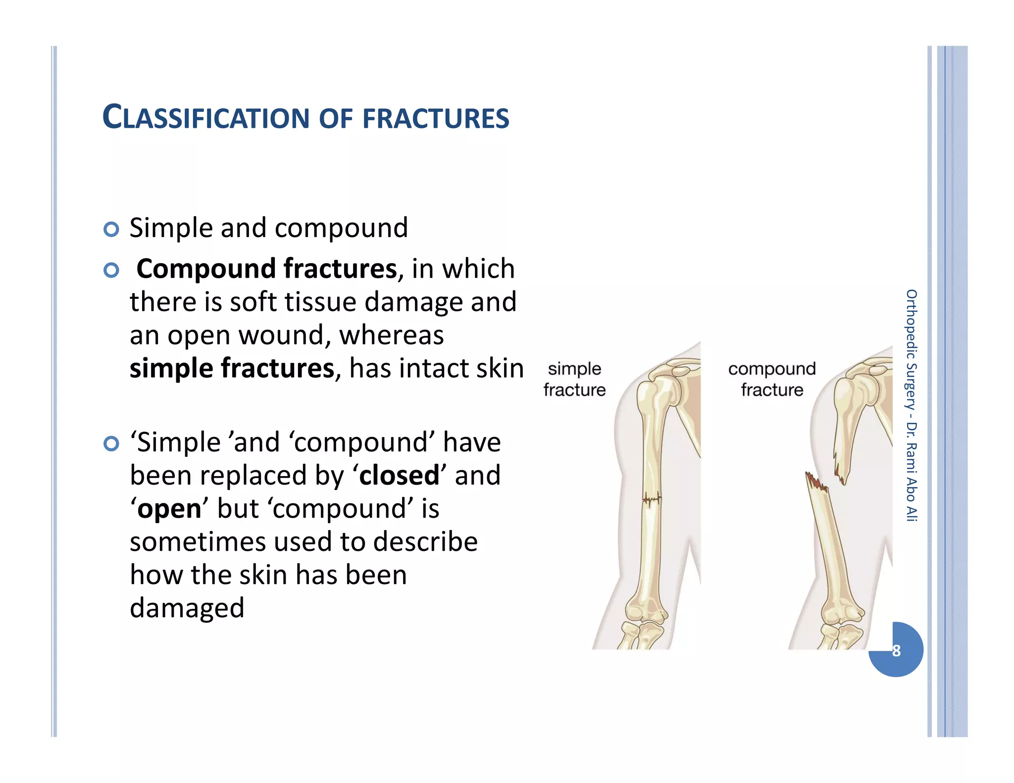
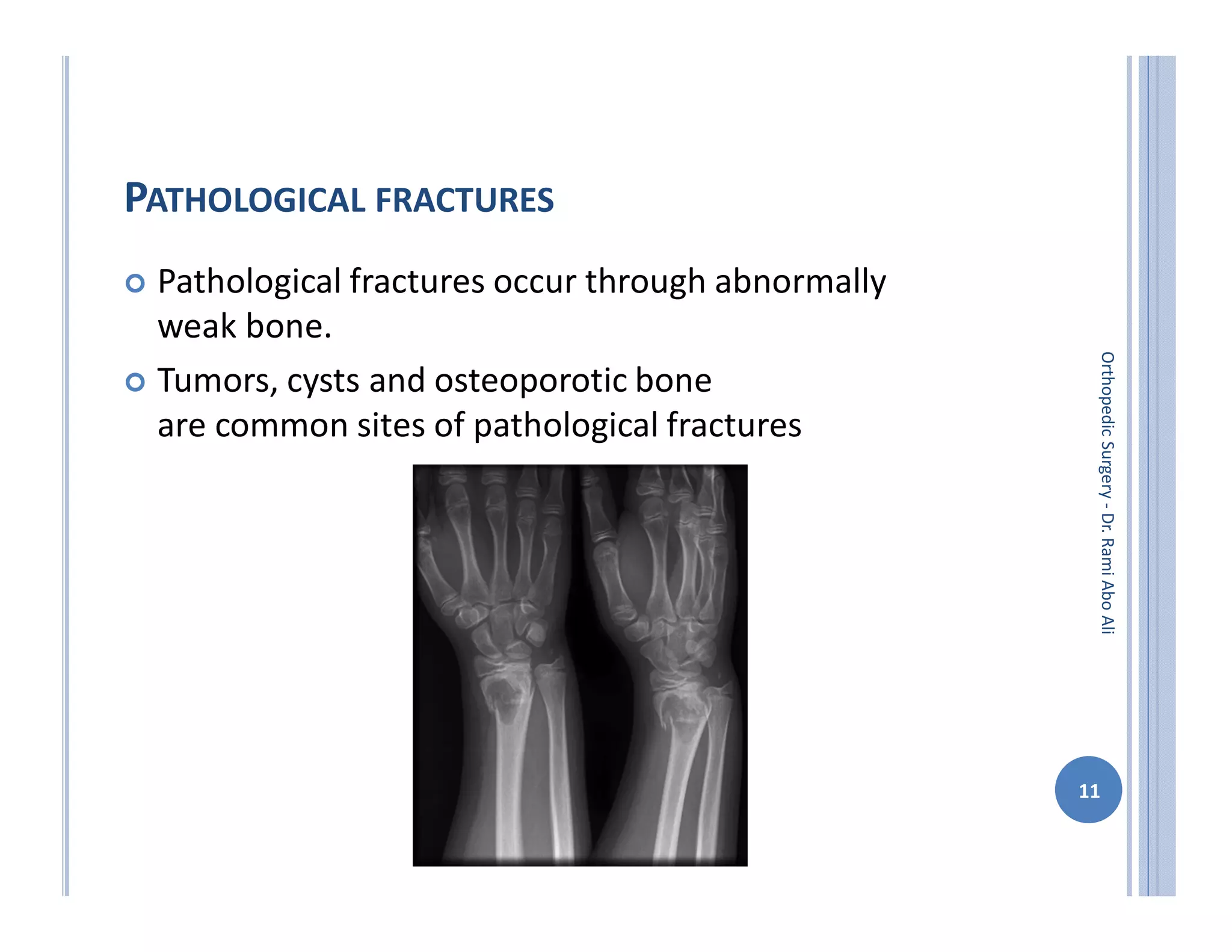
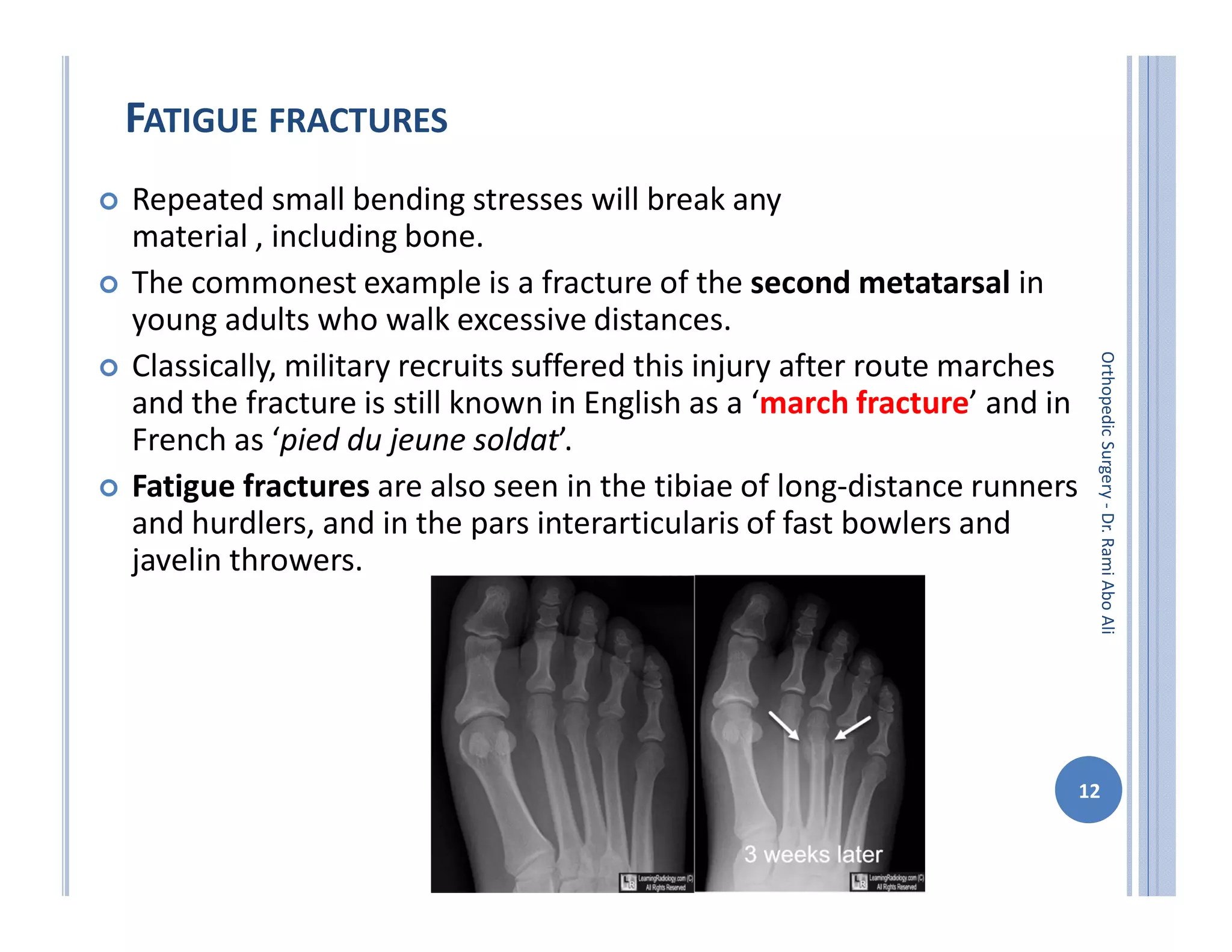
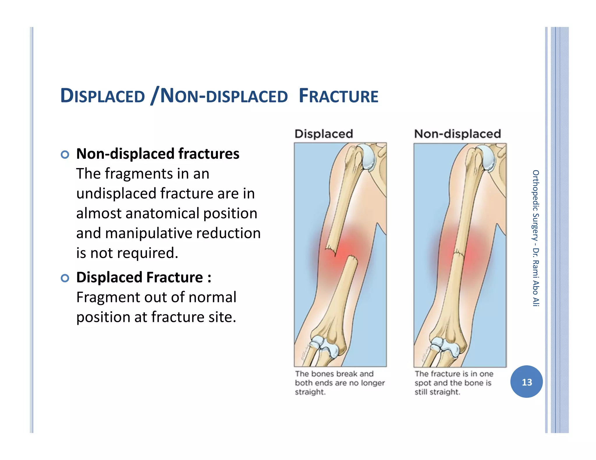
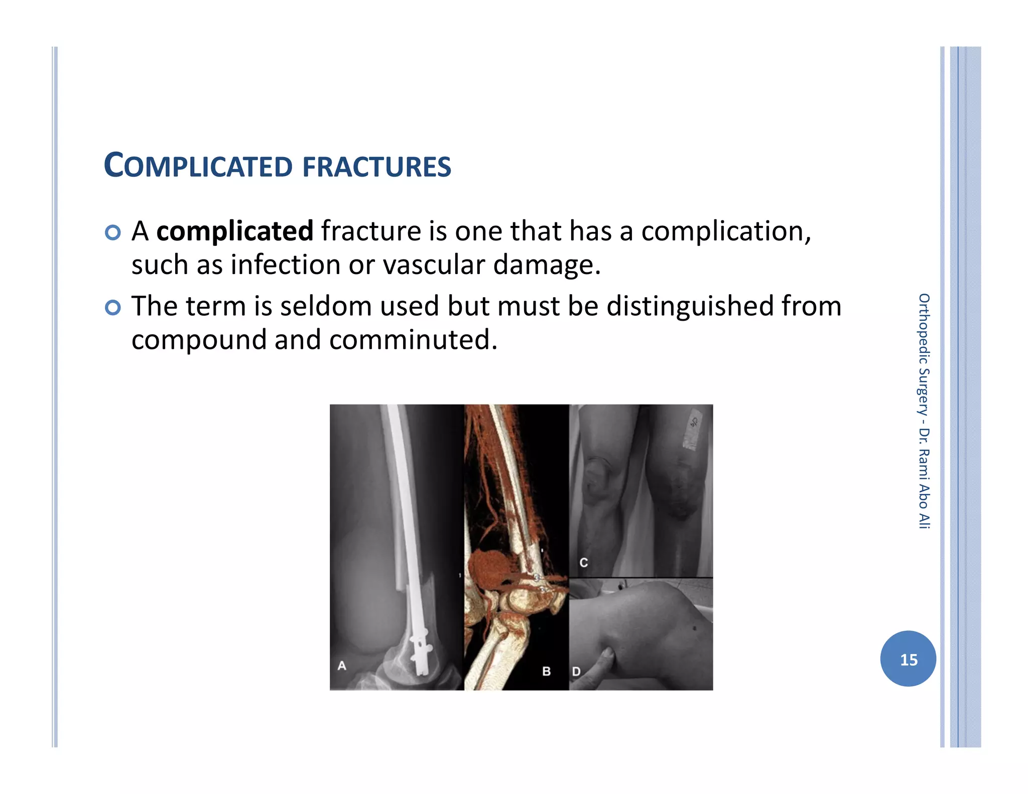
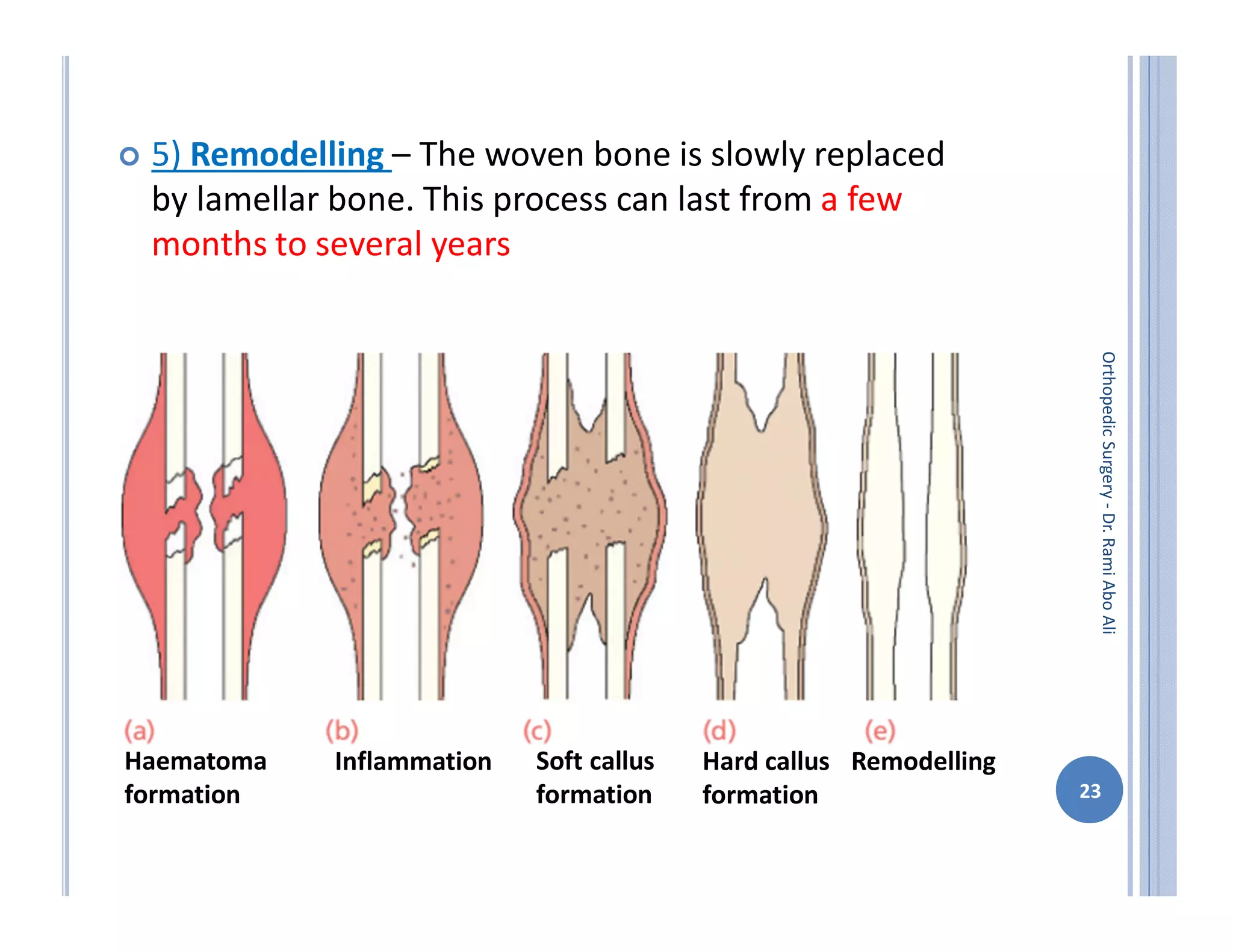
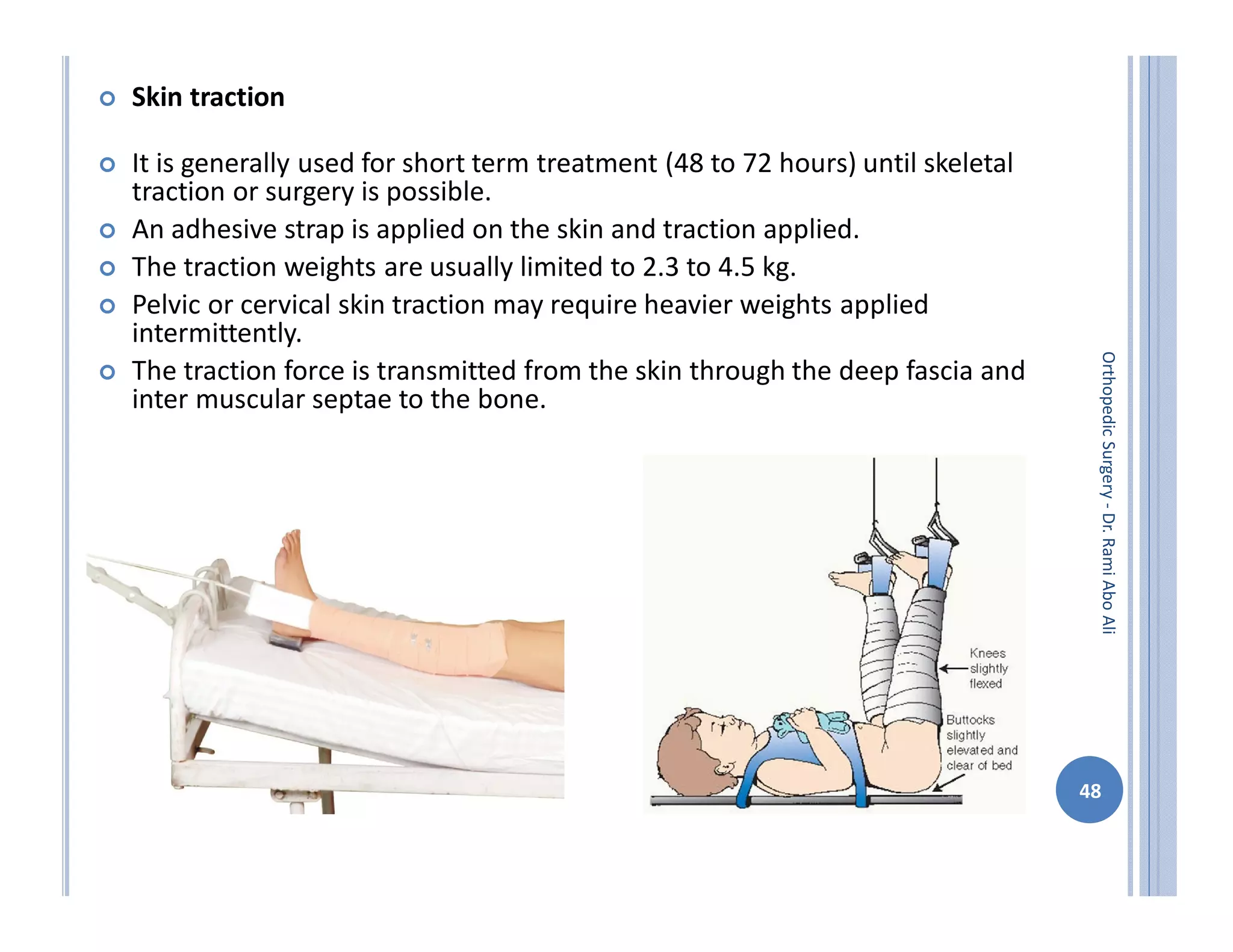
ORTHOPEDIC SURGERY
Dr. Rami Abo Ali
Orthopedic
Surgery
-
Dr.
Rami
Abo
Ali
1
GENERAL PRINCIPLES IN FRACTURES
Orthopedic
Surgery
-
Dr.
Rami
Abo
Ali
2
BONE FORMATION
 Bone formation (ossifi cation) occurs in 3 different ways:
enchondral, intramembranous, appositional
 Enchondral
 • Bone replaces a cartilage anlage (template). Osteoclasts
remove the cartilage, and osteoblasts make the new bone
matrix, which is then mineralized.
 • Typical in long bones (except clavicle).
 • Primary ossification centers (in shaft) typically develop in
prenatal period.
 • Secondary ossification centers occur at various times after
birth, usually in the epiphysis.
 • Longitudinal growth at the physis also occurs by enchondral
ossification.
 • Also found in fracture callus 3
Orthopedic
Surgery
-
Dr.
Rami
Abo
Ali
4
Orthopedic
Surgery
-
Dr.
Rami
Abo
Ali
Enchondral
BONE FORMATION
 Intramembranous
 • Bone develops directly from mesenchymal cells
without a cartilage anlage.
 • Mesenchymal cells differentiate into osteoblasts,
which produce bone.
 • Examples: flat bones (e.g., the cranium) and clavicle
 Appositional
 • Osteoblasts make new matrix/bone on top of existing
bone.
 • Example: periosteal-mediated bone diameter (width)
growth in long bones 5
Orthopedic
Surgery
-
Dr.
Rami
Abo
Ali
6
Orthopedic
Surgery
-
Dr.
Rami
Abo
Ali
Intramembranous
Appositional
FRACTURES
 Fracture definition: It is a break in the structural continuity of bone .
 How fracture happen?
1. from single traumatic incident.
2. repetitive stress .(stress fracture)
3. abnormal weakening of the bone (pathological fracture).
 Clinical manifestations
 Localized pain and tenderness
Decreased function
Inability to bear weight or use of the affected part
Edema and Swelling
Muscle Spasm
Deformity
Ecchymosis and Contusion
Loss of function
Crepitation
7
Orthopedic
Surgery
-
Dr.
Rami
Abo
Ali
CLASSIFICATION OF FRACTURES
 Simple and compound
 Compound fractures, in which
there is soft tissue damage and
an open wound, whereas
simple fractures, has intact skin
 ‘Simple ’and ‘compound’ have
been replaced by ‘closed’ and
‘open’ but ‘compound’ is
sometimes used to describe
how the skin has been
damaged
8
Orthopedic
Surgery
-
Dr.
Rami
Abo
Ali
SHAPE
 Fractures can also be classified according to the shape of
the fragments and this is helpful in deciding management
 Transverse fractures are the result of a direct blow
or a pure angular force applied to the bone
 Oblique or spiral fractures are caused by a violent
twisting movement about the long axis of the bone
 Comminuted. Applied to a fracture where the bone is
splintered into more than two fragments
 Crush fractures. A fracture in which cancellous bone is
squashed or crushed presents a difficult problem because
there are no fragments left to manipulate back into
position.
 Greenstick fractures. When a green stick breaks, it does
not snap cleanly but bends so that one ‘cortex’ buckles
while the other remains intact
9
Orthopedic
Surgery
-
Dr.
Rami
Abo
Ali
10
Orthopedic
Surgery
-
Dr.
Rami
Abo
Ali
PATHOLOGICAL FRACTURES
 Pathological fractures occur through abnormally
weak bone.
 Tumors, cysts and osteoporotic bone
are common sites of pathological fractures
11
Orthopedic
Surgery
-
Dr.
Rami
Abo
Ali
FATIGUE FRACTURES
 Repeated small bending stresses will break any
material , including bone.
 The commonest example is a fracture of the second metatarsal in
young adults who walk excessive distances.
 Classically, military recruits suffered this injury after route marches
and the fracture is still known in English as a ‘march fracture’ and in
French as ‘pied du jeune soldat’.
 Fatigue fractures are also seen in the tibiae of long-distance runners
and hurdlers, and in the pars interarticularis of fast bowlers and
javelin throwers.
12
Orthopedic
Surgery
-
Dr.
Rami
Abo
Ali
DISPLACED /NON-DISPLACED FRACTURE
 Non-displaced fractures
The fragments in an
undisplaced fracture are in
almost anatomical position
and manipulative reduction
is not required.
 Displaced Fracture :
Fragment out of normal
position at fracture site.
13
Orthopedic
Surgery
-
Dr.
Rami
Abo
Ali
STABLE AND UNSTABLE FRACTURES
 A stable fracture is one in which the two bones are lying
in a position from which they are unlikely to move.
 Stable fractures are often undisplaced but some are
stable even though the bone is misshapen.
 Unstable fracture is a fracture with an intrinsic tendency
to displace after reduction.
14
Orthopedic
Surgery
-
Dr.
Rami
Abo
Ali
COMPLICATED FRACTURES
 A complicated fracture is one that has a complication,
such as infection or vascular damage.
 The term is seldom used but must be distinguished from
compound and comminuted.
15
Orthopedic
Surgery
-
Dr.
Rami
Abo
Ali
PROXIMAL & DISTAL SEGMENT FRACTURES
 • Type A
– Extra-articular
• Type B
– Partial articular
• Type C
– Complete disruption
of the articular
surface from the
diaphysis
16
Orthopedic
Surgery
-
Dr.
Rami
Abo
Ali
DIAPHYSEAL FRACTURES
 • Type A
– Simple fractures with
two fragments
• Type B
– Wedge fractures
– After reduced, length
and alignment restored
• Type C
– Complex fractures with
no contact between
main fragments
17
Orthopedic
Surgery
-
Dr.
Rami
Abo
Ali
EPIPHYSEAL INJURY
 Injury to the epiphyses of growing bone can cause severe deformity in later
life if a bony bridge forms across the fracture site and prevents growth on
one side of the bone.
 The following five patterns of injury were described by Harris and Salter:
1. A fracture along the epiphyseal line.
2. Separation of the epiphysis with a triangular fragment of shaft attached
to it .
3. Fracture of the epiphysis, part of it remaining attached to the shaft.
4. A fracture line passing through both epiphysis and shaft.
5. A crushing injury . This is difficult to recognize at the time of injury, either
clinically or radiologically. This type of injury is more commonly associated
with later growth arrest or retardation
18
Orthopedic
Surgery
-
Dr.
Rami
Abo
Ali
HEALING BY DIRECT UNION
(PRIMARY BONE HEALING)
 If the fracture site is absolutely stable – for example, an
impacted fracture in cancellous bone, or a fracture held by a
metal plate with absolute stability – there is no stimulus for
callus.
 Instead, osteoblastic new bone formation occurs directly
between the fragments
 Gaps between the fracture surfaces are invaded by new
capillaries and osteoprogenitor cells growing in from the edges,
and new bone is laid down on the exposed surface (gap healing)
 Where the crevices are narrow (less than 200 µm), osteogensis
produces lamellar bone; wider gaps are filled with woven bone
first, which is then remodeled to lamellar bone
 By 3-4 weeks the fracture is solid enough to allow penetration
and bridging of the area by bone remodelling units, i.e.
osteoclastic ‘cutting cones’ followed by osteoblasts
19
Orthopedic
Surgery
-
Dr.
Rami
Abo
Ali
20
Orthopedic
Surgery
-
Dr.
Rami
Abo
Ali
HEALING BY CALLUS
(SECONDARY BONE HEALING)
 Secondary bone healing is the most common form of
healing in tubular bones; in the absence of rigid fixation
 It proceeds in five stages
 1) Haematoma formation – At the time of injury,
bleeding occurs from the bone and soft tissues.
 2) Inflammation – The inflammatory process starts
rapidly when the fracture haematoma forms and
cytokines are released, and lasts until fibrous tissue,
cartilage, or bone formation begins (1–7 days post
fracture). Osteoclasts are formed to remove the necrotic
ends of bony fragments.
21
Orthopedic
Surgery
-
Dr.
Rami
Abo
Ali
 3) Soft callus formation – After 2–3 weeks, the
first soft callus is formed. This is about the time when
the fragments can no longer move freely.
The strain applied to the cells in the fracture gap
modifies their growth factor expression and
progenitor cells are stimulated to become osteoblasts.
The cells form a cuff of woven bone periosteally . The
fracture can now still angulate but is stable in length.
 4 )Hard callus formation – When the fracture ends
are linked together, the hard callus starts and lasts
until the fragments are firmly united (3–4 months).
Bone callus forms at the periphery of the fracture
and progressively moves centrally
22
Orthopedic
Surgery
-
Dr.
Rami
Abo
Ali
 5) Remodelling – The woven bone is slowly replaced
by lamellar bone. This process can last from a few
months to several years
23
Orthopedic
Surgery
-
Dr.
Rami
Abo
Ali
Remodelling
Hard callus
formation
Soft callus
formation
Inflammation
Haematoma
formation
FACTORS AFFECTING FRACTURE HEALING
 Blood Supply
 Soft tissue injury
 Radiation
 Chemical or thermal burns
 Infection
 Anaemia & hypoxia
 Denervation
 Excessive compression
 Excessive Movement
 Inadequate immobilization
 Inadequate fixation or compliance
 Gap
 Intact fellow bone (as in fracture of tibia )
 Interposed soft tissue
 Distraction of bones
 Other
 Nutrition (Vit C required for normal collagen)
 Drugs (corticosteriods inhibit osteoblast differentiation)
24
Orthopedic
Surgery
-
Dr.
Rami
Abo
Ali
PRINCIPLES OF FRACTURE FIXATION
 The four principles of fracture fixation are :
1. Fracture reduction to restore anatomical relationships
2. Fracture fixation providing absolute or relative
stability as the “personality” of fracture, patient and
injury requires.
3. Preservation of blood supply to soft tissues and bone.
4. Early and safe mobilization of the injured part and the
patient as a whole.
25
Orthopedic
Surgery
-
Dr.
Rami
Abo
Ali
 There are two forms of displacement:
1. Translational displacement:
1. Medial or lateral and posterior or anterior
2. Shortening or lengthening
2. Rotational displacement:
1. Internal or external rotational malaligment
2. Valgus or varus malaligment
3. Flexion or extension malalignment
26
Orthopedic
Surgery
-
Dr.
Rami
Abo
Ali
 Aim of reduction :
 Anatomical reduction
 To restore the bony anatomy and morphology
 Intra-articular fractures
 Functional reduction
 To restore the relationship between the proximal and distal main
fragments of the fracture
 To restore the length, alignment and rotation
 Mechanical and anatomical axis as reference
 Often in diaphyseal fractures
27
Orthopedic
Surgery
-
Dr.
Rami
Abo
Ali
 Reduction methods
 The decision, which reduction method should be used, depends on
the location of the fracture:
 1. Meta- and diaphyseal fractures usually need functional reduction.
 2. Joint fractures need anatomical reduction
 Reduction of diaphyseal fractures
 The functional anatomy is restored (length, alignment, and rotational axis).
 The load-bearing axis of the extremity is restored (especially important in
the lower limb).
 An exception is the forearm which functions as a single articular unit.
 Reduction of articular fractures
 The joint surface is restored anatomically. Gaps and steps in the articular
surface must be avoided.
 “Steps” means that there is a difference between the levels of two main
articular fragments.
 “Gaps” means that there is some space between two adjacent main
articular fragments.
 The axial alignment is restored.
28
Orthopedic
Surgery
-
Dr.
Rami
Abo
Ali
 Goal of fracture fixation
 To maintain the reduction
 To create adequate stability which:
 Allows early and optimal function of the injured limb
 Minimizes pain
 Methods of reduction
 There are two reduction methods:
 Direct reduction where every fragment under direct vision is restored.
 Indirect reduction where the direction is done without direct view on the
fracture.
29
Orthopedic
Surgery
-
Dr.
Rami
Abo
Ali
Direct reduction
Indirect reduction
STABILITY
 Absolute stability
 no movement at fracture site, rigid
 Achieved through interfragmentary compression; e.g lag
screws, compression plates
 Often in intra-articular fractures
 No callus forms, fracture heals through direct healing
 Relative stability
 Movement at fracture site
 There is no interfragmentary compression at fracture site . It
is achieved by splinting or bridging, eg. elastic nails
 There is callus formation. Indirect bone healing is achieved.
30
Orthopedic
Surgery
-
Dr.
Rami
Abo
Ali
REDUCTION:
 IF fracture is displaced.
 Meant to realign fracture fragments.
 To minimize soft tissue injury.
 Can be considered definitive if fragments’ position is
accepted. If reduction is acceptable, we can put a cast
and that would be a definitive treatment. If not, we can
put a temporary splint and later on we can treat the
patient in a definitive way.
 Should be followed by immobilization.
 To maximize healing potential
 To ensure good function after healing 31
Orthopedic
Surgery
-
Dr.
Rami
Abo
Ali
Important points to remember:
1. Take consent from patient prior to
2. Patient must receive adequate analgesia prior to
reduction.
3. Most reductions occur under conscious sedation at
emergency.
4. Reduction must be followed by immobilization.
5. Nerve/Vascular status must be documented before and
after reduction and immobilization (before and after
reduction).
32
Orthopedic
Surgery
-
Dr.
Rami
Abo
Ali
Neurovascular assessment: 6Ps
pain, paralysis, paresthesias, pulselessness, pallor, and
pressure
EMERGENCY CARE
 • Begins at the site of the accident.
• It consists of ‘splint them where they lie’.
 Closed fracture
 Before splinting remove any ring or bangles worn by the patient.
 Almost any available object( for eg: folded news paper, magazine,
rigid cardboard, stick, umbrella, pillow etc.) can be used for splinting
at the site of the accident.
 Open fracture
 The bleeding from the wound is stopped by applying firm pressure
using a clean piece of cloth.
 Circular bandage can apply proximal to the wound in order to stop
bleeding.
 If the wound is very dirty, it is washed with clean tap water and
covered with a clean cloth.
 The fracture is splinted
33
Orthopedic
Surgery
-
Dr.
Rami
Abo
Ali
DEFINITIVE CARE
 The aim of treatment is rehabilitation of the limb
to pre-injury status.
 Anatomic realignment of bone fragments(reduction)
 Immobilization to maintain realignment
 Restoration of normal or near normal function of the
injured part
34
Orthopedic
Surgery
-
Dr.
Rami
Abo
Ali
METHODS OF TREATMENT
 Treatment by functional use of the limb: Some
fractures (eg: fractured ribs, scapula) need no reduction or
immobilization. These fractures unite despite functional use of
the body part. Analgesics are needed for the initial few days.
 Treatment by immobilization : Fractures without significant
displacement or fractures where the displacement is of no
concern are treated this way.
 Treatment by reduction followed by immobilization: It is
required for most displaced fractures. These otherwise result in
deformity, shortening etc.
 Open reduction and internal fixation: Some fractures , such as
intra- articular fractures, are best treated by open reduction and
internal fixation. 35
Orthopedic
Surgery
-
Dr.
Rami
Abo
Ali
 Fracture reduction
Reduction of a fracture can be carried out by
following methods
• Closed reduction
• Open reduction
• Continuous traction
36
Orthopedic
Surgery
-
Dr.
Rami
Abo
Ali
CLOSED REDUCTION
 Nonsurgical, manual realignment of bone fragments
to their previous anatomic position.
 Traction and counter traction are manually applied to
the bone fragments.
 Usually performed under local or general anesthesia.
 After reduction, traction, casting, external fixation,
splints or orthoses immobilize the injured part to
maintain alignment until healing occurs.
37
Orthopedic
Surgery
-
Dr.
Rami
Abo
Ali
OPEN REDUCTION
 Open reduction is the correction of bone alignment through a
surgical incision.
 It usually includes internal fixation of the fracture with the use
of wires, screws, pins, plates, intra medullary rods or nails.
 This techniques allows anatomic reduction and the creation of
highly stable constructs.
38
Orthopedic
Surgery
-
Dr.
Rami
Abo
Ali
 Indication Open reduction
1. Fracture that cannot be reduced except by operation
2. Fracture that are inherently unstable and prone to
displacement after reduction
3. Fracture that unite poorly and slowly
 Principally fracture of the femoral neck
4. Pathological fracture
 Bone disease may prevent healing
5. Multiple fracture
 Where early fixation reduced the risk of general
complication
6. Fracture in patient who present severe nursing
difficulty
39
Orthopedic
Surgery
-
Dr.
Rami
Abo
Ali
GOALS AND INDICATIONS FOR INTERNAL FIXATION
 Goals:
 Restoration of bony anatomy while respecting soft tissues
 Stable fixation
 Accelerated recovery
 More predictable and potentially faster healing
 Indications:
 Displaced intra-articular fractures
 Open fracture
 Polytrauma
 Associated neurovascular injury
 Failure of closed treatment
40
Orthopedic
Surgery
-
Dr.
Rami
Abo
Ali
IMMOBILIZATION
 Plaster casts or plastic functional braces – these hold the
bone in position until it has healed.
 Metal plates and screws – current procedures may use
minimally invasive techniques.
 Intra-medullary nails – internal metal rods are placed down
the center of long bones. Flexible wires may be used in
children.
 External fixators – these may be made of metal or carbon
fiber; they have steel pins that go into the bone directly
through the skin. They are a type of scaffolding outside the
body.
 Traction can be used for the treatment of any long bone
fracture or major joint dislocation using ropes, pulleys, and
weights
41
Orthopedic
Surgery
-
Dr.
Rami
Abo
Ali
 Strapping : The fractured part is strapped to an adjacent
part of the body.
Eg: phalanx fracture, where one finger is strapped to the
adjacent normal finger.
42
Orthopedic
Surgery
-
Dr.
Rami
Abo
Ali
 Sling : A fracture of the upper extremity is immobilized with
the help of a sling, mostly to relieve pain in cases where
strict immobilization is not necessary.
Eg : Triangular sling used for a fracture of the
clavicle
43
Orthopedic
Surgery
-
Dr.
Rami
Abo
Ali
 Casts immobilization
A cast is a temporary circumferential immobilization device.
It allows the patient to perform many normal activities of daily
living while providing sufficient immobilization to ensure stability.
Cast materials are natural (plaster of paris ) , synthetic acrylic,
latex free polymer or a hybrid of materials
44
Orthopedic
Surgery
-
Dr.
Rami
Abo
Ali
 Splints
• Splints are used for immobilizing fractures; either
temporary during transportation or for definitive
treatment.
45
Orthopedic
Surgery
-
Dr.
Rami
Abo
Ali
 Functional bracing
 Braces are used to provide support ,control movement ,
and prevent additional injury
46
Orthopedic
Surgery
-
Dr.
Rami
Abo
Ali
 Continuous Traction
 It is the application of a pulling force to an injured or diseased
part of the body or an extremity while counter traction pulls in
the opposite direction.
 Traction-Purposes
1. Prevent or reduce muscle spasm
2. Immobilize a joint or part of the body
3. Reduce a fracture or dislocation
4. Treat a pathologic condition.
 The two most common types of traction are
• Skin traction
• Skeletal traction
47
Orthopedic
Surgery
-
Dr.
Rami
Abo
Ali
 Skin traction
 It is generally used for short term treatment (48 to 72 hours) until skeletal
traction or surgery is possible.
 An adhesive strap is applied on the skin and traction applied.
 The traction weights are usually limited to 2.3 to 4.5 kg.
 Pelvic or cervical skin traction may require heavier weights applied
intermittently.
 The traction force is transmitted from the skin through the deep fascia and
inter muscular septae to the bone.
48
Orthopedic
Surgery
-
Dr.
Rami
Abo
Ali
 Skeletal Traction
 Provides long term pull that keeps the injured bones and joints aligned.
 Applied directly on the bone by inserting K-wire or Steinmann pin through the
bone to align and immobilize the injured body part.
 Used to align injured bones and joints or to treat joint contractures and
congenital hip dysplasia
 Weight for skeletal traction ranges from 2.3 to 20.4 kg. The use of too much
weight can result in delayed union or nonunion.
49
Orthopedic
Surgery
-
Dr.
Rami
Abo
Ali
 Types of Internal Fixation
 Pin & wire fixation.
 Screw fixation.
 Plate & screws fixation.
 Intra-medullary fixation.
50
Orthopedic
Surgery
-
Dr.
Rami
Abo
Ali
 Screw
• Interfragmentary screw (lag screw) are used for fixing small fragment onto the
main bone
 Wires
Kirschner wire often inserted percutaneously without exposing the fracture
Used in situation where fracture healing is predictably quick
 Pins and screws
• They are the simplest implants.
• often placed percutaneously.
• Krischner wires may be used temporarily and frequently for the stabilization of
small fragments.
• Screws can be used for inter fragmentary compression.
51
Orthopedic
Surgery
-
Dr.
Rami
Abo
Ali
 Plate & screws fixation
 Open Reductionand Internal fixation with Plates and screws
 Used for many fractures especially those involving joints
52
Orthopedic
Surgery
-
Dr.
Rami
Abo
Ali
 Intramedullary nail
 Suitable for long bones
 Nail is inserted onto
medullary canal to splint
the fracture
 Rotational fracture are
resisted by introducing
locking screw which
tranfix the bone cortices
and the nail proximal and
distal to the fracture.
53
Orthopedic
Surgery
-
Dr.
Rami
Abo
Ali
EXTERNAL FIXATORS
 The bone is transfixed above
and below the fracture with
screw or pins or tension wire
and these are then clamped
to a frame or connected to
each other by rigid bars
outside the skin
54
Orthopedic
Surgery
-
Dr.
Rami
Abo
Ali
REHABILITATION:
 To ensure return to function.
 Initiating motion (improve range of motion) should be
attempted as early as possible without jeopardizing
maintenance of reduction.
 Weight bearing restriction for short period (6-8 weeks).
Especially if the fracture is not stable. But after time you
have to start weight bearing because healing needs
stress
 Move unaffected areas immediately
55
Orthopedic
Surgery
-
Dr.
Rami
Abo
Ali
COMPLICATIONS OF FRACTURE HEALING
 • MALUNION
• DELAYED UNION
• NONUNION
 Malunion
A malunited Fracture is one that has healed with
the fragments in a non anatomical position.
Causes:
1) inaccurate reduction
2) ineffective immobilization
 Nonunion
• FDA defined nonunion as “established when a
minimum of 9 months has elapsed since fracture
with no visible progressive signs of
healing for 3 months”
• Every fracture has its own timetable (ie long
bone shaft fracture 6 months, femoral neck
fracture 3 months)
56
Orthopedic
Surgery
-
Dr.
Rami
Abo
Ali
 Types of nonunion
 septic nonunion
 caused by infection
 CRP test as the most accurate predictor of infection
 pseudoarthrosis
 hypertrophic nonunion
 caused by inadequate stability with adequate blood supply and
biology
 abundant callous formation without bridging bone
 typically heal once mechanical stability is improved
 atrophic nonunion
 caused by inadequate immobilization and inadequate blood supply
 oligotrophic nonunion
 produced by inadequate reduction with fracture fragment
displacement
57
Orthopedic
Surgery
-
Dr.
Rami
Abo
Ali
 Delayed union
a delayed union is generally defined as a failure to reach bony union by 6
months post-injury this also includes fractures that are taking longer than
expected to heal (ie. distal radial fractures)
 Systemic factors:
• Metabolic
• Nutritional status
• General health
• Activity level
• Tobacco and alcohol use
 Local factors
• Open
• Infected
• Segmental (impaired blood supply)
• Comminuted
• Insecurely fixed
• Immobilized for an insufficient time
• Treated by ill-advised open reduction
• Distracted by (traction/plate and screws)
• Irradiated bone
• Delayed weight-bearing > 6 weeks
• Soft tissue injury > method of initial treatment 58
Orthopedic
Surgery
-
Dr.
Rami
Abo
Ali
59
Orthopedic
Surgery
-
Dr.
Rami
Abo
Ali

Orthopedic surgery 2nd general principles in fractures

  • 1.
    ORTHOPEDIC SURGERY Dr. RamiAbo Ali Orthopedic Surgery - Dr. Rami Abo Ali 1
  • 2.
    GENERAL PRINCIPLES INFRACTURES Orthopedic Surgery - Dr. Rami Abo Ali 2
  • 3.
    BONE FORMATION  Boneformation (ossifi cation) occurs in 3 different ways: enchondral, intramembranous, appositional  Enchondral  • Bone replaces a cartilage anlage (template). Osteoclasts remove the cartilage, and osteoblasts make the new bone matrix, which is then mineralized.  • Typical in long bones (except clavicle).  • Primary ossification centers (in shaft) typically develop in prenatal period.  • Secondary ossification centers occur at various times after birth, usually in the epiphysis.  • Longitudinal growth at the physis also occurs by enchondral ossification.  • Also found in fracture callus 3 Orthopedic Surgery - Dr. Rami Abo Ali
  • 4.
  • 5.
    BONE FORMATION  Intramembranous • Bone develops directly from mesenchymal cells without a cartilage anlage.  • Mesenchymal cells differentiate into osteoblasts, which produce bone.  • Examples: flat bones (e.g., the cranium) and clavicle  Appositional  • Osteoblasts make new matrix/bone on top of existing bone.  • Example: periosteal-mediated bone diameter (width) growth in long bones 5 Orthopedic Surgery - Dr. Rami Abo Ali
  • 6.
  • 7.
    FRACTURES  Fracture definition:It is a break in the structural continuity of bone .  How fracture happen? 1. from single traumatic incident. 2. repetitive stress .(stress fracture) 3. abnormal weakening of the bone (pathological fracture).  Clinical manifestations  Localized pain and tenderness Decreased function Inability to bear weight or use of the affected part Edema and Swelling Muscle Spasm Deformity Ecchymosis and Contusion Loss of function Crepitation 7 Orthopedic Surgery - Dr. Rami Abo Ali
  • 8.
    CLASSIFICATION OF FRACTURES Simple and compound  Compound fractures, in which there is soft tissue damage and an open wound, whereas simple fractures, has intact skin  ‘Simple ’and ‘compound’ have been replaced by ‘closed’ and ‘open’ but ‘compound’ is sometimes used to describe how the skin has been damaged 8 Orthopedic Surgery - Dr. Rami Abo Ali
  • 9.
    SHAPE  Fractures canalso be classified according to the shape of the fragments and this is helpful in deciding management  Transverse fractures are the result of a direct blow or a pure angular force applied to the bone  Oblique or spiral fractures are caused by a violent twisting movement about the long axis of the bone  Comminuted. Applied to a fracture where the bone is splintered into more than two fragments  Crush fractures. A fracture in which cancellous bone is squashed or crushed presents a difficult problem because there are no fragments left to manipulate back into position.  Greenstick fractures. When a green stick breaks, it does not snap cleanly but bends so that one ‘cortex’ buckles while the other remains intact 9 Orthopedic Surgery - Dr. Rami Abo Ali
  • 10.
  • 11.
    PATHOLOGICAL FRACTURES  Pathologicalfractures occur through abnormally weak bone.  Tumors, cysts and osteoporotic bone are common sites of pathological fractures 11 Orthopedic Surgery - Dr. Rami Abo Ali
  • 12.
    FATIGUE FRACTURES  Repeatedsmall bending stresses will break any material , including bone.  The commonest example is a fracture of the second metatarsal in young adults who walk excessive distances.  Classically, military recruits suffered this injury after route marches and the fracture is still known in English as a ‘march fracture’ and in French as ‘pied du jeune soldat’.  Fatigue fractures are also seen in the tibiae of long-distance runners and hurdlers, and in the pars interarticularis of fast bowlers and javelin throwers. 12 Orthopedic Surgery - Dr. Rami Abo Ali
  • 13.
    DISPLACED /NON-DISPLACED FRACTURE Non-displaced fractures The fragments in an undisplaced fracture are in almost anatomical position and manipulative reduction is not required.  Displaced Fracture : Fragment out of normal position at fracture site. 13 Orthopedic Surgery - Dr. Rami Abo Ali
  • 14.
    STABLE AND UNSTABLEFRACTURES  A stable fracture is one in which the two bones are lying in a position from which they are unlikely to move.  Stable fractures are often undisplaced but some are stable even though the bone is misshapen.  Unstable fracture is a fracture with an intrinsic tendency to displace after reduction. 14 Orthopedic Surgery - Dr. Rami Abo Ali
  • 15.
    COMPLICATED FRACTURES  Acomplicated fracture is one that has a complication, such as infection or vascular damage.  The term is seldom used but must be distinguished from compound and comminuted. 15 Orthopedic Surgery - Dr. Rami Abo Ali
  • 16.
    PROXIMAL & DISTALSEGMENT FRACTURES  • Type A – Extra-articular • Type B – Partial articular • Type C – Complete disruption of the articular surface from the diaphysis 16 Orthopedic Surgery - Dr. Rami Abo Ali
  • 17.
    DIAPHYSEAL FRACTURES  •Type A – Simple fractures with two fragments • Type B – Wedge fractures – After reduced, length and alignment restored • Type C – Complex fractures with no contact between main fragments 17 Orthopedic Surgery - Dr. Rami Abo Ali
  • 18.
    EPIPHYSEAL INJURY  Injuryto the epiphyses of growing bone can cause severe deformity in later life if a bony bridge forms across the fracture site and prevents growth on one side of the bone.  The following five patterns of injury were described by Harris and Salter: 1. A fracture along the epiphyseal line. 2. Separation of the epiphysis with a triangular fragment of shaft attached to it . 3. Fracture of the epiphysis, part of it remaining attached to the shaft. 4. A fracture line passing through both epiphysis and shaft. 5. A crushing injury . This is difficult to recognize at the time of injury, either clinically or radiologically. This type of injury is more commonly associated with later growth arrest or retardation 18 Orthopedic Surgery - Dr. Rami Abo Ali
  • 19.
    HEALING BY DIRECTUNION (PRIMARY BONE HEALING)  If the fracture site is absolutely stable – for example, an impacted fracture in cancellous bone, or a fracture held by a metal plate with absolute stability – there is no stimulus for callus.  Instead, osteoblastic new bone formation occurs directly between the fragments  Gaps between the fracture surfaces are invaded by new capillaries and osteoprogenitor cells growing in from the edges, and new bone is laid down on the exposed surface (gap healing)  Where the crevices are narrow (less than 200 µm), osteogensis produces lamellar bone; wider gaps are filled with woven bone first, which is then remodeled to lamellar bone  By 3-4 weeks the fracture is solid enough to allow penetration and bridging of the area by bone remodelling units, i.e. osteoclastic ‘cutting cones’ followed by osteoblasts 19 Orthopedic Surgery - Dr. Rami Abo Ali
  • 20.
  • 21.
    HEALING BY CALLUS (SECONDARYBONE HEALING)  Secondary bone healing is the most common form of healing in tubular bones; in the absence of rigid fixation  It proceeds in five stages  1) Haematoma formation – At the time of injury, bleeding occurs from the bone and soft tissues.  2) Inflammation – The inflammatory process starts rapidly when the fracture haematoma forms and cytokines are released, and lasts until fibrous tissue, cartilage, or bone formation begins (1–7 days post fracture). Osteoclasts are formed to remove the necrotic ends of bony fragments. 21 Orthopedic Surgery - Dr. Rami Abo Ali
  • 22.
     3) Softcallus formation – After 2–3 weeks, the first soft callus is formed. This is about the time when the fragments can no longer move freely. The strain applied to the cells in the fracture gap modifies their growth factor expression and progenitor cells are stimulated to become osteoblasts. The cells form a cuff of woven bone periosteally . The fracture can now still angulate but is stable in length.  4 )Hard callus formation – When the fracture ends are linked together, the hard callus starts and lasts until the fragments are firmly united (3–4 months). Bone callus forms at the periphery of the fracture and progressively moves centrally 22 Orthopedic Surgery - Dr. Rami Abo Ali
  • 23.
     5) Remodelling– The woven bone is slowly replaced by lamellar bone. This process can last from a few months to several years 23 Orthopedic Surgery - Dr. Rami Abo Ali Remodelling Hard callus formation Soft callus formation Inflammation Haematoma formation
  • 24.
    FACTORS AFFECTING FRACTUREHEALING  Blood Supply  Soft tissue injury  Radiation  Chemical or thermal burns  Infection  Anaemia & hypoxia  Denervation  Excessive compression  Excessive Movement  Inadequate immobilization  Inadequate fixation or compliance  Gap  Intact fellow bone (as in fracture of tibia )  Interposed soft tissue  Distraction of bones  Other  Nutrition (Vit C required for normal collagen)  Drugs (corticosteriods inhibit osteoblast differentiation) 24 Orthopedic Surgery - Dr. Rami Abo Ali
  • 25.
    PRINCIPLES OF FRACTUREFIXATION  The four principles of fracture fixation are : 1. Fracture reduction to restore anatomical relationships 2. Fracture fixation providing absolute or relative stability as the “personality” of fracture, patient and injury requires. 3. Preservation of blood supply to soft tissues and bone. 4. Early and safe mobilization of the injured part and the patient as a whole. 25 Orthopedic Surgery - Dr. Rami Abo Ali
  • 26.
     There aretwo forms of displacement: 1. Translational displacement: 1. Medial or lateral and posterior or anterior 2. Shortening or lengthening 2. Rotational displacement: 1. Internal or external rotational malaligment 2. Valgus or varus malaligment 3. Flexion or extension malalignment 26 Orthopedic Surgery - Dr. Rami Abo Ali
  • 27.
     Aim ofreduction :  Anatomical reduction  To restore the bony anatomy and morphology  Intra-articular fractures  Functional reduction  To restore the relationship between the proximal and distal main fragments of the fracture  To restore the length, alignment and rotation  Mechanical and anatomical axis as reference  Often in diaphyseal fractures 27 Orthopedic Surgery - Dr. Rami Abo Ali
  • 28.
     Reduction methods The decision, which reduction method should be used, depends on the location of the fracture:  1. Meta- and diaphyseal fractures usually need functional reduction.  2. Joint fractures need anatomical reduction  Reduction of diaphyseal fractures  The functional anatomy is restored (length, alignment, and rotational axis).  The load-bearing axis of the extremity is restored (especially important in the lower limb).  An exception is the forearm which functions as a single articular unit.  Reduction of articular fractures  The joint surface is restored anatomically. Gaps and steps in the articular surface must be avoided.  “Steps” means that there is a difference between the levels of two main articular fragments.  “Gaps” means that there is some space between two adjacent main articular fragments.  The axial alignment is restored. 28 Orthopedic Surgery - Dr. Rami Abo Ali
  • 29.
     Goal offracture fixation  To maintain the reduction  To create adequate stability which:  Allows early and optimal function of the injured limb  Minimizes pain  Methods of reduction  There are two reduction methods:  Direct reduction where every fragment under direct vision is restored.  Indirect reduction where the direction is done without direct view on the fracture. 29 Orthopedic Surgery - Dr. Rami Abo Ali Direct reduction Indirect reduction
  • 30.
    STABILITY  Absolute stability no movement at fracture site, rigid  Achieved through interfragmentary compression; e.g lag screws, compression plates  Often in intra-articular fractures  No callus forms, fracture heals through direct healing  Relative stability  Movement at fracture site  There is no interfragmentary compression at fracture site . It is achieved by splinting or bridging, eg. elastic nails  There is callus formation. Indirect bone healing is achieved. 30 Orthopedic Surgery - Dr. Rami Abo Ali
  • 31.
    REDUCTION:  IF fractureis displaced.  Meant to realign fracture fragments.  To minimize soft tissue injury.  Can be considered definitive if fragments’ position is accepted. If reduction is acceptable, we can put a cast and that would be a definitive treatment. If not, we can put a temporary splint and later on we can treat the patient in a definitive way.  Should be followed by immobilization.  To maximize healing potential  To ensure good function after healing 31 Orthopedic Surgery - Dr. Rami Abo Ali
  • 32.
    Important points toremember: 1. Take consent from patient prior to 2. Patient must receive adequate analgesia prior to reduction. 3. Most reductions occur under conscious sedation at emergency. 4. Reduction must be followed by immobilization. 5. Nerve/Vascular status must be documented before and after reduction and immobilization (before and after reduction). 32 Orthopedic Surgery - Dr. Rami Abo Ali Neurovascular assessment: 6Ps pain, paralysis, paresthesias, pulselessness, pallor, and pressure
  • 33.
    EMERGENCY CARE  •Begins at the site of the accident. • It consists of ‘splint them where they lie’.  Closed fracture  Before splinting remove any ring or bangles worn by the patient.  Almost any available object( for eg: folded news paper, magazine, rigid cardboard, stick, umbrella, pillow etc.) can be used for splinting at the site of the accident.  Open fracture  The bleeding from the wound is stopped by applying firm pressure using a clean piece of cloth.  Circular bandage can apply proximal to the wound in order to stop bleeding.  If the wound is very dirty, it is washed with clean tap water and covered with a clean cloth.  The fracture is splinted 33 Orthopedic Surgery - Dr. Rami Abo Ali
  • 34.
    DEFINITIVE CARE  Theaim of treatment is rehabilitation of the limb to pre-injury status.  Anatomic realignment of bone fragments(reduction)  Immobilization to maintain realignment  Restoration of normal or near normal function of the injured part 34 Orthopedic Surgery - Dr. Rami Abo Ali
  • 35.
    METHODS OF TREATMENT Treatment by functional use of the limb: Some fractures (eg: fractured ribs, scapula) need no reduction or immobilization. These fractures unite despite functional use of the body part. Analgesics are needed for the initial few days.  Treatment by immobilization : Fractures without significant displacement or fractures where the displacement is of no concern are treated this way.  Treatment by reduction followed by immobilization: It is required for most displaced fractures. These otherwise result in deformity, shortening etc.  Open reduction and internal fixation: Some fractures , such as intra- articular fractures, are best treated by open reduction and internal fixation. 35 Orthopedic Surgery - Dr. Rami Abo Ali
  • 36.
     Fracture reduction Reductionof a fracture can be carried out by following methods • Closed reduction • Open reduction • Continuous traction 36 Orthopedic Surgery - Dr. Rami Abo Ali
  • 37.
    CLOSED REDUCTION  Nonsurgical,manual realignment of bone fragments to their previous anatomic position.  Traction and counter traction are manually applied to the bone fragments.  Usually performed under local or general anesthesia.  After reduction, traction, casting, external fixation, splints or orthoses immobilize the injured part to maintain alignment until healing occurs. 37 Orthopedic Surgery - Dr. Rami Abo Ali
  • 38.
    OPEN REDUCTION  Openreduction is the correction of bone alignment through a surgical incision.  It usually includes internal fixation of the fracture with the use of wires, screws, pins, plates, intra medullary rods or nails.  This techniques allows anatomic reduction and the creation of highly stable constructs. 38 Orthopedic Surgery - Dr. Rami Abo Ali
  • 39.
     Indication Openreduction 1. Fracture that cannot be reduced except by operation 2. Fracture that are inherently unstable and prone to displacement after reduction 3. Fracture that unite poorly and slowly  Principally fracture of the femoral neck 4. Pathological fracture  Bone disease may prevent healing 5. Multiple fracture  Where early fixation reduced the risk of general complication 6. Fracture in patient who present severe nursing difficulty 39 Orthopedic Surgery - Dr. Rami Abo Ali
  • 40.
    GOALS AND INDICATIONSFOR INTERNAL FIXATION  Goals:  Restoration of bony anatomy while respecting soft tissues  Stable fixation  Accelerated recovery  More predictable and potentially faster healing  Indications:  Displaced intra-articular fractures  Open fracture  Polytrauma  Associated neurovascular injury  Failure of closed treatment 40 Orthopedic Surgery - Dr. Rami Abo Ali
  • 41.
    IMMOBILIZATION  Plaster castsor plastic functional braces – these hold the bone in position until it has healed.  Metal plates and screws – current procedures may use minimally invasive techniques.  Intra-medullary nails – internal metal rods are placed down the center of long bones. Flexible wires may be used in children.  External fixators – these may be made of metal or carbon fiber; they have steel pins that go into the bone directly through the skin. They are a type of scaffolding outside the body.  Traction can be used for the treatment of any long bone fracture or major joint dislocation using ropes, pulleys, and weights 41 Orthopedic Surgery - Dr. Rami Abo Ali
  • 42.
     Strapping :The fractured part is strapped to an adjacent part of the body. Eg: phalanx fracture, where one finger is strapped to the adjacent normal finger. 42 Orthopedic Surgery - Dr. Rami Abo Ali
  • 43.
     Sling :A fracture of the upper extremity is immobilized with the help of a sling, mostly to relieve pain in cases where strict immobilization is not necessary. Eg : Triangular sling used for a fracture of the clavicle 43 Orthopedic Surgery - Dr. Rami Abo Ali
  • 44.
     Casts immobilization Acast is a temporary circumferential immobilization device. It allows the patient to perform many normal activities of daily living while providing sufficient immobilization to ensure stability. Cast materials are natural (plaster of paris ) , synthetic acrylic, latex free polymer or a hybrid of materials 44 Orthopedic Surgery - Dr. Rami Abo Ali
  • 45.
     Splints • Splintsare used for immobilizing fractures; either temporary during transportation or for definitive treatment. 45 Orthopedic Surgery - Dr. Rami Abo Ali
  • 46.
     Functional bracing Braces are used to provide support ,control movement , and prevent additional injury 46 Orthopedic Surgery - Dr. Rami Abo Ali
  • 47.
     Continuous Traction It is the application of a pulling force to an injured or diseased part of the body or an extremity while counter traction pulls in the opposite direction.  Traction-Purposes 1. Prevent or reduce muscle spasm 2. Immobilize a joint or part of the body 3. Reduce a fracture or dislocation 4. Treat a pathologic condition.  The two most common types of traction are • Skin traction • Skeletal traction 47 Orthopedic Surgery - Dr. Rami Abo Ali
  • 48.
     Skin traction It is generally used for short term treatment (48 to 72 hours) until skeletal traction or surgery is possible.  An adhesive strap is applied on the skin and traction applied.  The traction weights are usually limited to 2.3 to 4.5 kg.  Pelvic or cervical skin traction may require heavier weights applied intermittently.  The traction force is transmitted from the skin through the deep fascia and inter muscular septae to the bone. 48 Orthopedic Surgery - Dr. Rami Abo Ali
  • 49.
     Skeletal Traction Provides long term pull that keeps the injured bones and joints aligned.  Applied directly on the bone by inserting K-wire or Steinmann pin through the bone to align and immobilize the injured body part.  Used to align injured bones and joints or to treat joint contractures and congenital hip dysplasia  Weight for skeletal traction ranges from 2.3 to 20.4 kg. The use of too much weight can result in delayed union or nonunion. 49 Orthopedic Surgery - Dr. Rami Abo Ali
  • 50.
     Types ofInternal Fixation  Pin & wire fixation.  Screw fixation.  Plate & screws fixation.  Intra-medullary fixation. 50 Orthopedic Surgery - Dr. Rami Abo Ali
  • 51.
     Screw • Interfragmentaryscrew (lag screw) are used for fixing small fragment onto the main bone  Wires Kirschner wire often inserted percutaneously without exposing the fracture Used in situation where fracture healing is predictably quick  Pins and screws • They are the simplest implants. • often placed percutaneously. • Krischner wires may be used temporarily and frequently for the stabilization of small fragments. • Screws can be used for inter fragmentary compression. 51 Orthopedic Surgery - Dr. Rami Abo Ali
  • 52.
     Plate &screws fixation  Open Reductionand Internal fixation with Plates and screws  Used for many fractures especially those involving joints 52 Orthopedic Surgery - Dr. Rami Abo Ali
  • 53.
     Intramedullary nail Suitable for long bones  Nail is inserted onto medullary canal to splint the fracture  Rotational fracture are resisted by introducing locking screw which tranfix the bone cortices and the nail proximal and distal to the fracture. 53 Orthopedic Surgery - Dr. Rami Abo Ali
  • 54.
    EXTERNAL FIXATORS  Thebone is transfixed above and below the fracture with screw or pins or tension wire and these are then clamped to a frame or connected to each other by rigid bars outside the skin 54 Orthopedic Surgery - Dr. Rami Abo Ali
  • 55.
    REHABILITATION:  To ensurereturn to function.  Initiating motion (improve range of motion) should be attempted as early as possible without jeopardizing maintenance of reduction.  Weight bearing restriction for short period (6-8 weeks). Especially if the fracture is not stable. But after time you have to start weight bearing because healing needs stress  Move unaffected areas immediately 55 Orthopedic Surgery - Dr. Rami Abo Ali
  • 56.
    COMPLICATIONS OF FRACTUREHEALING  • MALUNION • DELAYED UNION • NONUNION  Malunion A malunited Fracture is one that has healed with the fragments in a non anatomical position. Causes: 1) inaccurate reduction 2) ineffective immobilization  Nonunion • FDA defined nonunion as “established when a minimum of 9 months has elapsed since fracture with no visible progressive signs of healing for 3 months” • Every fracture has its own timetable (ie long bone shaft fracture 6 months, femoral neck fracture 3 months) 56 Orthopedic Surgery - Dr. Rami Abo Ali
  • 57.
     Types ofnonunion  septic nonunion  caused by infection  CRP test as the most accurate predictor of infection  pseudoarthrosis  hypertrophic nonunion  caused by inadequate stability with adequate blood supply and biology  abundant callous formation without bridging bone  typically heal once mechanical stability is improved  atrophic nonunion  caused by inadequate immobilization and inadequate blood supply  oligotrophic nonunion  produced by inadequate reduction with fracture fragment displacement 57 Orthopedic Surgery - Dr. Rami Abo Ali
  • 58.
     Delayed union adelayed union is generally defined as a failure to reach bony union by 6 months post-injury this also includes fractures that are taking longer than expected to heal (ie. distal radial fractures)  Systemic factors: • Metabolic • Nutritional status • General health • Activity level • Tobacco and alcohol use  Local factors • Open • Infected • Segmental (impaired blood supply) • Comminuted • Insecurely fixed • Immobilized for an insufficient time • Treated by ill-advised open reduction • Distracted by (traction/plate and screws) • Irradiated bone • Delayed weight-bearing > 6 weeks • Soft tissue injury > method of initial treatment 58 Orthopedic Surgery - Dr. Rami Abo Ali
  • 59.