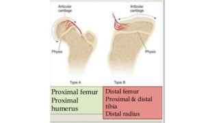
Epiphyseal injury
Importance
• The most predictable complication of epiphyseal injury is growth
disturbance
• And so could be prevented if diagnosed properly and managed
accordingly
• Each epiphysis has its own epiphysial plate through which skeletal
growth occurs
• So it is important that a distinction be made between the epiphysis
and epiphysial plate
Types of epiphysis
• Two types
• Pressure epiphysis
• Traction epiphysis
• Pressure epiphysis
• Location – at the end of a long bone
• Subjected to pressure transmitted through the joint
• Could be considered articular epiphysis
• Provides longitudinal growth of long bone
• Pressure epiphysis itself could be divided into two depending on whether
their nutrient vessels enter epiphysis directly like lower femoral epiphysis or
indirectly like upper femoral epiphysis
• Traction epiphysis
• Location – site of origin or insertion of major muscles or muscles group
• Subjected to traction rather than pressure
• Does not enter into joint formation , so extra articular
• Does not contribute to longitudinal growth of long bone
• Examples : trochanters of femur
• Normal epiphysial plate has four distinct layers
• Resting cells
• Proliferating cells
• Layer of hypertrophy
• Endochondral ossification
Space between cells filled with cartilage matrix ( intercellular substance)
Made up of collagen embedded with chondroitin sulphate
It is the inter cellular substance which provides strength to epiphysial plate
• Collagen fibres are arranged longitudinally in the matrix
• First two layers , matrix is abundant and the plate is strong
• In third layer ( hypertrophy layer ) matrix is scanty and so plate is
weak.
• It is layer of hypertrophy which is a commen site of epiphysial injury
Mechanism of nutrition in epiphysial plates
• Two separate systems of blood vessels
• Epiphysial system
• Metaphyseal system
•
• Epiphyseal system – vessels penetrate the plate of epiphysis and end in
capillary tufts or loops in a layer of resting cells
• Metaphyseal system – arises in marrow of shaft and ends in loops in layer
of endochondral ossification
• So the epiphysial system is responsible for nutrition of proliferating cells
• And metaphysial system is responsible for nutrition of endochondral
ossification
Etiology
• Trauma ( shearing / avulsion / crushing )
• Pathological separation of epiphysis ( rickets , scurvy , osteomyelitis )
• Iatrogenic damage to epiphysial plates
• Epiphysial injury mainly divided into two groups
• Fracture that cross the epiphysial plate
• Epiphysial separation
• Fracture crossing the epiphysial plate
• With or without longitudinal displacement
• When there is no displacement of fragment but fracture crossing the plate
will lead to premature closure of epiphysis and ultimately leads to shortening
• When displacement is present it will be combined with shortening and
angular deformity
• Epiphysial separation :
• Healing of epiphysis separation is rapid if there is no vascular damage
• Prognosis is not good if it damages nutrient vessels to epiphysis
• Such as avascular necrosis of femoral head in slipped capital femoral epiphysis
Classification of epiphysial plate injuries
• Salter Harris classification
• Type 1 : complete separation of epiphysis from metaphysis
• More common in birth injuries or pathological separation as in scurvy, rickets
• Type 2 : commonest type of epiphysial plate injuries
• Line of separation extends along the epiphyseal plate to a variable distance
and then out through a metaphysis
• Producing a triangular shaped metaphyseal fragment called Thurston Holland
sign
• Type 3: fracture which is intra articular extends from the joint surface to
the weak zone of epiphyseal plate and then extends along the plate to its
periphery
• Type 4 : fracture which is intra articular extends from the joint surface
through epiphysis across ful thickness of epiphyseal plate and through a
portion of metaphysis
• Producing a complete split
• Commonest example of this type is fracture of lateral condyle humerus
• Type 5
• Relatively uncommon injury
• Occurs due to severe crushing force applied through epiphysis to epiphyseal
plate
• More common in ankle and knee
• Injury might be misdiagnosed on xray
• Type 6 Injury by RANG
• Injury to perichondrial ring
• Type 7 Ogden – isolated epiphysis injury
• Type 8 – isolated injury to metaphysis
• Type 9 – avulsion injury to periosteum
Factors in prognosis
• Type of injury
• Type 1,2,3 have a good prognosis provided the intact blood supply
• Type 4 has a bad prognosis unless the plate is completely realigned
• Type 5 has the worst prognosis
• Age of the child
• Depends on the amount of growth expected in particular epiphyseal plate for
the remaining years of growth
• Younger the child , poor the prognosis
• Blood supply
• Examples femoral head necrosis
• Method of reduction :
• Undue forceful manipulation further damages the plate
• Mostly carried out after tenth day of injury
• Damage by instruments
• Screws or wires traversing the plate
• Open or closed injury :
• Open injuries of epiphysis uncommon
• But when associated , outcome is worst
Principles of treatment
• Time of reduction :
• Best time to reduce an epiphyseal plate injury is the day of injury
• Reduction becomes progressively more difficult with each passing day
• After 10 days , forceful manipulation is required and which may Further
damage the plate and so should be avoided
• And wiser to accept an imperfect reduction than to risk the danger of either
forceful manipulation or open reduction
• Method of reduction
• Closed or open
• Majority type 1&2 reduced by closed means
• Type 3 may require open reduction to obtain smooth joint surface
• Type 4 mostly requires open reduction always
•
• Follow up : growth disturbance may be delayed and so minimum of
one year follow up is must
• Common type of injury at upper radial epiphysis is type 2
• Produced by valgus force on the elbow , resulting in angulation of
radial head
• Reduction can be achieved by holding the extended elbow in varus
and directly pressing over the radial head
• If angulation is > 15° after closed reduction , supination – pronation
may get affected and open reduction is indicated
• Radial head should not be excised in children , if done loss of radius
growth wll produce progressive radial deviation at the wrist joint and
valgus deformity at the elbow
• Fracture of lateral condyle humerus (capitellum ) is type 4 injury
• Weekly x-rays needed to check the alignment
• When it’s displaced , open reduction is must
• Elbow is immobilised for 3 weeks in 90° flexion
• If not reduced properly – may lead to over growth of radial head /
valgus deformity / tardy ulnar nerve palsy
• Largest of pressure epiphysis – lower femoral epiphysis
• Serious injury of lower femur epiphysis is hyper extension type with
forward displacement of epiphysis
• Posterior corner of metaphysis may injur popliteal vessels
• Reduction is achieved by traction on semibent knee , pushing the
proximal fragment forward and then flexing the knee
• Reduction is most stable in 90° knee flexion
• Total period of immobilisation 3 weeks
Traction epiphysis injuries
• Medial epicondyle humerus injury : common flexor muscles avulsion
• Severe valgus injury
• Tibial tubercle : traction epiphysis of quadriceps
• Lesser trochanter : iliopsoas inserted to lesser tubercle
• Sudden abduction and extension of hip may result in lesser trochanteric
avulsion
• Anterior superior iliac spine – traction epiphysis of sartorius
• Anterior inferior iliac spine – traction epiphysis of rectus femoris
Thank you

Epiphyseal injuries

  • 1.
  • 2.
    Importance • The mostpredictable complication of epiphyseal injury is growth disturbance • And so could be prevented if diagnosed properly and managed accordingly
  • 3.
    • Each epiphysishas its own epiphysial plate through which skeletal growth occurs • So it is important that a distinction be made between the epiphysis and epiphysial plate
  • 4.
    Types of epiphysis •Two types • Pressure epiphysis • Traction epiphysis • Pressure epiphysis • Location – at the end of a long bone • Subjected to pressure transmitted through the joint • Could be considered articular epiphysis • Provides longitudinal growth of long bone • Pressure epiphysis itself could be divided into two depending on whether their nutrient vessels enter epiphysis directly like lower femoral epiphysis or indirectly like upper femoral epiphysis
  • 5.
    • Traction epiphysis •Location – site of origin or insertion of major muscles or muscles group • Subjected to traction rather than pressure • Does not enter into joint formation , so extra articular • Does not contribute to longitudinal growth of long bone • Examples : trochanters of femur
  • 8.
    • Normal epiphysialplate has four distinct layers • Resting cells • Proliferating cells • Layer of hypertrophy • Endochondral ossification Space between cells filled with cartilage matrix ( intercellular substance) Made up of collagen embedded with chondroitin sulphate It is the inter cellular substance which provides strength to epiphysial plate
  • 9.
    • Collagen fibresare arranged longitudinally in the matrix • First two layers , matrix is abundant and the plate is strong • In third layer ( hypertrophy layer ) matrix is scanty and so plate is weak. • It is layer of hypertrophy which is a commen site of epiphysial injury
  • 10.
    Mechanism of nutritionin epiphysial plates • Two separate systems of blood vessels • Epiphysial system • Metaphyseal system • • Epiphyseal system – vessels penetrate the plate of epiphysis and end in capillary tufts or loops in a layer of resting cells • Metaphyseal system – arises in marrow of shaft and ends in loops in layer of endochondral ossification • So the epiphysial system is responsible for nutrition of proliferating cells • And metaphysial system is responsible for nutrition of endochondral ossification
  • 12.
    Etiology • Trauma (shearing / avulsion / crushing ) • Pathological separation of epiphysis ( rickets , scurvy , osteomyelitis ) • Iatrogenic damage to epiphysial plates
  • 13.
    • Epiphysial injurymainly divided into two groups • Fracture that cross the epiphysial plate • Epiphysial separation • Fracture crossing the epiphysial plate • With or without longitudinal displacement • When there is no displacement of fragment but fracture crossing the plate will lead to premature closure of epiphysis and ultimately leads to shortening • When displacement is present it will be combined with shortening and angular deformity
  • 14.
    • Epiphysial separation: • Healing of epiphysis separation is rapid if there is no vascular damage • Prognosis is not good if it damages nutrient vessels to epiphysis • Such as avascular necrosis of femoral head in slipped capital femoral epiphysis
  • 15.
    Classification of epiphysialplate injuries • Salter Harris classification • Type 1 : complete separation of epiphysis from metaphysis • More common in birth injuries or pathological separation as in scurvy, rickets • Type 2 : commonest type of epiphysial plate injuries • Line of separation extends along the epiphyseal plate to a variable distance and then out through a metaphysis • Producing a triangular shaped metaphyseal fragment called Thurston Holland sign
  • 17.
    • Type 3:fracture which is intra articular extends from the joint surface to the weak zone of epiphyseal plate and then extends along the plate to its periphery • Type 4 : fracture which is intra articular extends from the joint surface through epiphysis across ful thickness of epiphyseal plate and through a portion of metaphysis • Producing a complete split • Commonest example of this type is fracture of lateral condyle humerus
  • 18.
    • Type 5 •Relatively uncommon injury • Occurs due to severe crushing force applied through epiphysis to epiphyseal plate • More common in ankle and knee • Injury might be misdiagnosed on xray
  • 19.
    • Type 6Injury by RANG • Injury to perichondrial ring • Type 7 Ogden – isolated epiphysis injury • Type 8 – isolated injury to metaphysis • Type 9 – avulsion injury to periosteum
  • 20.
    Factors in prognosis •Type of injury • Type 1,2,3 have a good prognosis provided the intact blood supply • Type 4 has a bad prognosis unless the plate is completely realigned • Type 5 has the worst prognosis • Age of the child • Depends on the amount of growth expected in particular epiphyseal plate for the remaining years of growth • Younger the child , poor the prognosis
  • 21.
    • Blood supply •Examples femoral head necrosis • Method of reduction : • Undue forceful manipulation further damages the plate • Mostly carried out after tenth day of injury • Damage by instruments • Screws or wires traversing the plate
  • 22.
    • Open orclosed injury : • Open injuries of epiphysis uncommon • But when associated , outcome is worst
  • 23.
    Principles of treatment •Time of reduction : • Best time to reduce an epiphyseal plate injury is the day of injury • Reduction becomes progressively more difficult with each passing day • After 10 days , forceful manipulation is required and which may Further damage the plate and so should be avoided • And wiser to accept an imperfect reduction than to risk the danger of either forceful manipulation or open reduction
  • 24.
    • Method ofreduction • Closed or open • Majority type 1&2 reduced by closed means • Type 3 may require open reduction to obtain smooth joint surface • Type 4 mostly requires open reduction always • • Follow up : growth disturbance may be delayed and so minimum of one year follow up is must
  • 26.
    • Common typeof injury at upper radial epiphysis is type 2 • Produced by valgus force on the elbow , resulting in angulation of radial head • Reduction can be achieved by holding the extended elbow in varus and directly pressing over the radial head • If angulation is > 15° after closed reduction , supination – pronation may get affected and open reduction is indicated • Radial head should not be excised in children , if done loss of radius growth wll produce progressive radial deviation at the wrist joint and valgus deformity at the elbow
  • 28.
    • Fracture oflateral condyle humerus (capitellum ) is type 4 injury • Weekly x-rays needed to check the alignment • When it’s displaced , open reduction is must • Elbow is immobilised for 3 weeks in 90° flexion • If not reduced properly – may lead to over growth of radial head / valgus deformity / tardy ulnar nerve palsy
  • 30.
    • Largest ofpressure epiphysis – lower femoral epiphysis • Serious injury of lower femur epiphysis is hyper extension type with forward displacement of epiphysis • Posterior corner of metaphysis may injur popliteal vessels • Reduction is achieved by traction on semibent knee , pushing the proximal fragment forward and then flexing the knee • Reduction is most stable in 90° knee flexion • Total period of immobilisation 3 weeks
  • 31.
    Traction epiphysis injuries •Medial epicondyle humerus injury : common flexor muscles avulsion • Severe valgus injury • Tibial tubercle : traction epiphysis of quadriceps • Lesser trochanter : iliopsoas inserted to lesser tubercle • Sudden abduction and extension of hip may result in lesser trochanteric avulsion • Anterior superior iliac spine – traction epiphysis of sartorius • Anterior inferior iliac spine – traction epiphysis of rectus femoris
  • 32.